



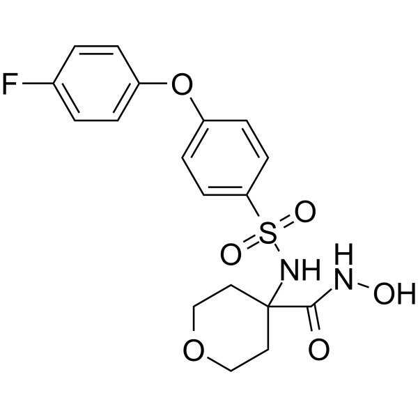
CP-544439
CP-544439 is a potent and orally active matrix metalloproteinase-13 (MMP-13) inhibitor with an IC50 of 0.75 nM.
Nur für Forschungszwecke. Wir verkaufen nicht an Patienten.
- CAS. Nr.: 230954-09-3
- Formel: C18H19FN2O6S
- Molecular Weight:410.42
-
Speicherung:
Please store the product under the recommended conditions in the Certificate of Analysis.

To place orders, for customer services and technical support, please contact: MedChemExpress USA
Tel: 609-228-6898 E-mail: [email protected] [email protected]


-
 Biologische Aktivität
-
Chemical Information
- Protokoll
- Reinheit & Dokumentation
- Verweise
-
 Help & FAQs




Biologische Aktivität
Beschreibung
|
MMP-13 0.75 nM (IC50) |
MMP-12 0.24 nM (IC50) |
MMP-1 420 nM (IC50) |
MMP-2 1.6 nM (IC50) |
MMP-3 16 nM (IC50) |
MMP-8 1.4 nM (IC50) |
MMP-9 12 nM (IC50) |
MMP-14 7.4 nM (IC50) |
In Vivo
MedChemExpress (MCE) has not independently confirmed the accuracy of these methods. They are for reference only.


Chemical Information
-
CAS. Nr. 230954-09-3
-
Molecular Weight 410.42
-
Formel C18H19FN2O6S
-
SMILES
O=C(NO)C1(CCOCC1)NS(=O)(C2=CC=C(C=C2)OC3=CC=C(C=C3)F)=O
-
Versand
Room temperature in continental US; may vary elsewhere.
-
Speicherung
Please store the product under the recommended conditions in the Certificate of Analysis.


Reinheit & Dokumentation
Verweise
[1]. Dalvie D, et al. Metabolism distribution and excretion of a matrix metalloproteinase-13 inhibitor, 4-[4-(4-fluorophenoxy)-benzenesulfonylamino]tetrahydropyran-4-carboxylic acid hydroxyamide (CP-544439), in rats and dogs: assessment of the metabolic profil [Content Brief]
[2]. Reiter LA, et al. Pyran-containing sulfonamide hydroxamic acids: potent MMP inhibitors that spare MMP-1. Bioorg Med Chem Lett. 2004;14(13):3389-3395. [Content Brief]

Calculators
Konzentration (Stammlösung) × Volumen (Stammlösung) = Konzentration (Ziellösung) × Volumen (Ziellösung)


CP-544439 Related Classifications
HY-107013 Related Classifications
 Powered by Bioz