



Collagen, Type I, from bovine achilles tendon
Based on 1 publication(s) in Google Scholar
Collagen, Type I, from bovine achilles tendon are extracellular matrix proteins and show many applications in food, cosmetic, pharmaceutical and biomedical industries. This product is enzymatically hydrolyzed collagen peptide.
For research use only. We do not sell to patients.
- CAS No.: 9007-34-5
-
Storage:
Please store the product under the recommended conditions in the Certificate of Analysis.

To place orders, for customer services and technical support, please contact: MedChemExpress USA
Tel: 609-228-6898 E-mail: [email protected] [email protected]


-
 Biological Activity
-
Chemical Information
-
Solvent & Solubility
- Protocols
- Purity & Documentation
- References
-
 Help & FAQs


-
HY-D0842
BSA ProteinGlobular Protein -

HY-10219
RapamycinmTOR Inhibitor -

HY-15371
ForskolinAdenylate Cyclase Activator -
HY-P3160
FibronectinTissue Injury Biomarker -

HY-NP010
Type I Collagen, from rat tailCollagen -
HY-NP003
Collagen, Type II, from bovine snout membraneType II Collagen -

HY-10071
Y-27632ROCK Inhibitor -

HY-10182
LaduviglusibGSK-3 Inhibitor -
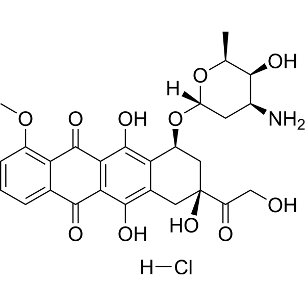
HY-15142
Doxorubicin hydrochlorideTopoisomerase Inhibitor -
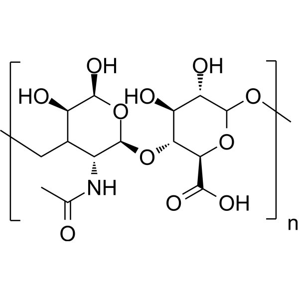
HY-B0633A
Hyaluronic acidGlycosaminoglycan -
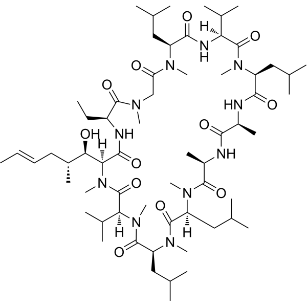
HY-B0579
Cyclosporin ACalcineurin Inhibitor -
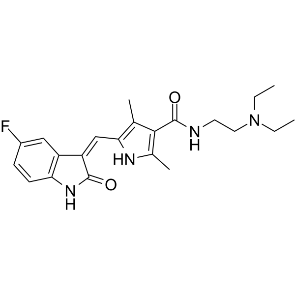
HY-10255A
SunitinibMulti-targeted RTK Inhibitor
Publications Citing Use of MedChemExpress (MCE) Collagen, Type I, from bovine achilles tendon
More

Biological Activity
Description
In Vivo
Collagen (hydrolyzed form, 3.8-38 mg/150 mg mixture would, p.o., daily, 7-16 weeks) shows chondroprotective and anti-inflammatory effects in murine posttraumatic osteoarthritis[3].
MedChemExpress (MCE) has not independently confirmed the accuracy of these methods. They are for reference only.


Chemical Information
-
CAS No. 9007-34-5
-
Appearance Solid
-
Color White to light yellow
-
SMILES
[Collagen, Type I, from bovine achilles tendon]
-
Shipping
Room temperature in continental US; may vary elsewhere.
-
Storage
Please store the product under the recommended conditions in the Certificate of Analysis.


Publications (1)
-
Journal Impact Factor
-
Most Recent


Solvent & Solubility
In Vitro:
H2O : ≥ 100 mg/mL
* "≥" means soluble, but saturation unknown.


Purity & Documentation
-
Data Sheet (264 KB)
-
SDS (393 KB)
- English - EN (393 KB)
- Français - FR (393 KB)
- Deutsch - DE (393 KB)
- Norwegian - NO (393 KB)
- Español - ES (393 KB)
- Swedish - SV (393 KB)
- Italian - IT (393 KB)
- Korean - KR (393 KB)
- Portuguese - PT (393 KB)
-
Handling Instructions (2659 KB)
References
[1]. Sorushanova A, et al. The Collagen Suprafamily: From Biosynthesis to Advanced Biomaterial Development. Adv Mater. 2019 Jan;31(1):e1801651. [Content Brief]
[2]. Guillerminet F, et al. Hydrolyzed collagen improves bone status and prevents bone loss in ovariectomized C3H/HeN mice. Osteoporos Int. 2012 Jul;23(7):1909-19. [Content Brief]
[3]. Dar QA, et al. Daily oral consumption of hydrolyzed type 1 collagen is chondroprotective and anti-inflammatory in murine posttraumatic osteoarthritis. PLoS One. 2017 Apr 6;12(4):e0174705. [Content Brief]

Calculators
Concentration (start) × Volume (start) = Concentration (final) × Volume (final)


Collagen, Type I, from bovine achilles tendon Related Classifications
HY-P2738 Related Classifications
 Powered by Bioz