



FHD-609
Based on 3 publication(s) in Google Scholar
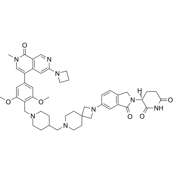
FHD-609 is a BRD9 PROTAC degrader. FHD-609 induces BRD9 degradation, downregulates primary transcripts of HBG/HBD, Myc expression and proliferation gene sets, reduces the levels of synovial sarcoma tumor proliferation markers, and inhibits tumor growth. FHD-609 can be used in research related to advanced synovial sarcoma, SMARCB1-deficient tumors and adrenocortical carcinoma.
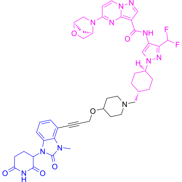
(Pink: BRD9 ligand (HY-49393); Blue: Cereblon ligand (HY-168308); Black: linker (HY-168309)).
For research use only. We do not sell to patients.
- Purity : 98.95%
- CAS No.: 2676211-64-4
- Formula: C47H56N8O6
- Molecular Weight:829.00
-
Storage:Powder -20°C, 3 years , 4°C, 2 years ; In solvent -80°C, 6 months , -20°C, 1 month

To place orders, for customer services and technical support, please contact: MedChemExpress USA
Tel: 609-228-6898 E-mail: [email protected] [email protected]


-
 Biological Activity
-
Chemical Information
-
Solvent & Solubility
- Protocols
- Purity & Documentation
- References
-
 Help & FAQs


-
HY-145483
KT-474PROTAC IRAK4 Degrader -
HY-158101
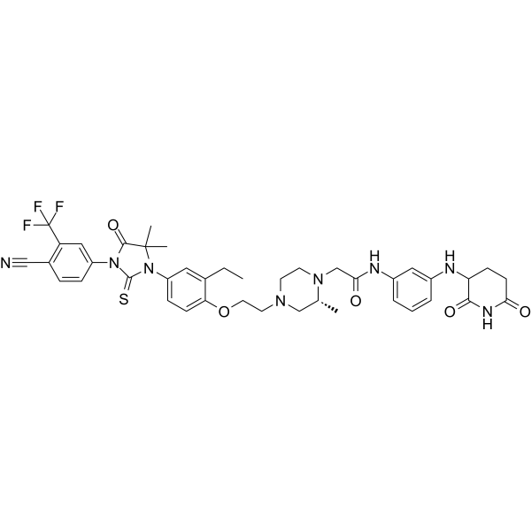
BMS-986365PROTAC AR Degrader -
HY-18975
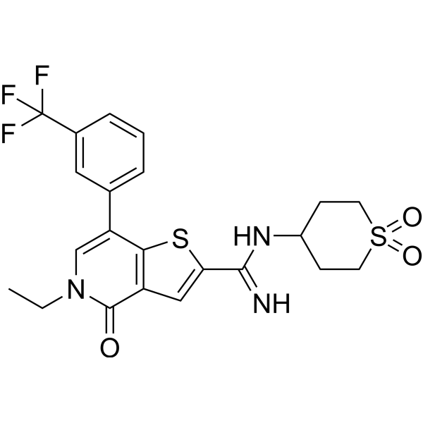
I-BRD9Chemical Probe -
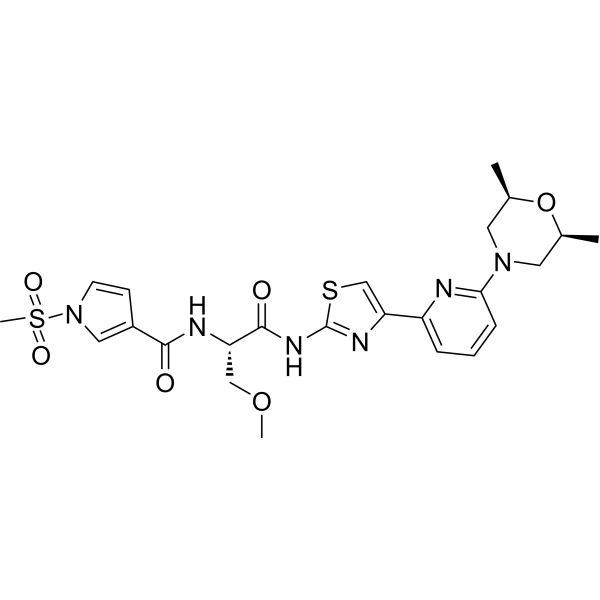
HY-144835
CamibirstatBRG1/BRM ATPase Inhibitor -
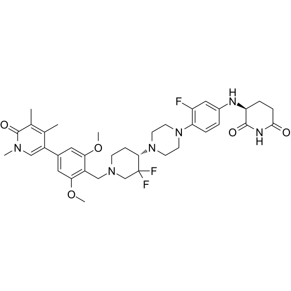
HY-145925B
CFT8634BRD9 PROTAC Degrader -
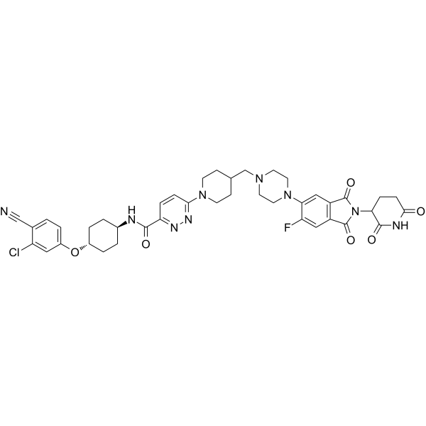
HY-138641
BavdegalutamideAR PROTAC Degrader -
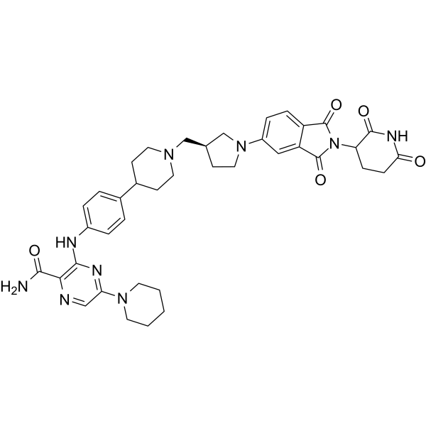
HY-153220
ZelebrudomideBTK PROTAC Degrader -
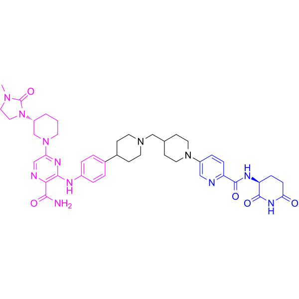
HY-153321
BexobrutidegBTK Degrader -
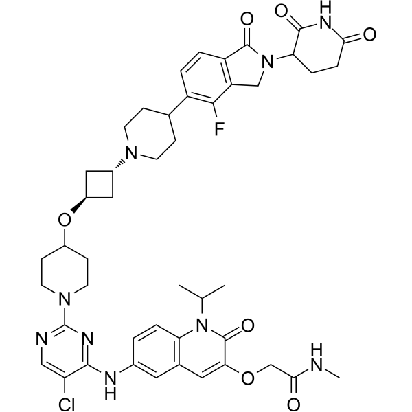
HY-158105
ARV-393BCL6 PROTAC Degrader -
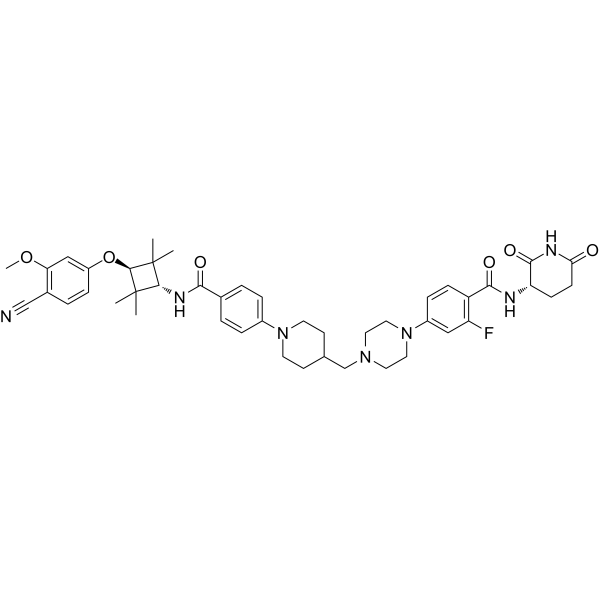
HY-153342
LuxdegalutamidePROTAC -
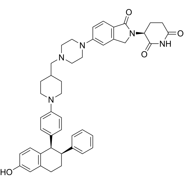
HY-138642
VepdegestrantPROTAC ER Degrader -
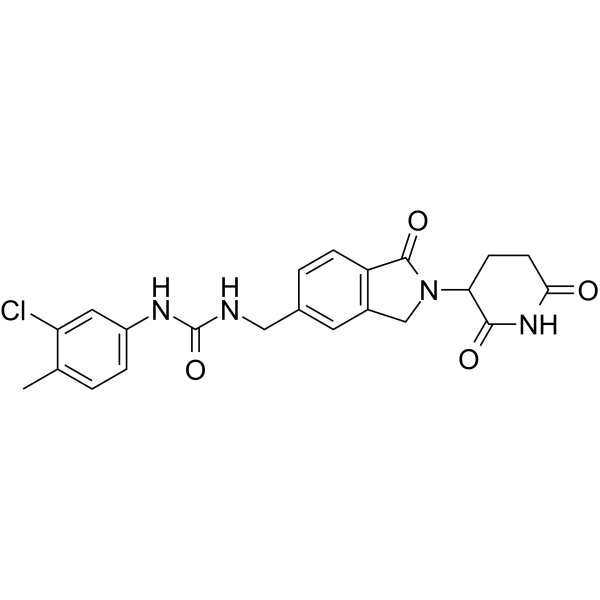
HY-101488
CC-885CRBN Modulator/GSPT1 Molecular Glue Degrader
Publications Citing Use of MedChemExpress (MCE) FHD-609
More Customer Validation & Images

-
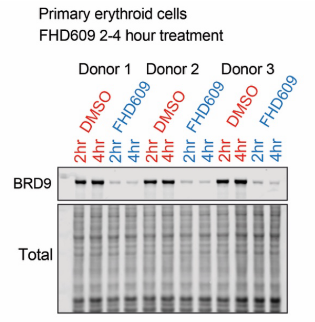
WB
-
RT-PCR
All PROTACs Isoforms
More

Biological Activity
Description
|
BRD9 |
HBG |
HBD |
In Vitro
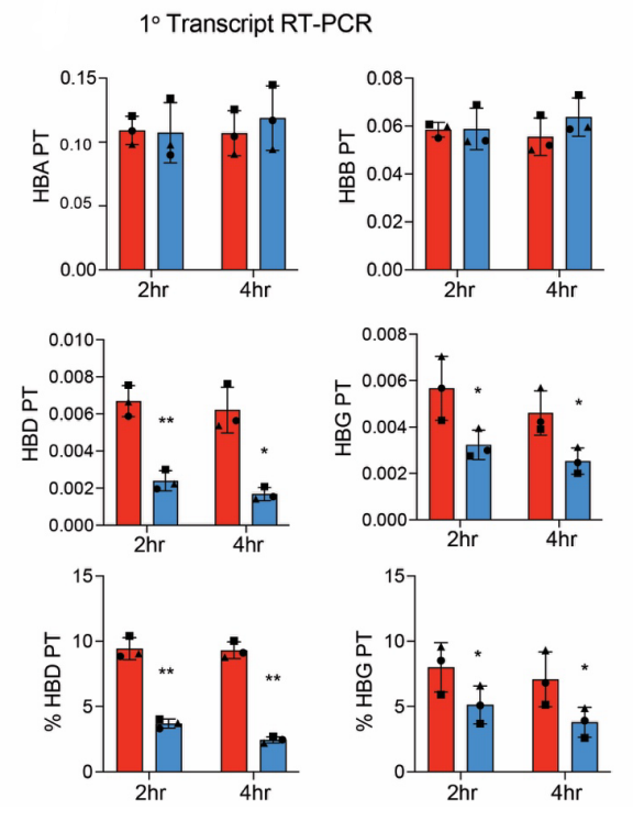
Acute treatment of primary human erythroid cultures with FHD-609 (10 nM; 2 h) directly reduces the ratio of HBD and HBG primary transcripts[1].
FHD-609 (5-500 nM; 4 h) dose-dependently reduces the relative mean fluorescence intensity of BRD9 in human peripheral blood mononuclear cells[4].
FHD-609 (0.5 μM; up to 8 h) has an epimerization half-life of 3.7 h in 0.1 M deuterated phosphate buffer at 37 °C, and it tends to adopt a linear conformation[5].
MedChemExpress (MCE) has not independently confirmed the accuracy of these methods. They are for reference only. Further protocols information, click here.
In Vivo
FHD-609 (0.1-3 mg/kg; intravenous injection; single administration once every two weeks for a total of 5 times) induces rapid, dose-dependent degradation of BRD9 in PBMC immune cell subsets of CD-1 mice, and BRD9 levels gradually recover over time after the final administration[4].
FHD-609 (intravenous injection; single dose, once every two weeks for a total of 3 doses, once every two weeks for a total of 5 doses, dosage 0.1-3 mg/kg) dose-dependently reduces the expression level of MYC in SYO-1 CDX tumors, downregulates the Myc target gene set, and decreases proliferation markers (c-Myc, Ki67), while the expression levels of SSX-SS18 and TLE1 remain unchanged[4].
MedChemExpress (MCE) has not independently confirmed the accuracy of these methods. They are for reference only.
-
Animal Model:BALB/c nude (immunodeficient; SYO-1 cell line-derived xenograft model)[4]
-
Dosage:Potent antitumor activity (3.5 mg/kg; 0.7 mg/kg; 0.5 mg/kg)
Dose-dependent downregulation of MYC expression (0.1 mg/kg; 3 mg/kg) -
Administration:Potent antitumor activity (i.p.; weekly (3.5 mg/kg, 0.7 mg/kg); daily (0.5 mg/kg); 25 days)
Dose-dependent downregulation of MYC expression (i.v.; single dose, 3 biweekly doses, 5 biweekly doses) -
Result:Reduced tumor volume to <500 mm3 at 25 days post-treatment initiation (3.5 mg/kg i.p. weekly).
Reduced tumor volume to ~1000 mm3 at 25 days post-treatment initiation (0.7 mg/kg i.p. weekly).
Reduced tumor volume to <500 mm3 at 25 days post-treatment initiation (0.5 mg/kg i.p. daily).
Induced complete BRD9 degradation in tumor lysates collected 4 hours after the last dose (0.7 mg/kg or 3.5 mg/kg weekly).
Induced partial recovery of BRD9 levels in tumor lysates collected 7 days after the last dose (0.7 mg/kg or 3.5 mg/kg weekly).
Reduced MYC expression log2 fold-change relative to vehicle by -0.25 (0.1 mg/kg single dose), -0.73 (3 mg/kg single dose), -0.62 (0.1 mg/kg 3 doses), -1.02 (3 mg/kg 3 doses), -0.78 (0.1 mg/kg 5 doses), and -1.50 (3 mg/kg 5 doses), all with FDR-adjusted p-values ≤10-4.
Downregulated HALLMARK_MYC_TARGETS_V1 gene set with normalized enrichment scores (NES) ranging from -3.46 to -3.02 across all treatment groups.
Reduced c-Myc H-score to 143 (0.1 mg/kg 5 doses) and 102 (3 mg/kg 5 doses) compared to vehicle control H-score of 161.
Reduced Ki67 H-score to 148 (0.1 mg/kg 5 doses) and 118 (3 mg/kg 5 doses) compared to vehicle control H-score of 183.
Induced near-complete loss of BRD9 IHC staining in tumors treated with 3 mg/kg 5 doses.
Left SSX-SS18 and TLE1 staining unchanged relative to vehicle.
-
Animal Model:CD-1 (immunocompetent; naïve)[4]
-
Dosage:0.1 mg/kg; 3 mg/kg
-
Administration:i.v.; single dose, 5 biweekly doses
-
Result:Reduced relative BRD9 mean fluorescence intensity (MFI) normalized to vehicle to ~0 in B cells by 3 hours post-dose (0.1 mg/kg single dose), recovered to ~0.2 by 72 hours, and returned to vehicle levels by 504 hours.
Reduced relative BRD9 MFI normalized to vehicle to ~0 in B cells by 3 hours post-last dose (0.1 mg/kg 5 BIW doses), recovered to ~0.2 by 72 hours, and returned to vehicle levels by 504 hours.
Reduced relative BRD9 MFI normalized to vehicle to ~0 in B cells by 3 hours post-dose (3 mg/kg single dose), remained suppressed through 72 hours, and returned to vehicle levels by 504 hours.
Reduced relative BRD9 MFI normalized to vehicle to ~0 in B cells by 3 hours post-last dose (3 mg/kg 5 BIW doses), remained suppressed through 72 hours, was still ~0 at 168 hours post-last dose, and returned to vehicle levels by 504 hours.
Induced similar patterns of BRD9 degradation and recovery across B cells, CD4+ T cells, CD8+ T cells, monocytes, and NK cells.
Clinical Trial
| NCT Number | Sponsor | Condition | Start Date |
Phase

|
|---|---|---|---|---|
| NCT01329991 | Plexxikon| | 2011-05 | PHASE1 |


Chemical Information
-
CAS No. 2676211-64-4
-
Appearance Solid
-
Molecular Weight 829.00
-
Formula C47H56N8O6
-
Color White to off-white
-
SMILES
COC1=C(CN2CCC(CN3CCC4(CN(C5=CC(C(N([C@@H]6C(NC(CC6)=O)=O)C7)=O)=C7C=C5)C4)CC3)CC2)C(OC)=CC(C(C8=C9C=NC(N%10CCC%10)=C8)=CN(C)C9=O)=C1
-
Shipping
Room temperature in continental US; may vary elsewhere.
-
Storage
Powder -20°C 3 years 4°C 2 years In solvent -80°C 6 months -20°C 1 month


Publications (3)
-
Journal Impact Factor
-
Most Recent
-
Blood
Highly discriminative globin gene activation by the non-canonical BAF chromatin remodeling complex. [Abstract]2025 Nov 14:blood.2025029429. PMID: 41237357
FHD-609 purchased from MedChemExpress. Usage Cited in: Blood. 2025 Nov 14:blood.2025029429. [Abstract]
FHD-609 (10 nM; 2-4 h) degraded BRD9 in primary erythroid cultures.
FHD-609 purchased from MedChemExpress. Usage Cited in: Blood. 2025 Nov 14:blood.2025029429. [Abstract]
FHD-609 (10 nM; 2-4 h) decreased HBD and HBG primary transcripts in primary erythroid cells.
-
Anal Chem
Hydrogen/Deuterium Exchange for Chiral Stability Assessment in Acidic Methine-Containing Compounds. [Abstract]2025 Dec 2;97(47):26097-26107. PMID: 41243541 -


Solvent & Solubility
In Vitro:
DMSO : 100 mg/mL (120.63 mM; Need ultrasonic; Hygroscopic DMSO has a significant impact on the solubility of product, please use newly opened DMSO)
Please refer to the solubility information to select the appropriate solvent. Once prepared, please aliquot and store the solution to prevent product inactivation from repeated freeze-thaw cycles.
Storage method and period of stock solution: -80°C, 6 months; -20°C, 1 month. When stored at -80°C, please use it within 6 months. When stored at -20°C, please use it within 1 month.
Please refer to the solubility information to select the appropriate solvent. Once prepared, please aliquot and store the solution to prevent product inactivation from repeated freeze-thaw cycles.
Storage method and period of stock solution: -80°C, 6 months; -20°C, 1 month. When stored at -80°C, please use it within 6 months. When stored at -20°C, please use it within 1 month.
Concentration (start) × Volume (start) = Concentration (final) × Volume (final)
In Vivo:
Select the appropriate dissolution method based on your experimental animal and administration route.
- For the following dissolution methods, please ensure to first prepare a clear stock solution using an In Vitro approach and then sequentially add co-solvents:
- To ensure reliable experimental results, the clarified stock solution can be appropriately stored based on storage conditions. As for the working solution for In Vivo experiments, it is recommended to prepare freshly and use it on the same day.
- The percentages shown for the solvents indicate their volumetric ratio in the final prepared solution. If precipitation or phase separation occurs during preparation, heat and/or sonication can be used to aid dissolution.
Add each solvent one by one: 10% DMSO 90% (20% SBE-β-CD in Saline)
Solubility: ≥ 2.5 mg/mL (3.02 mM); Clear solution
This protocol yields a clear solution of ≥ 2.5 mg/mL (saturation unknown).
Taking 1 mL working solution as an example, add 100 μL DMSO stock solution (25.0 mg/mL) to 900 μL 20% SBE-β-CD in Saline, and mix evenly.
Preparation of 20% SBE-β-CD in Saline (4°C, storage for one week): 2 g SBE-β-CD powder is dissolved in 10 mL Saline, completely dissolve until clear.
In Vivo Dissolution Calculator
Please enter the basic information of animal experiments:
-
-
-
-
Recommended: Prepare an additional quantity of animals to account for potential losses during experiments.
Please enter your animal formula composition:
-
%DMSO +
Recommended: Keep the proportion of DMSO in working solution below 2% if your animal is weak.
-
%+
-
+%Tween-80 + +
-
%Saline +
The co-solvents required include: DMSO, . All of co-solvents are available by MedChemExpress (MCE). , Tween 80. All of co-solvents are available by MedChemExpress (MCE).
Working solution concentration: 0.22 mg/mL
Method for preparing stock solution: mg drug dissolved in μL DMSO. Stock solution concentration: mg/mL.
1. Take μL DMSO stock solution;
2. Add μL .
μL , mix evenly;
3. Then add μL Tween 80, mix evenly;
4. Then add μL
Please ensure that the stock solution in the first step is dissolved to a clear state, and add co-solvents in sequence. You can use ultrasonic heating (ultrasonic cleaner, recommended frequency 20-40 kHz), vortexing, etc. to assist dissolution.

Protocols
-
RNA extraction experimental
By lysing cells, releasing RNA, and removing impurities such as proteins and DNA, high-purity RNA products are finally obtained. The commonly used traditional method is the guanidine isothiocyanate/phenol/chloroform method (Trizol), which is suitable for a variety of animal materials including animal tissues, microorganisms, cultured cells, etc., and most plant materials.
-
Patient-Derived Xenograft (PDX)
Patient-derived xenograft (PDX) models are generated by engrafting primary human tumor tissue directly into immunodeficient mice, allowing in vivo propagation of patient tumor biology without initial in vitro adaptation. These models are used to preserve key histopathological and molecular characteristics of the original tumor and enable assessment of tumor growth dynamics and therapeutic response in a living organism. The biological readout is tumor engraftment and subsequent growth in the murine host, which reflects the ability of human tumor cells to survive, vascularize, and expand in an immunocompromised microenvironment.
-
Patient-Derived Orthotopic Xenograft (PDOX)
Patient-derived orthotopic xenograft (PDOX) modeling implants fresh patient tumor tissue or patient-derived tumor cells into the anatomically corresponding organ or tissue site of immunodeficient mice, usually by surgical orthotopic implantation, to preserve patient tumor histology, local microenvironmental context, invasion, metastatic behavior, and treatment-response features better than subcutaneous implantation. PDOX readouts include tumor engraftment, orthotopic tumor growth, local invasion, metastasis, recurrence after resection, histologic similarity to the donor tumor, biomarker retention, molecular concordance, survival, and response or resistance to therapy. PDOX models are used for preclinical drug testing and individualized therapy evaluation, but engraftment success varies by tumor type and specimen quality.
-
Subcutaneous Cell-Line-Derived Xenograft
Subcutaneous cell-line-derived xenograft (CDX) models are established by implanting cultured human cancer cell lines into immunodeficient mice, where the injected cells form localized tumors that can be monitored in vivo as a measure of tumorigenic potential, growth kinetics, and treatment response. These models are widely used in oncology research because they allow reproducible tumor formation and enable comparative assessment of tumor growth between different cell lines or genetic manipulations in a controlled in vivo microenvironment. Subcutaneous implantation of cancer cells in immunodeficient mice is a standard approach for evaluating tumor growth behavior and therapeutic response across multiple cancer types, including prostate, esophageal, pancreatic, and colon cancer models.
-
Orthotopic Cell-Line Xenograft
Orthotopic cell-line xenograft models involve implantation of human cancer cell lines into the anatomically corresponding organ of immunodeficient mice to reproduce tumor growth within a native microenvironment, enabling more clinically relevant tumor behavior compared with subcutaneous models. These models are widely used because orthotopic placement better recapitulates tumor progression, including invasion and metastatic spread, which are often underrepresented in heterotopic implantation systems. Compared with conventional xenografts, orthotopic implantation is described as more technically complex but provides improved simulation of tumor-microenvironment interactions and metastatic behavior, making it particularly valuable for translational oncology research. Surgical orthotopic implantation approaches have been emphasized as enabling faithful reproduction of clinical cancer features, including metastasis and disease progression patterns that align with the tumor’s organ of origi
-
Cell Viability Determination by MTT Colorimetric Assay
The following protocol uses the MTT colorimetric assay as a classic literature-established method for assessing cell viability/metabolic activity in cultured mammalian cells. MTT[3-(4,5-dimethylthiazol-2-yl)-2,5-diphenyltetrazolium bromide] is reduced by metabolically active cells to a colored formazan product; the amount of formazan is quantified spectrophotometrically and provides an indirect measure of metabolically active viable cells. Importantly, MTT reduction reflects cellular oxidoreductase/metabolic activity rather than an absolute direct count of living cells, so changes in cellular metabolism can alter the signal independently of cell number.


Purity & Documentation
-
Data Sheet (281 KB)
-
SDS (254 KB)
- English - EN (254 KB)
- Français - FR (254 KB)
- Deutsch - DE (254 KB)
- Norwegian - NO (254 KB)
- Español - ES (254 KB)
- Swedish - SV (254 KB)
- Italian - IT (254 KB)
- Korean - KR (254 KB)
- Portuguese - PT (254 KB)
-
Handling Instructions (2659 KB)
References
[1]. Bagchi A, et al. Highly discriminative globin gene activation by the noncanonical BAF chromatin remodeling complex. Blood. 2026 Feb 5;147(6):675-688. [Content Brief]
[2]. Livingston JA, et al. A Phase I Study of FHD-609, a Heterobifunctional Degrader of Bromodomain-Containing Protein 9, in Patients with Advanced Synovial Sarcoma or SMARCB1-Deficient Tumors. Clinical cancer research : an official journal of the American Association for Cancer Research. 2025 Feb 17;31(4):628-638. [Content Brief]
[5]. Yu ZJ, et al. Hydrogen/Deuterium Exchange for Chiral Stability Assessment in Acidic Methine-Containing Compounds. Analytical chemistry. 2025 Dec 02;97(47):26097-26107. [Content Brief]
[6]. Hescheler DA, et al. Targeted Therapy for Adrenocortical Carcinoma: A Genomic-Based Search for Available and Emerging Options. Cancers. 2022 May 31;14(11):2721. [Content Brief]


Complete Stock Solution Preparation Table
Please refer to the solubility information to select the appropriate solvent. Once prepared, please aliquot and store the solution to prevent product inactivation from repeated freeze-thaw cycles.
Storage method and period of stock solution: -80°C, 6 months; -20°C, 1 month. When stored at -80°C, please use it within 6 months. When stored at -20°C, please use it within 1 month.
| Optional Solvent | Concentration Solvent Mass | 1 mg | 5 mg | 10 mg | 25 mg |
|---|---|---|---|---|---|
| DMSO | 1 mM | 1.2063 mL | 6.0314 mL | 12.0627 mL | 30.1568 mL |
| 5 mM | 0.2413 mL | 1.2063 mL | 2.4125 mL | 6.0314 mL | |
| 10 mM | 0.1206 mL | 0.6031 mL | 1.2063 mL | 3.0157 mL | |
| 15 mM | 0.0804 mL | 0.4021 mL | 0.8042 mL | 2.0105 mL | |
| 20 mM | 0.0603 mL | 0.3016 mL | 0.6031 mL | 1.5078 mL | |
| 25 mM | 0.0483 mL | 0.2413 mL | 0.4825 mL | 1.2063 mL | |
| 30 mM | 0.0402 mL | 0.2010 mL | 0.4021 mL | 1.0052 mL | |
| 40 mM | 0.0302 mL | 0.1508 mL | 0.3016 mL | 0.7539 mL | |
| 50 mM | 0.0241 mL | 0.1206 mL | 0.2413 mL | 0.6031 mL | |
| 60 mM | 0.0201 mL | 0.1005 mL | 0.2010 mL | 0.5026 mL | |
| 80 mM | 0.0151 mL | 0.0754 mL | 0.1508 mL | 0.3770 mL | |
| 100 mM | 0.0121 mL | 0.0603 mL | 0.1206 mL | 0.3016 mL |


FHD-609 Related Classifications
HY-153367 Related Classifications
 Powered by Bioz