



Talmapimod
Based on 3 publication(s) in Google Scholar
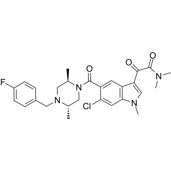
Talmapimod (SCIO-469) is an orally active and selective inhibitor of p38α MAPK with an IC50 of 9 nM. Talmapimod inhibits the secretion of inflammatory factors (such as TNFα, IL-1β, IL-6, and VEGF) by suppressing the p38α MAPK pathway, and it also inhibits angiogenesis and osteoclast activation. Talmapimod inhibits the growth of multiple myeloma cells and induces apoptosis. Talmapimod can be used to study various hematological malignancies (such as multiple myeloma, myelodysplastic syndrome).
Para uso exclusivo en investigación. No vendemos a pacientes.
- Pureza : 98.68%
- No. CAS: 309913-83-5
- Fòrmula: C27H30ClFN4O3
- Peso molecular:513.00
-
Almacenamiento:Powder -20°C, 3 years , 4°C, 2 years ; In solvent -80°C, 2 years , -20°C, 1 year

To place orders, for customer services and technical support, please contact: MedChemExpress USA
Tel: 609-228-6898 E-mail: [email protected] [email protected]


-
 Actividad biológica
-
Chemical Information
-
Solvente y solubilidad
- Protocolo
- Pureza y Documentación
- Referencias
-
 Help & FAQs


-
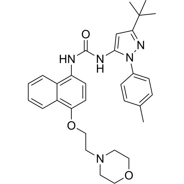
HY-10320
Doramapimodp38 MAPK Inhibitor -
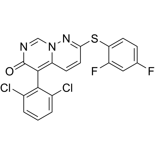
HY-10328
Neflamapimodp38α Inhibitor -
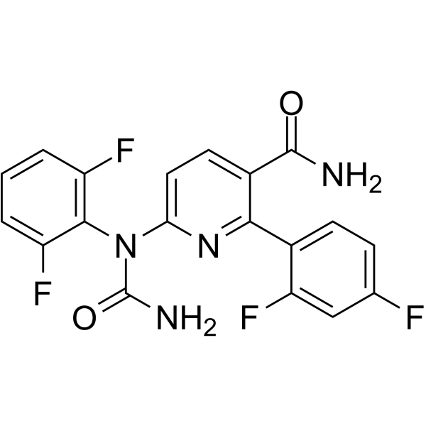
HY-10401
VX-702p38α MAPK Inhibitor -
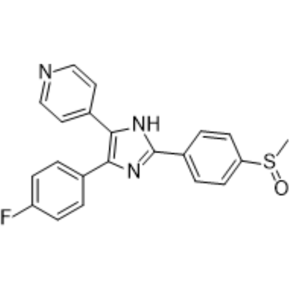
HY-10256
Adezmapimodp38 MAPK Inhibitor -
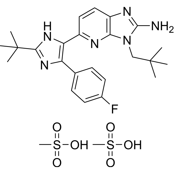
HY-13241
Ralimetinib dimesylateApoptosis Inducer -
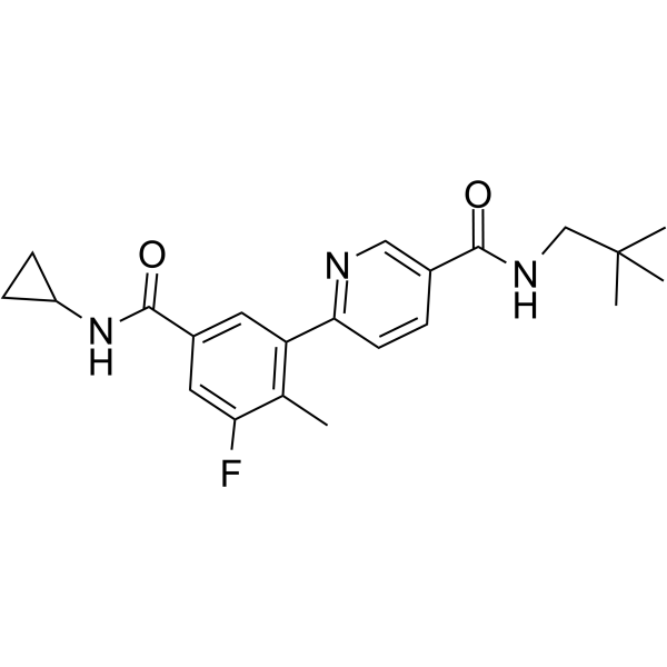
HY-10402
Losmapimodp38α/β MAPK Inhibitor -
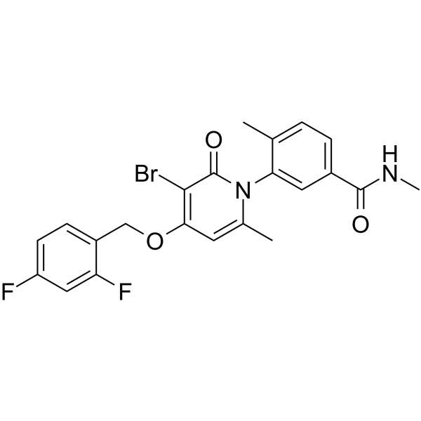
HY-10403
PH-797804p38α/β Inhibitor -
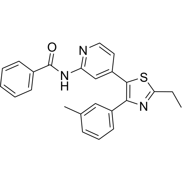
HY-10456
TAK-715p38 MAPK Inhibitor -
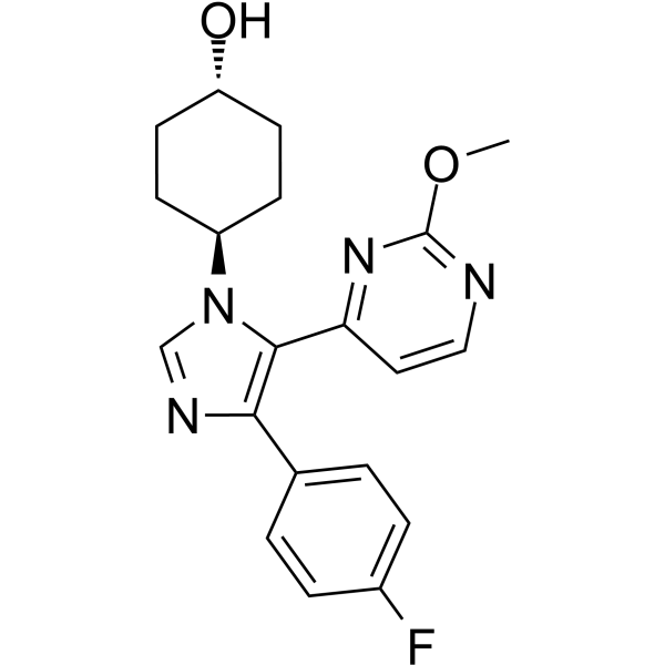
HY-11068
SB 239063p38 MAPK Inhibitor -
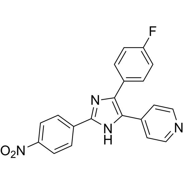
HY-10578
PD 169316p38 MAP Kinase Inhibitor -
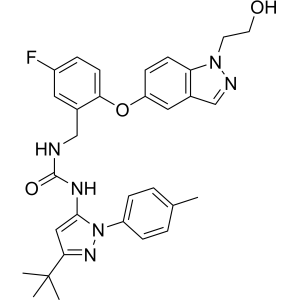
HY-16782
PexmetinibTie-2/p38 MAPK Inhibitor -
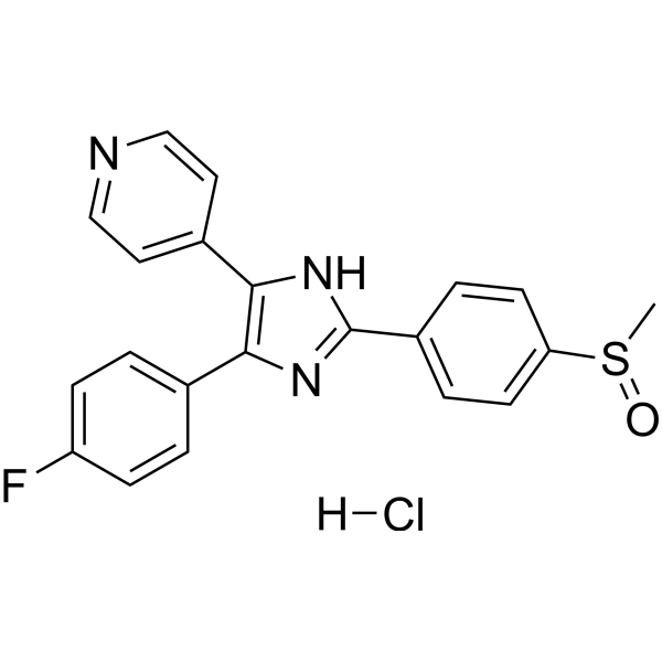
HY-10256A
Adezmapimod hydrochloridep38 MAPK Inhibitor
Publications Citing Use of MedChemExpress (MCE) Talmapimod
MoreVer todos los productos específicos de isoformas TNF Receptor
MoreVer todos los productos específicos de isoformas VEGFR
More

Actividad biológica
Descripciòn
IC50 & Target
[1]|
p38α 9 nM (IC50) |
p38β 90 nM (IC50) |
Cellular Effect
|
Cell Line

|
Type | Value | Description | References |
|---|---|---|---|---|
| PBMC | EC50 |
20 nM
Compound: SCIO-469
|
Antiinflammatory activity in human PBMC assessed as inhibition of LPS-induced IL1-beta production
Antiinflammatory activity in human PBMC assessed as inhibition of LPS-induced IL1-beta production
|
[PMID: 20071169] |
| PBMC | EC50 |
20 nM
Compound: SCIO-469
|
Antiinflammatory activity in human PBMC assessed as inhibition of LPS-induced IL-6 production
Antiinflammatory activity in human PBMC assessed as inhibition of LPS-induced IL-6 production
|
[PMID: 20071169] |
| PBMC | EC50 |
20 nM
Compound: SCIO-469
|
Antiinflammatory activity in human PBMC assessed as inhibition of LPS-induced IL-8 production
Antiinflammatory activity in human PBMC assessed as inhibition of LPS-induced IL-8 production
|
[PMID: 20071169] |
| PBMC | EC50 |
20 nM
Compound: SCIO-469
|
Antiinflammatory activity in human PBMC assessed as inhibition of LPS-induced TNFalpha production
Antiinflammatory activity in human PBMC assessed as inhibition of LPS-induced TNFalpha production
|
[PMID: 20071169] |
In Vitro
Talmapimod (SCIO-469) (100-200 nM; 1 hour) inhibits phosphorylation of p38 MAPK in MM cells[1].
Talmapimod inhibits LPS-induced TNF-a production in human whole blood[2].
Talmapimod decreases constitutive p38alpha MAPK phosphorylation of both 5T2MM and 5T33MM cells[3].
MedChemExpress (MCE) has not independently confirmed the accuracy of these methods. They are for reference only.
-
Cell Line:MM.1S, U266, RPMI8226, MM.1R, and RPMI-Dox40 cell lines
-
Concentration:100, 200 nM
-
Incubation Time:1 hour
-
Result:Strongly inhibits phosphorylation of p38 MAPK.
In Vivo
Talmapimod inhibits multiple myeloma growth and prevents bone disease in the 5T2MM and 5T33MM models[3].
Talmapimod (10-90 mg/kg; p.o.; twice daily orally for 14 days) dose-dependently reduced tumor growth and also dose-dependently reduced weight of the palpable tumors at termination[4].
MedChemExpress (MCE) has not independently confirmed the accuracy of these methods. They are for reference only.
-
Animal Model:Six-week-old male triple immune-deficient BNX mice (RPMI-8226 MM palpable tumors)[4]
-
Dosage:P.o.; twice daily orally for 14 days
-
Administration:10, 30, 90 mg/kg
-
Result:Dose-dependently reduced tumor growth.
- Human pluripotent stem cell endothelial-cell differentiation
- Detection of Apoptosis
- TUNEL staining for apoptotic DNA fragmentation
- Vascular/Branching Fractal Analysis
- Annexin V plus membrane-impermeant dye apoptosis staining
- Endothelial Tube Formation Assay
- Apoptosis Solutions
- Endothelial Cell Migration/Angiogenic Sprouting Assay
- Pyroptosis Solutions
- Osteoclast differentiation from monocyte/macrophage precursors
Ensayo clínico
| NCT Number | Sponsor | Condition | Start Date |
Phase
|
|---|---|---|---|---|
| NCT01329991 | Plexxikon| | 2011-05 | PHASE1 |


Chemical Information
-
No. CAS 309913-83-5
-
Appearance Solid
-
Peso molecular 513.00
-
Fòrmula C27H30ClFN4O3
-
Color Pink to red
-
SMILES
O=C(N(C)C)C(C1=CN(C)C2=C1C=C(C(N3[C@H](C)CN(CC4=CC=C(F)C=C4)[C@@H](C)C3)=O)C(Cl)=C2)=O
-
Synonyms
SCIO-469
-
Envío
Room temperature in continental US; may vary elsewhere.
-
Almacenamiento
Powder -20°C 3 years 4°C 2 years In solvent -80°C 2 years -20°C 1 year


Publications (3)
-
Journal Impact Factor
-
Most Recent
-
Cell
2020 Aug 6;182(3):685-712.e19. PMID: 32645325 -
Sci Transl Med
PP2A inhibition is a druggable MEK inhibitor resistance mechanism in KRAS-mutant lung cancer cells. [Abstract]2018 Jul 18;10(450):eaaq1093. PMID: 30021885 -
Cell Biol Toxicol
The 14-3-3η/GSK-3β/β-catenin complex regulates EndMT induced by 27-hydroxycholesterol in HUVECs and promotes the migration of breast cancer cells. [Abstract]2021 Aug;37(4):515-529. PMID: 33131013


Solvente y solubilidad
In Vitro:
DMSO : ≥ 100 mg/mL (194.93 mM; Hygroscopic DMSO has a significant impact on the solubility of product, please use newly opened DMSO)
* "≥" means soluble, but saturation unknown.
Please refer to the solubility information to select the appropriate solvent. Once prepared, please aliquot and store the solution to prevent product inactivation from repeated freeze-thaw cycles.
Storage method and period of stock solution: -80°C, 2 years; -20°C, 1 year. When stored at -80°C, please use it within 2 years. When stored at -20°C, please use it within 1 year.
Please refer to the solubility information to select the appropriate solvent. Once prepared, please aliquot and store the solution to prevent product inactivation from repeated freeze-thaw cycles.
Storage method and period of stock solution: -80°C, 2 years; -20°C, 1 year. When stored at -80°C, please use it within 2 years. When stored at -20°C, please use it within 1 year.
Concentration (start) × Volume (start) = Concentration (final) × Volume (final)
In Vivo:
Select the appropriate dissolution method based on your experimental animal and administration route.
- For the following dissolution methods, please ensure to first prepare a clear stock solution using an In Vitro approach and then sequentially add co-solvents:
- To ensure reliable experimental results, the clarified stock solution can be appropriately stored based on storage conditions. As for the working solution for In Vivo experiments, it is recommended to prepare freshly and use it on the same day.
- The percentages shown for the solvents indicate their volumetric ratio in the final prepared solution. If precipitation or phase separation occurs during preparation, heat and/or sonication can be used to aid dissolution.
Add each solvent one by one: 10% DMSO 40% PEG300 5% Tween-80 45% Saline
Solubility: 2.5 mg/mL (4.87 mM); Suspended solution; Need ultrasonic
This protocol yields a suspended solution of 2.5 mg/mL. Suspended solution can be used for oral and intraperitoneal injection.
Taking 1 mL working solution as an example, add 100 μL DMSO stock solution (25.0 mg/mL) to 400 μL PEG300, and mix evenly; then add 50 μL Tween-80 and mix evenly; then add 450 μL Saline to adjust the volume to 1 mL.
Preparation of Saline: Dissolve 0.9 g sodium chloride in ddH₂O and dilute to 100 mL to obtain a clear Saline solution.
Add each solvent one by one: 10% DMSO 90% (20% SBE-β-CD in Saline)
Solubility: ≥ 2.5 mg/mL (4.87 mM); Clear solution
This protocol yields a clear solution of ≥ 2.5 mg/mL (saturation unknown).
Taking 1 mL working solution as an example, add 100 μL DMSO stock solution (25.0 mg/mL) to 900 μL 20% SBE-β-CD in Saline, and mix evenly.
Preparation of 20% SBE-β-CD in Saline (4°C, storage for one week): 2 g SBE-β-CD powder is dissolved in 10 mL Saline, completely dissolve until clear.
In Vivo Dissolution Calculator
Please enter the basic information of animal experiments:
-
-
-
-
Recommended: Prepare an additional quantity of animals to account for potential losses during experiments.
Please enter your animal formula composition:
-
%DMSO +
Recommended: Keep the proportion of DMSO in working solution below 2% if your animal is weak.
-
%+
-
+%Tween-80 + +
-
%Saline +
The co-solvents required include: DMSO, . All of co-solvents are available by MedChemExpress (MCE). , Tween 80. All of co-solvents are available by MedChemExpress (MCE).
Working solution concentration: 0.22 mg/mL
Method for preparing stock solution: mg drug dissolved in μL DMSO. Stock solution concentration: mg/mL.
1. Take μL DMSO stock solution;
2. Add μL .
μL , mix evenly;
3. Then add μL Tween 80, mix evenly;
4. Then add μL
Please ensure that the stock solution in the first step is dissolved to a clear state, and add co-solvents in sequence. You can use ultrasonic heating (ultrasonic cleaner, recommended frequency 20-40 kHz), vortexing, etc. to assist dissolution.


Pureza y Documentación
-
Ficha de datos (279 KB)
-
SDS (252 KB)
- English - EN (252 KB)
- Français - FR (252 KB)
- Deutsch - DE (252 KB)
- Norwegian - NO (252 KB)
- Español - ES (252 KB)
- Swedish - SV (252 KB)
- Italian - IT (252 KB)
- Korean - KR (252 KB)
- Portuguese - PT (252 KB)
-
Instrucciones de manejo (2659 KB)
Referencias
[1]. Hideshima T et al. p38 MAPK inhibition enhances PS-341 (bortezomib)-induced cytotoxicity against multiple myeloma cells. Oncogene. 2004 Nov 18, 23(54), 8766-76. [Content Brief]
[2]. Navas T, et al. Inhibition of p38alpha MAPK disrupts the pathological loop of proinflammatory factor production in the myelodysplastic syndrome bone marrow microenvironment. Leuk Lymphoma. 2008 Oct;49(10):1963-75. [Content Brief]
[3]. Vanderkerken K et al. Inhibition of p38alpha mitogen-activated protein kinase prevents the development of osteolytic bone disease,reduces tumor burden, and increases survival in murine models of multiple myeloma. Cancer Res. 2007 May 15;67(10):4572-7. [Content Brief]
[4]. Medicherla S, et al. p38alpha-selective MAP kinase inhibitor reduces tumor growth in mouse xenograft models of multiple myeloma. Anticancer Res. 2008 Nov-Dec;28(6A):3827-33. [Content Brief]


Complete Stock Solution Preparation Table
Please refer to the solubility information to select the appropriate solvent. Once prepared, please aliquot and store the solution to prevent product inactivation from repeated freeze-thaw cycles.
Storage method and period of stock solution: -80°C, 2 years; -20°C, 1 year. When stored at -80°C, please use it within 2 years. When stored at -20°C, please use it within 1 year.
| Optional Solvent | Concentration Solvent Mass | 1 mg | 5 mg | 10 mg | 25 mg |
|---|---|---|---|---|---|
| DMSO | 1 mM | 1.9493 mL | 9.7466 mL | 19.4932 mL | 48.7329 mL |
| 5 mM | 0.3899 mL | 1.9493 mL | 3.8986 mL | 9.7466 mL | |
| 10 mM | 0.1949 mL | 0.9747 mL | 1.9493 mL | 4.8733 mL | |
| 15 mM | 0.1300 mL | 0.6498 mL | 1.2995 mL | 3.2489 mL | |
| 20 mM | 0.0975 mL | 0.4873 mL | 0.9747 mL | 2.4366 mL | |
| 25 mM | 0.0780 mL | 0.3899 mL | 0.7797 mL | 1.9493 mL | |
| 30 mM | 0.0650 mL | 0.3249 mL | 0.6498 mL | 1.6244 mL | |
| 40 mM | 0.0487 mL | 0.2437 mL | 0.4873 mL | 1.2183 mL | |
| 50 mM | 0.0390 mL | 0.1949 mL | 0.3899 mL | 0.9747 mL | |
| 60 mM | 0.0325 mL | 0.1624 mL | 0.3249 mL | 0.8122 mL | |
| 80 mM | 0.0244 mL | 0.1218 mL | 0.2437 mL | 0.6092 mL | |
| 100 mM | 0.0195 mL | 0.0975 mL | 0.1949 mL | 0.4873 mL |


Talmapimod Related Classifications
HY-10406 Related Classifications
 Powered by Bioz