



CM-272
Based on 3 publication(s) in Google Scholar
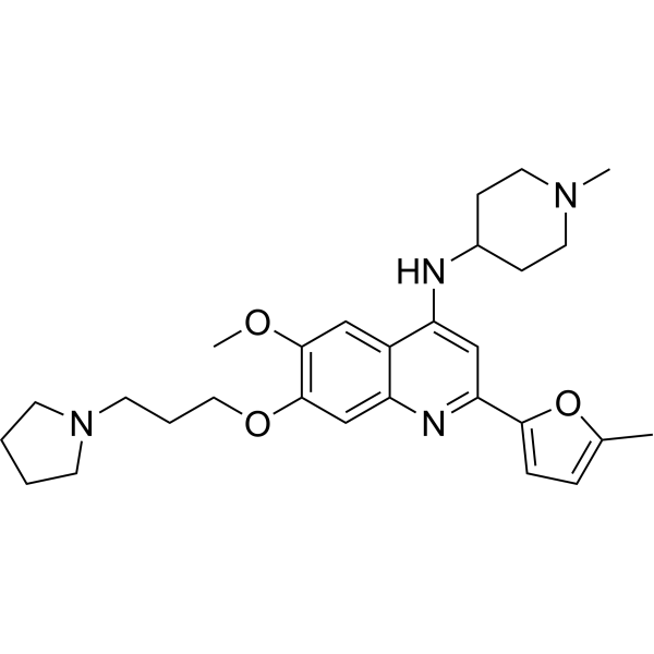
CM-272 is a first-in-class, potent, selective, substrate-competitive and reversible dual G9a/DNA methyltransferases (DNMTs) inhibitor with antitumor activities. CM-272 inhibits G9a, DNMT1, DNMT3A, DNMT3B and GLP with IC50s of 8 nM, 382 nM, 85 nM, 1200 nM and 2 nM, respectively. CM-272 inhibits cell proliferation and promotes apoptosis, inducing IFN-stimulated genes and immunogenic cell death.
For research use only. We do not sell to patients.
- Purity : 99.55%
- CAS No.: 1846570-31-7
- Formula: C28H38N4O3
- Molecular Weight:478.63
-
Storage:Powder -20°C, 3 years , 4°C, 2 years ; In solvent -80°C, 6 months , -20°C, 1 month

To place orders, for customer services and technical support, please contact: MedChemExpress USA
Tel: 609-228-6898 E-mail: [email protected] [email protected]


-
 Biological Activity
-
Chemical Information
-
Solvent & Solubility
- Protocols
- Purity & Documentation
- References
-
 Help & FAQs


-
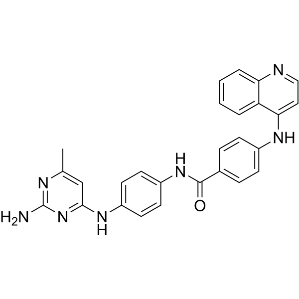
HY-13962
SGI-1027Apoptosis Inducer -
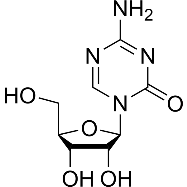
HY-10586
5-AzacytidineDNMT Inhibitor -
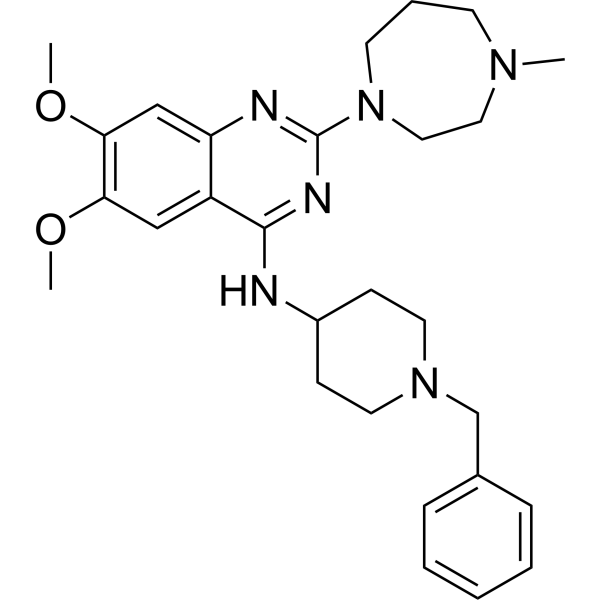
HY-10587
BIX-01294G9a/GLP Inhibitor -
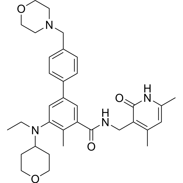
HY-13803
TazemetostatEZH2 Inhibitor -
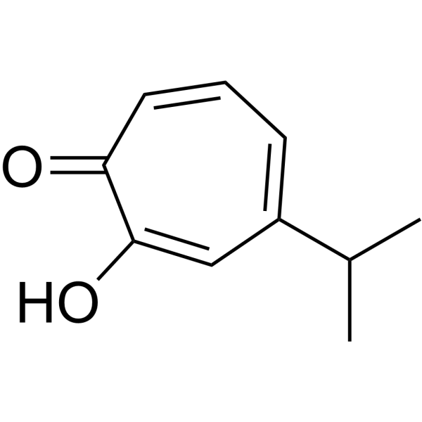
HY-B2230
HinokitiolVirus Protease Inhibitor -
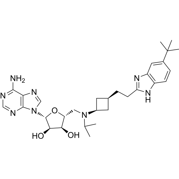
HY-15593
PinometostatDOT1L Inhibitor -
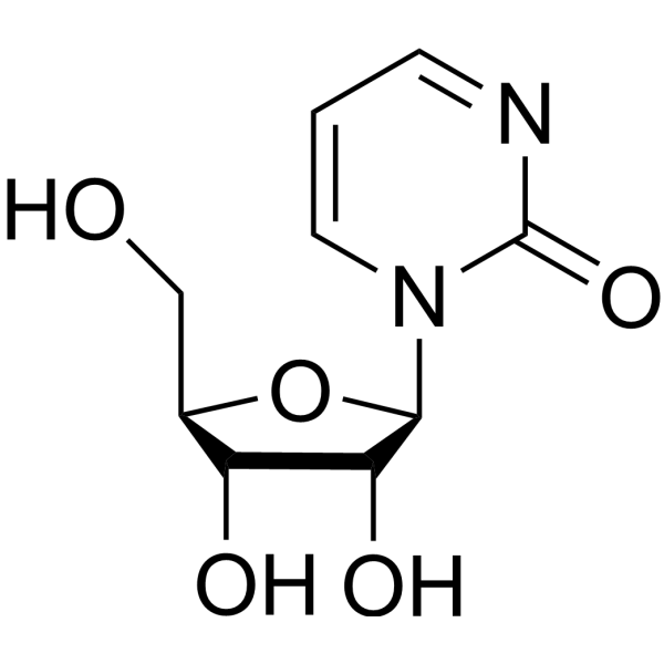
HY-13420
ZebularineDNA Methylation Inhibitor -
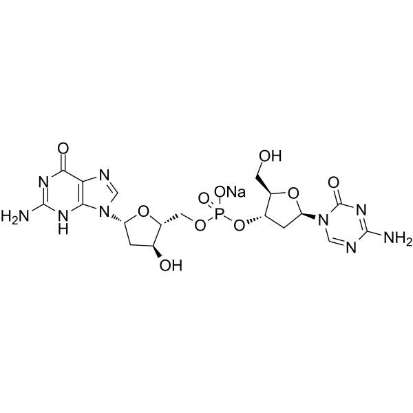
HY-15229
Guadecitabine sodiumDNMT Inhibitor -

HY-B2194
γ-OryzanolDNA Methyltransferase Inhibitor -
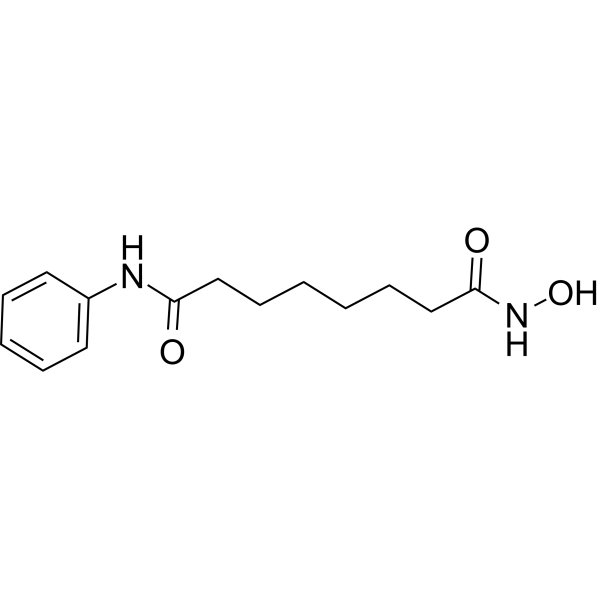
HY-10221
VorinostatHDAC Inhibitor -
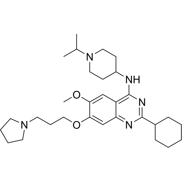
HY-15273
UNC0638G9a/GLP Inhibitor -
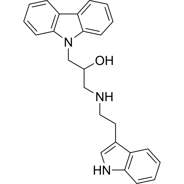
HY-12746
DC-05DNA Methyltransferase Inhibitor
Publications Citing Use of MedChemExpress (MCE) CM-272
More Customer Validation & Images

-
Others

-
WB

-
In Vivo Efficacy Study

-
In Vivo Efficacy Study

-
Histological Imaging/Staining

All Histone Methyltransferase Isoforms
MoreAll DNA Methyltransferase Isoforms
More

Biological Activity
Description
IC50 & Target
[1]|
G9a 8 nM (IC50) |
EHMT1/GLP/KMT1D 2 nM (IC50) |
DNMT1 382 nM (IC50) |
DNMT3A 85 nM (IC50) |
DNMT3B 1200 nM (IC50) |
Cellular Effect
|
Cell Line

|
Type | Value | Description | References |
|---|---|---|---|---|
| MCF-10A | EC50 |
0.2 μM
Compound: 2;CM-272
|
Antitumor activity against human spontaneously immortalized CDH-/- MCF10A cells assessed as cell numbers measured after 48 hrs
Antitumor activity against human spontaneously immortalized CDH-/- MCF10A cells assessed as cell numbers measured after 48 hrs
|
[PMID: 34878770] |
| MCF-10A | EC50 |
0.5 μM
Compound: 2;CM-272
|
Antitumor activity against human spontaneously immortalized wild type MCF-10A cells assessed as cell numbers measured after 48 hrs
Antitumor activity against human spontaneously immortalized wild type MCF-10A cells assessed as cell numbers measured after 48 hrs
|
[PMID: 34878770] |
| MM1.S | GI50 |
1041 nM
Compound: 5; CM-272
|
Antiproliferative activity against human MM1.S cells assessed as inhibition of cell proliferation measured up to 72 hrs by CellTiter96 Aqueous one reagent based assay
Antiproliferative activity against human MM1.S cells assessed as inhibition of cell proliferation measured up to 72 hrs by CellTiter96 Aqueous one reagent based assay
|
[PMID: 33661013] |
| MV4-11 | GI50 |
269 nM
Compound: 12; CM-272
|
Growth inhibition of human MV4-11 cells after 48 hrs by MTS assay
Growth inhibition of human MV4-11 cells after 48 hrs by MTS assay
|
[PMID: 29953809] |
| OCI-Ly10 | GI50 |
455 nM
Compound: 12; CM-272
|
Growth inhibition of human OCI-LY10 cells after 48 hrs by MTS assay
Growth inhibition of human OCI-LY10 cells after 48 hrs by MTS assay
|
[PMID: 29953809] |
| OCI-Ly10 | GI50 |
455 nM
Compound: 5
|
Antiproliferative activity against human OCI-LY10 cells after 48 hrs by MTS assay
Antiproliferative activity against human OCI-LY10 cells after 48 hrs by MTS assay
|
[PMID: 29890830] |
| OCI-Ly3 | GI50 |
409 nM
Compound: 12; CM-272
|
Growth inhibition of human OCI-LY3 cells after 48 hrs by MTS assay
Growth inhibition of human OCI-LY3 cells after 48 hrs by MTS assay
|
[PMID: 29953809] |
| OCI-Ly3 | GI50 |
409 nM
Compound: 5
|
Antiproliferative activity against human OCI-LY3 cells after 48 hrs by MTS assay
Antiproliferative activity against human OCI-LY3 cells after 48 hrs by MTS assay
|
[PMID: 29890830] |
In Vitro
CM-272 (100-1000 nM; 12-72 hours; CEMO-1, MV4-11 and OCI-Ly10 cell lines) treatment inhibits cell proliferation in a dose- and time-dependent manner[1].
CM-272 (100-1000 nM; 24 hours; CEMO-1, MV4-11 and OCI-Ly10 cell lines) treatment blocks cell cycle progression[1].
CM-272 (100-1000 nM; 12-72 hours; CEMO-1, MV4-11, and OCI-Ly10 cell lines) treatment induces apoptosis in ALL, AML and DLBCL cell lines in a dose- and time-dependent manner[1].
CM-272 after 48 h of treatment CEMO-1 acute lymphoblastic leukaemia (ALL) cell line, MV4-11 acute myeloid leukaemia (AML) cell line, and OCI-Ly10 diffuse large B-cell lymphoma (DLBCL) cell line, the GI50 values of 218 nM, 269 nM and 455 nM, respectively, and is associated with a decrease in global levels of H3K9me2 and 5mC[1].
The therapeutic activity of CM-272 relies on the early activation of the type I IFN response in tumor cells, potentially leading to the induction of cell-autonomous immunogenic death in tumor cells[1].
MedChemExpress (MCE) has not independently confirmed the accuracy of these methods. They are for reference only.
-
Cell Line:CEMO-1, MV4-11 and OCI-Ly10 cell lines
-
Concentration:125 nM, 250 nM, 500 nM (CEMO-1 cells); 135 nM, 270 nM, 540 nM (MV4-11 cells); 100 nM, 400 nM, 1000 nM (OCI-Ly10 cells)
-
Incubation Time:12 hours, 24 hours, 48 hours and 72 hours
-
Result:Inhibited cell proliferation in a dose- and time-dependent manner.
-
Cell Line:CEMO-1, MV4-11 and OCI-Ly10 cell lines
-
Concentration:125 nM, 250 nM, 500 nM (CEMO-1 cells); 135 nM, 270 nM, 540 nM (MV4-11 cells); 100 nM, 400 nM, 1000 nM (OCI-Ly10 cells)
-
Incubation Time:24 hours
-
Result:Blocked cell cycle progression.
-
Cell Line:CEMO-1, MV4-11 and OCI-Ly10 cell lines
-
Concentration:125 nM, 250 nM, 500 nM (CEMO-1 cells); 135 nM, 270 nM, 540 nM (MV4-11 cells); 100 nM, 400 nM, 1000 nM (OCI-Ly10 cells)
-
Incubation Time:12 hours, 24 hours, 48 hours and 72 hours
-
Result:Induced apoptosis in ALL, AML and DLBCL cell lines in a dose- and time-dependent manner.
In Vivo
MedChemExpress (MCE) has not independently confirmed the accuracy of these methods. They are for reference only.
-
Animal Model:Female BALB/Ca-Rag2−/−γc−/− mice (6–8-week-old) with CEMO-1 cells[1]
-
Dosage:2.5 mg/kg
-
Administration:Intravenous injection; daily; for 28 days
-
Result:Induced a statistically significant increase in overall survival (OS) in mice.
- Mammalian live/dead viability and cytotoxicity staining
- Annexin V plus membrane-impermeant dye apoptosis staining
- BrdU Incorporation Assay
- Somatic Cell Culture
- CFSE Dye Dilution Proliferation Assay
- Dye-dilution cell tracking and proliferation staining
- CCK-8/WST-8 Cell Proliferation Assay
- Protocol for Cell Cycle
- Cell Counting-Based Growth Curve Assay
- Colony Formation (Clonogenic) Assay
- Cell Viability Determination by MTT Colorimetric Assay
- EdU Incorporation Assay (Click Chemistry-Based DNA Synthesis Measurement)
- Ki-67 Immunostaining Proliferation Assay
- Detection of Apoptosis
- TUNEL staining for apoptotic DNA fragmentation
- PCNA Immunodetection Proliferation Assay
- Cell Cytotoxicity Assay
- Protocol for Cell Counting and Cell Density Analysis
- Apoptosis Solutions
- MTT Cell Proliferation Assay


Chemical Information
-
CAS No. 1846570-31-7
-
Appearance Solid
-
Molecular Weight 478.63
-
Formula C28H38N4O3
-
Color Off-white to light yellow
-
SMILES
CN(CC1)CCC1NC2=CC(C3=CC=C(C)O3)=NC4=CC(OCCCN5CCCC5)=C(OC)C=C42
-
Shipping
Room temperature in continental US; may vary elsewhere.
-
Storage
Powder -20°C 3 years 4°C 2 years In solvent -80°C 6 months -20°C 1 month


Publications (3)
-
Journal Impact Factor
-
Most Recent
-
Adv Mater
2024 Aug;36(31):e2402456. PMID: 38810924
CM-272 purchased from MedChemExpress. Usage Cited in: Adv Mater. 2024 Aug;36(31):e2402456. [Abstract]
Schematic illustration for the effects of CM-272-loaded NPs in tumor cells.
CM-272 purchased from MedChemExpress. Usage Cited in: Adv Mater. 2024 Aug;36(31):e2402456. [Abstract]
Incubation with free CM-272 (100 nM, 48 h), 272@MSNs, or 272@MM resulted in decreased levels of H3K9me2 in 4T1 cells.
CM-272 purchased from MedChemExpress. Usage Cited in: Adv Mater. 2024 Aug;36(31):e2402456. [Abstract]
The unloaded SIANPs (uSIANPs), CM-272 (5 mg/kg) + IBR, single drug-loaded NPs (272@MMP or IBR@OM), or dual drug-loaded SIANPs were intravenously administered in the 4T1 tumor-bearing mice every three days.
-
Biochim Biophys Acta Gen Subj
G9a and DNMT1 inhibition modulates CDKN1A promoter methylation and the cell cycle leading to improvement in kidney fibrosis. [Abstract]2023 Sep;1867(9):130417. PMID: 37356504
CM-272 purchased from MedChemExpress. Usage Cited in: Biochim Biophys Acta Gen Subj. 2023 Sep;1867(9):130417. [Abstract]
In in vivo experiments, mice underwent UUO surgery and were subsequently treated with CM272 (2.5 mg/kg) or vehicle for one week.
CM-272 purchased from MedChemExpress. Usage Cited in: Biochim Biophys Acta Gen Subj. 2023 Sep;1867(9):130417. [Abstract]
Renal injury scores and Masson staining results for detecting collagen deposition indicated that mice treated with CM272 (2.5 mg/kg) exhibited a milder degree of renal fibrosis.

CM-272 purchased from MedChemExpress. Usage Cited in: Biochim Biophys Acta Gen Subj. 2023 Sep;1867(9):130417. [Abstract]
Dual immunofluorescence staining showed that CM272 (2.5 mg/kg) also reduced the expression of α-SMA, G9a, and DNMT1.
-


Solvent & Solubility
In Vitro:
DMSO : 125 mg/mL (261.16 mM; Need ultrasonic; Hygroscopic DMSO has a significant impact on the solubility of product, please use newly opened DMSO)
Please refer to the solubility information to select the appropriate solvent. Once prepared, please aliquot and store the solution to prevent product inactivation from repeated freeze-thaw cycles.
Storage method and period of stock solution: -80°C, 6 months; -20°C, 1 month. When stored at -80°C, please use it within 6 months. When stored at -20°C, please use it within 1 month.
Please refer to the solubility information to select the appropriate solvent. Once prepared, please aliquot and store the solution to prevent product inactivation from repeated freeze-thaw cycles.
Storage method and period of stock solution: -80°C, 6 months; -20°C, 1 month. When stored at -80°C, please use it within 6 months. When stored at -20°C, please use it within 1 month.
Concentration (start) × Volume (start) = Concentration (final) × Volume (final)
In Vivo:
Select the appropriate dissolution method based on your experimental animal and administration route.
- For the following dissolution methods, please ensure to first prepare a clear stock solution using an In Vitro approach and then sequentially add co-solvents:
- To ensure reliable experimental results, the clarified stock solution can be appropriately stored based on storage conditions. As for the working solution for In Vivo experiments, it is recommended to prepare freshly and use it on the same day.
- The percentages shown for the solvents indicate their volumetric ratio in the final prepared solution. If precipitation or phase separation occurs during preparation, heat and/or sonication can be used to aid dissolution.
Add each solvent one by one: 10% DMSO 40% PEG300 5% Tween-80 45% Saline
Solubility: ≥ 2.08 mg/mL (4.35 mM); Clear solution
This protocol yields a clear solution of ≥ 2.08 mg/mL (saturation unknown).
Taking 1 mL working solution as an example, add 100 μL DMSO stock solution (20.8 mg/mL) to 400 μL PEG300, and mix evenly; then add 50 μL Tween-80 and mix evenly; then add 450 μL Saline to adjust the volume to 1 mL.
Preparation of Saline: Dissolve 0.9 g sodium chloride in ddH₂O and dilute to 100 mL to obtain a clear Saline solution.
In Vivo Dissolution Calculator
Please enter the basic information of animal experiments:
-
-
-
-
Recommended: Prepare an additional quantity of animals to account for potential losses during experiments.
Please enter your animal formula composition:
-
%DMSO +
Recommended: Keep the proportion of DMSO in working solution below 2% if your animal is weak.
-
%+
-
+%Tween-80 + +
-
%Saline +
The co-solvents required include: DMSO, . All of co-solvents are available by MedChemExpress (MCE). , Tween 80. All of co-solvents are available by MedChemExpress (MCE).
Working solution concentration: 0.22 mg/mL
Method for preparing stock solution: mg drug dissolved in μL DMSO. Stock solution concentration: mg/mL.
1. Take μL DMSO stock solution;
2. Add μL .
μL , mix evenly;
3. Then add μL Tween 80, mix evenly;
4. Then add μL
Please ensure that the stock solution in the first step is dissolved to a clear state, and add co-solvents in sequence. You can use ultrasonic heating (ultrasonic cleaner, recommended frequency 20-40 kHz), vortexing, etc. to assist dissolution.


Purity & Documentation
-
Data Sheet (278 KB)
-
SDS (396 KB)
- English - EN (396 KB)
- Français - FR (396 KB)
- Deutsch - DE (396 KB)
- Norwegian - NO (396 KB)
- Español - ES (396 KB)
- Swedish - SV (396 KB)
- Italian - IT (396 KB)
- Korean - KR (396 KB)
- Portuguese - PT (396 KB)
-
Handling Instructions (2659 KB)
References


Complete Stock Solution Preparation Table
Please refer to the solubility information to select the appropriate solvent. Once prepared, please aliquot and store the solution to prevent product inactivation from repeated freeze-thaw cycles.
Storage method and period of stock solution: -80°C, 6 months; -20°C, 1 month. When stored at -80°C, please use it within 6 months. When stored at -20°C, please use it within 1 month.
| Optional Solvent | Concentration Solvent Mass | 1 mg | 5 mg | 10 mg | 25 mg |
|---|---|---|---|---|---|
| DMSO | 1 mM | 2.0893 mL | 10.4465 mL | 20.8930 mL | 52.2324 mL |
| 5 mM | 0.4179 mL | 2.0893 mL | 4.1786 mL | 10.4465 mL | |
| 10 mM | 0.2089 mL | 1.0446 mL | 2.0893 mL | 5.2232 mL | |
| 15 mM | 0.1393 mL | 0.6964 mL | 1.3929 mL | 3.4822 mL | |
| 20 mM | 0.1045 mL | 0.5223 mL | 1.0446 mL | 2.6116 mL | |
| 25 mM | 0.0836 mL | 0.4179 mL | 0.8357 mL | 2.0893 mL | |
| 30 mM | 0.0696 mL | 0.3482 mL | 0.6964 mL | 1.7411 mL | |
| 40 mM | 0.0522 mL | 0.2612 mL | 0.5223 mL | 1.3058 mL | |
| 50 mM | 0.0418 mL | 0.2089 mL | 0.4179 mL | 1.0446 mL | |
| 60 mM | 0.0348 mL | 0.1741 mL | 0.3482 mL | 0.8705 mL | |
| 80 mM | 0.0261 mL | 0.1306 mL | 0.2612 mL | 0.6529 mL | |
| 100 mM | 0.0209 mL | 0.1045 mL | 0.2089 mL | 0.5223 mL |


CM-272 Related Classifications
HY-101925 Related Classifications
 Powered by Bioz