



Bafilomycin C1
Based on 1 publication(s) in Google Scholar
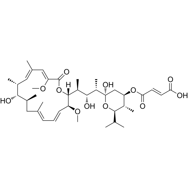
Bafilomycin C1 is a macrolide antibiotic isolated from Streptomyces sp. Bafilomycin C1 is a potent, specific and reversible inhibitor of vacuolar-type H+-ATPases (V-ATPases). Bafilomycin C1 inhibits growth of gram-positive bacteria and fungi. Bafilomycin C1 induces cell apoptosis and can be used for the study of hepatocellular carcinoma (HCC).
商品は「研究用試薬」です。人や動物の医療用・臨床診断用・食品用の製品ではありません。
研究用途以外に使用した場合、当社は一切の責任を負いかねます。
- 純度 : 99.9%
- CAS 番号: 88979-61-7
- 分子式: C39H60O12
- 分子量:720.89
-
保管条件:
-20°C, sealed storage, away from moisture
* In solvent : -80°C, 6 months; -20°C, 1 month (sealed storage, away from moisture)

To place orders, for customer services and technical support, please contact: MedChemExpress USA
Tel: 609-228-6898 E-mail: [email protected] [email protected]


-
 生物活性
生物活性
-
化学情報
-
溶剤 & 溶解度
- プロトコル
- 純度とドキュメンテーション
- 参考文献
-
 Help & FAQs


-

HY-13259
MG-132Proteasome Inhibitor -
HY-13821
EpoxomicinProteasome Inhibitor -

HY-N6782
OligomycinH+-ATP-Synthase Inhibitor -
HY-108970
Bafilomycin DV-ATPase Inhibitor -
HY-N0877
BufalinNa+/K+ ATPase Inhibitor -
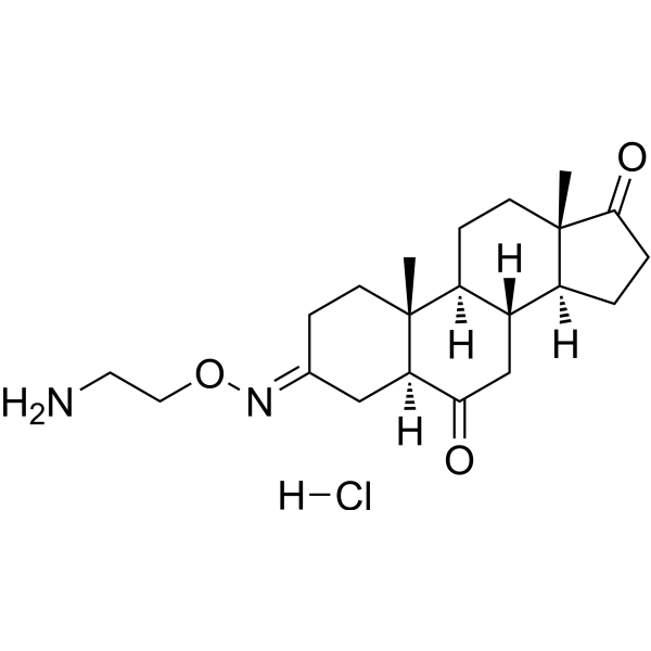
HY-15718A
Istaroxime hydrochlorideCalcium Channel Activator -
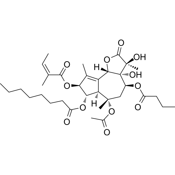
HY-13433
ThapsigarginER Stress Inducer -
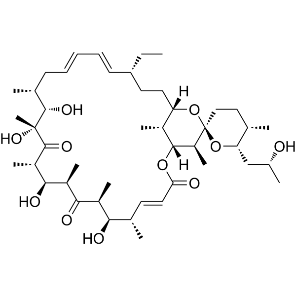
HY-16589
Oligomycin AMitochondrial ATP Synthase Inhibitor -
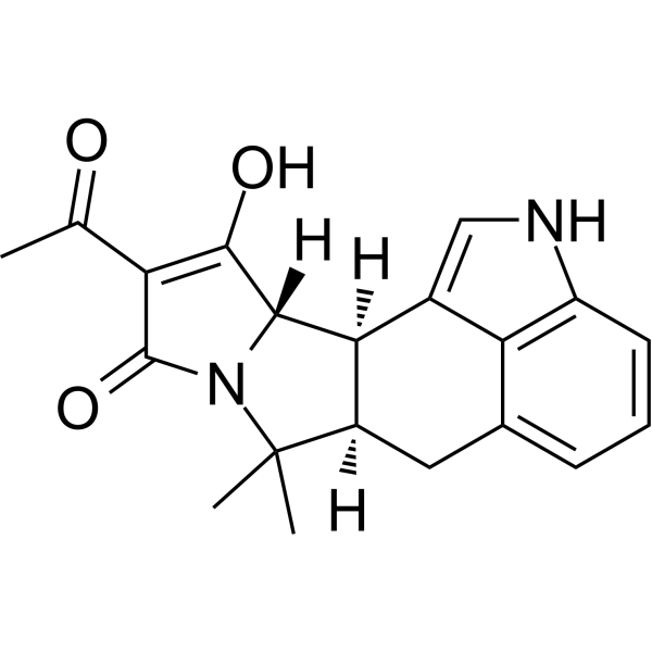
HY-N6771
Cyclopiazonic acidECAs Inhibitor -
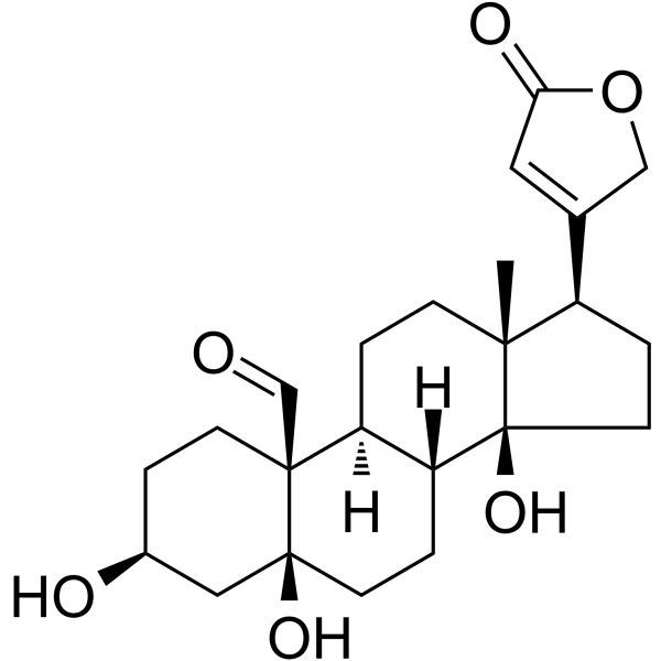
HY-114252
StrophanthidinCardiac Glycoside -
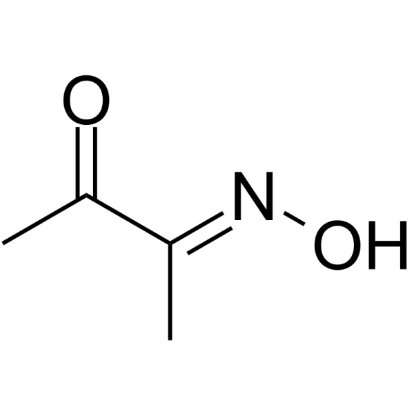
HY-Y0413
Biacetyl monoximeATPase Inhibitor
MedChemExpress(MCE)の使用を引用している文献 Bafilomycin C1
MoreAntibiotic アイソフォーム固有の製品をすべて表示
More

生物活性
製品説明
IC50 & Target
|
Macrolide |
Cellular Effect
|
Cell Line

|
Type | Value | Description | References |
|---|---|---|---|---|
| BGC-823 | IC50 |
1.83 μM
Compound: 7
|
Cytotoxicity against human BGC823 cells assessed as reduction in cell viability by MTT assay
Cytotoxicity against human BGC823 cells assessed as reduction in cell viability by MTT assay
|
[PMID: 26933756] |
| Caco-2 | IC50 |
1.89 μM
Compound: 7
|
Cytotoxicity against human Caco2 cells assessed as reduction in cell viability by MTT assay
Cytotoxicity against human Caco2 cells assessed as reduction in cell viability by MTT assay
|
[PMID: 26933756] |
| NCI-H460 | IC50 |
3.43 μM
Compound: 7
|
Cytotoxicity against human NCI-H460 cells assessed as reduction in cell viability by MTT assay
Cytotoxicity against human NCI-H460 cells assessed as reduction in cell viability by MTT assay
|
[PMID: 26933756] |
| SMMC-7721 | IC50 |
0.54 μM
Compound: 7
|
Cytotoxicity against human SMMC7721 cells assessed as reduction in cell viability by MTT assay
Cytotoxicity against human SMMC7721 cells assessed as reduction in cell viability by MTT assay
|
[PMID: 26933756] |
体外実験
Bafilomycin C1 (0.33-10 μM; 6 days) inhibits the growth and proliferation of SMMC7721 and HepG2 cells in a timeand dose-dependent manner[2].Bafilomycin C1 (0.33-3.3 μM; 24 hours) decreases cyclin D3, cyclin E1, CDK2, CDK4, and CDK6 expression in both mRNA and protein expression in SMMC7721 cells[2].Bafilomycin C1 (3.3-10 μM; 24 hours) causes morphological alterations and increases the population of apoptotic cells by Hoechst 33258 (HY-15558) staining compared to vehicle[2].
MedChemExpress (MCE) has not independently confirmed the accuracy of these methods. They are for reference only. Further protocols information, click here.
-
Cell Line:SMMC7721 cell and HepG2 cell
-
Concentration:0.33 μM, 1.1 μM, and 3.3 μM for SMMC77211.1 μM, 3.3 μM, and 10.0 μM for HepG2
-
Incubation Time:6 days
-
Result:Retarded the cell growth.
-
Cell Line:SMMC7721 cells
-
Concentration:3.3 μM
-
Incubation Time:24 hours
-
Result:Decreaed cyclin D3/E1,CDK2/4/6 protein expression and increased p21.
-
Cell Line:SMMC7721 and HepG2 cells
-
Concentration:3.3 μM; 10 μM
-
Incubation Time:24 hours
-
Result:Induced apoptosis in SMMC7721 and HepG2 cells.
体内実験
MedChemExpress (MCE) has not independently confirmed the accuracy of these methods. They are for reference only.
-
Animal Model:BALB/c nude mice (weighing 18-20 g) subcutaneous injected by SMMC7721 cell suspension (5×10⁶ cells/100 μL)[2]
-
Dosage:0.2 mg/kg
-
Administration:Subcutaneous injection; 20 days
-
Result:Suppressed tumor growth of SMMC7721 tumor xenografts.


化学情報
-
CAS 番号 88979-61-7
-
性状 Solid
-
分子量 720.89
-
分子式 C39H60O12
-
Color White to off-white
-
SMILES
C[C@H]([C@@](C[C@H]([C@@H]1C)OC(/C=C/C(O)=O)=O)(O[C@@H]1C(C)C)O)[C@H](O)[C@@H]([C@](OC(/C(OC)=C/C(C)=C/[C@H]2C)=O)([H])[C@H](/C=C/C=C(C[C@@H]([C@@H]2O)C)\C)OC)C
-
Structure Classification
-
Initial Source
Streptomyces sp
-
輸送条件
Room temperature in continental US; may vary elsewhere.
-
保管条件
-20°C, sealed storage, away from moisture
* In solvent : -80°C, 6 months; -20°C, 1 month (sealed storage, away from moisture)


Publications (1)
-
Journal Impact Factor
-
Most Recent


溶剤 & 溶解度
体外:
DMSO : 5 mg/mL (6.94 mM; Need ultrasonic and warming; Hygroscopic DMSO has a significant impact on the solubility of product, please use newly opened DMSO)
Please refer to the solubility information to select the appropriate solvent. Once prepared, please aliquot and store the solution to prevent product inactivation from repeated freeze-thaw cycles.
Storage method and period of stock solution: -80°C, 6 months; -20°C, 1 month (sealed storage, away from moisture). When stored at -80°C, please use it within 6 months. When stored at -20°C, please use it within 1 month.
Please refer to the solubility information to select the appropriate solvent. Once prepared, please aliquot and store the solution to prevent product inactivation from repeated freeze-thaw cycles.
Storage method and period of stock solution: -80°C, 6 months; -20°C, 1 month (sealed storage, away from moisture). When stored at -80°C, please use it within 6 months. When stored at -20°C, please use it within 1 month.
濃度 (開始) × 体積 (開始) = 濃度 (終了) × 体積 (終了)
体内:
The following protocol is derived from the literature and is for reference only. It is recommended to first try a small sample.
In Vivo Dissolution Calculator
Please enter the basic information of animal experiments:
-
-
-
-
Recommended: Prepare an additional quantity of animals to account for potential losses during experiments.
Working solution concentration: 0.22 mg/mL

プロトコル
-
Detection of Apoptosis
Apoptosis, also called programmed cell death, is generally characterized by distinct morphological characteristics.
-
TUNEL staining for apoptotic DNA fragmentation
TUNEL staining detects DNA strand breaks by using terminal deoxynucleotidyl transferase to add labeled nucleotides to exposed 3′-OH DNA termini, generating either microscopic staining in fixed cells or tissue sections, or fluorescence/cytometric signal in cell suspensions. TUNEL positivity reflects DNA fragmentation but should not be interpreted alone as definitive apoptosis, because TUNEL can also label necrotic, autolytic, mechanically damaged, or DNA-repair-associated DNA breaks.
-
Annexin V plus membrane-impermeant dye apoptosis staining
Annexin V-based apoptosis assays rely on the detection of phosphatidylserine (PS) externalization from the inner leaflet of the plasma membrane to the outer leaflet, an early biochemical hallmark of apoptosis. Fluorescently labeled Annexin V binds PS in a calcium-dependent manner, enabling identification of early apoptotic cells by flow cytometry or fluorescence microscopy. When combined with a membrane-impermeant DNA-binding dye (e. g. , propidium iodide), this approach allows discrimination between viable (Annexin V−/dye−), early apoptotic (Annexin V+/dye−), and late apoptotic or necrotic (Annexin V+/dye+) cell populations by assessing membrane integrity and PS exposure.
-
Apoptosis Solutions
Apoptosis is a regulated, generally non-lytic cell-death pathway that removes unwanted, damaged, infected, or abnormal cells through coordinated morphological changes, caspase activation, DNA fragmentation, and membrane remodeling. The intrinsic apoptosis pathway is controlled mainly by mitochondrial outer membrane permeabilization, BCL-2 family proteins, cytochrome c release, apoptosome formation, caspase-9 activation, and downstream executioner caspase-3/7 activation. The extrinsic apoptosis pathway is initiated by death receptors such as Fas, TNFR, and TRAIL receptors, which recruit adaptor proteins and activate caspase-8 before engaging executioner caspases or mitochondrial amplification through BID cleavage. Apoptosis is linked to many phenotypes, including cancer cell killing, tissue homeostasis, immune regulation, neurodegeneration, infection response, and treatment-induced cytotoxicity; unresolved questions include how apoptosis interacts with necroptosis, pyroptosis, ferroptos
-
Gram Staining of Tissue Sections
Gram staining of tissue sections is a histochemical technique used to differentiate Gram-positive and Gram-negative bacteria within histological specimens based on differences in bacterial cell wall structure and dye retention, adapted from classical bacteriological Gram staining into tissue-compatible “histological Gram stain” variants. In tissue applications, modifications of the Brown-Hopps and Brown-Brenn methods are commonly used to improve differentiation of microorganisms embedded within host connective tissue and to reduce overstaining or loss of Gram-negative signal, which are known limitations of earlier approaches. The principle relies on crystal violet-iodine complex retention in Gram-positive organisms and subsequent decolorization and counterstaining steps that allow contrast visualization of Gram-negative organisms against tissue background.
-
Liver Cancer Modeling
Liver cancer can be classified into primary liver cancer and secondary liver cancer. Secondary liver cancer is the metastatic liver cancer. Primary liver cancer includes hepatocellular carcinoma (HCC), intrahepatic cholangiocarcinoma (ICC) and fibrolamellar HCC, of which HCC is the most common form, accounting for approximately 90% of primary liver cancers[1]. HCC mouse models include chemical agent-induced models, transplanted tumor models, and genetic engineered models.


純度とドキュメンテーション
-
データシート (277 KB)
-
SDS (420 KB)
- English - EN (420 KB)
- Français - FR (420 KB)
- Deutsch - DE (420 KB)
- Norwegian - NO (420 KB)
- Español - ES (420 KB)
- Swedish - SV (420 KB)
- Italian - IT (420 KB)
- Korean - KR (420 KB)
- Portuguese - PT (420 KB)
-
取扱説明書 (2659 KB)
参考文献
[1]. E J Bowman, et al. Bafilomycins: A Class of Inhibitors of Membrane ATPases From Microorganisms, Animal Cells, and Plant Cells. Proc Natl Acad Sci U S A. 1988 Nov;85(21):7972-6. [Content Brief]
[2]. Xiaoxiao Gao,et al.Bafilomycin C1 Induces G0/G1 Cell-Cycle Arrest and Mitochondrial-Mediated Apoptosis in Human Hepatocellular Cancer SMMC7721 Cells. J Antibiot (Tokyo). 2018 Sep;71(9):808-817. [Content Brief]


Complete Stock Solution Preparation Table
Please refer to the solubility information to select the appropriate solvent. Once prepared, please aliquot and store the solution to prevent product inactivation from repeated freeze-thaw cycles.
Storage method and period of stock solution: -80°C, 6 months; -20°C, 1 month (sealed storage, away from moisture). When stored at -80°C, please use it within 6 months. When stored at -20°C, please use it within 1 month.
| Optional Solvent | Concentration Solvent Mass | 1 mg | 5 mg | 10 mg | 25 mg |
|---|---|---|---|---|---|
| DMSO | 1 mM | 1.3872 mL | 6.9359 mL | 13.8717 mL | 34.6794 mL |
| 5 mM | 0.2774 mL | 1.3872 mL | 2.7743 mL | 6.9359 mL |


Bafilomycin C1 関連分類
HY-130173 関連分類
 Powered by Bioz