



CALP1
Based on 3 publication(s) in Google Scholar
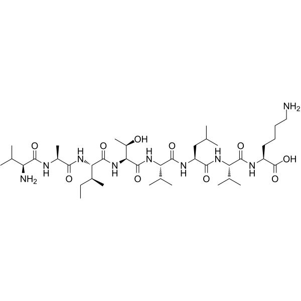
CALP1 is a calmodulin (CaM) agonist (Kd of 88 µM) with binding to the CaM EF-hand/Ca2+-binding site. CALP1 blocks calcium influx and apoptosis (IC50 of 44.78 µM) through inhibition of calcium channel opening. CALP1 blocks glutamate receptor channels and blocks a store-operated nonselective cation channel. CALP1 activates CaM-dependent phosphodiesterase activity.
商品は「研究用試薬」です。人や動物の医療用・臨床診断用・食品用の製品ではありません。
研究用途以外に使用した場合、当社は一切の責任を負いかねます。
- 純度 : 98.44%
- CAS 番号: 145224-99-3
- 分子式: C40H75N9O10
- 分子量:842.08
-
保管条件:
Sealed storage, away from moisture.
Powder -80°C, 2 years , -20°C, 1 year* In solvent : -80°C, 6 months; -20°C, 1 month (sealed storage, away from moisture)

To place orders, for customer services and technical support, please contact: MedChemExpress USA
Tel: 609-228-6898 E-mail: [email protected] [email protected]


-
 生物活性
生物活性
-
化学情報
-
溶剤 & 溶解度
- プロトコル
- 純度とドキュメンテーション
- 参考文献
-
 Help & FAQs


-

HY-P1074
SNX-482CaV2.3 Channel Blocker -
HY-100912
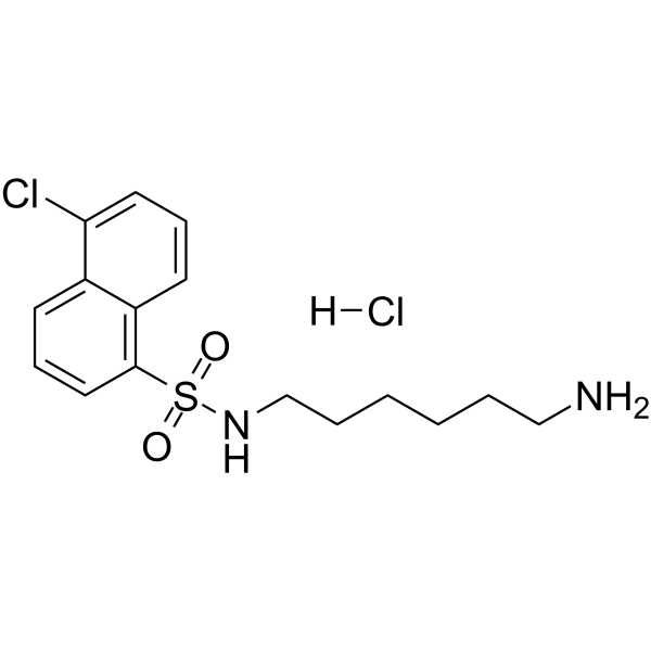
W-7 hydrochlorideCalmodulin Antagonist -

HY-P1080
ω-Agatoxin IVAP/Q Type Ca2+ Channel Blocker -
HY-17404
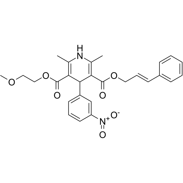
CilnidipineCa Channel Blocker -
HY-P1075
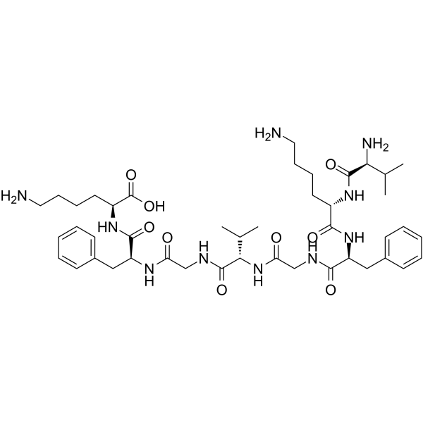
CALP3Ca2+ Channel Blocker -
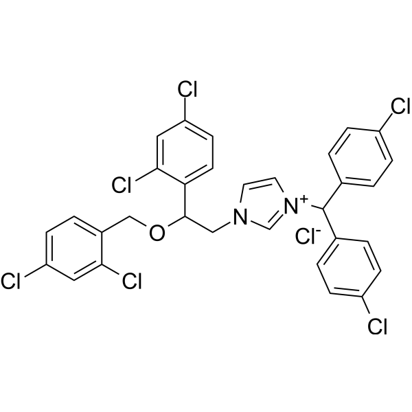
HY-103319
Calmidazolium chlorideCalmodulin Antagonist -

HY-P1073
ProTx-INaV/Cav Inhibitor -
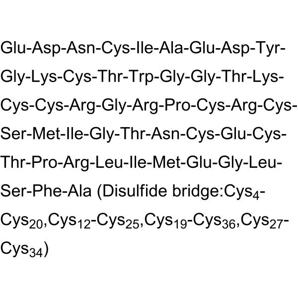
HY-P1079
ω-Agatoxin TKP/Q Type Ca2+ Channel Blocker -
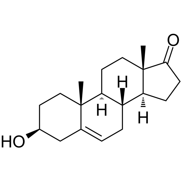
HY-14650
DHEASteroid Hormone -
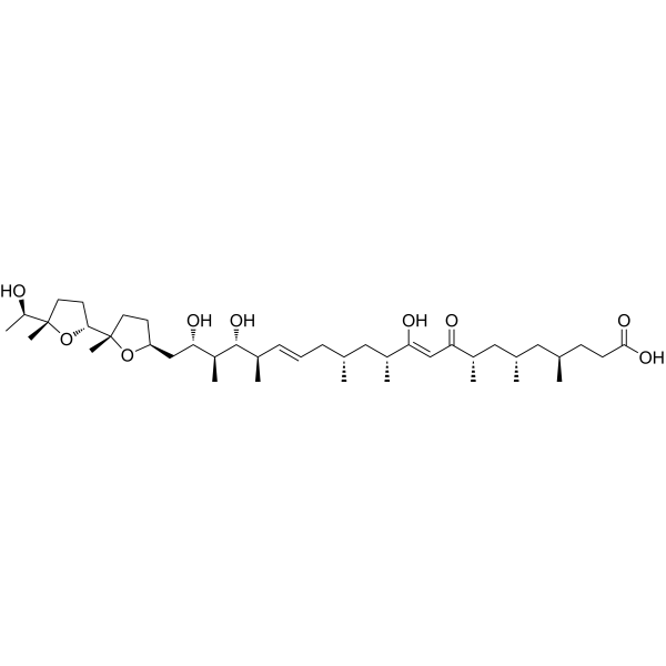
HY-13434
IonomycinCalcium Ionophore -
HY-N6687
CalcimycinCation Ionophore -

HY-P0214
Autocamtide-2-related inhibitory peptideCaMKII Inhibitor
MedChemExpress(MCE)の使用を引用している文献 CALP1
More

生物活性
製品説明
IC50 & Target
Kd: 88 µM (Calmodulin)[4]


化学情報
-
CAS 番号 145224-99-3
-
性状 Solid
-
分子量 842.08
-
分子式 C40H75N9O10
-
Color White to off-white
-
配列
Val-Ala-Ile-Thr-Val-Leu-Val-Lys
-
シーケンスの短縮
VAITVLVK
-
輸送条件
Room temperature in continental US; may vary elsewhere.
-
保管条件
Sealed storage, away from moisture
Powder -80°C 2 years -20°C 1 year * In solvent : -80°C, 6 months; -20°C, 1 month (sealed storage, away from moisture)


Publications (3)
-
Journal Impact Factor
-
Most Recent
-
Phytomedicine
Bioactive equivalent combinatorial components of Xiao-Xu-Ming decoction inhibit the calmodulin-mediated MLCK/MLC axis to attenuate coronary artery spasm. [Abstract]2025 Jul:142:156713. PMID: 40349422 -
Int J Nanomedicine
Electromechanical Regulation Underlying Protein Nanoparticle-Induced Osmotic Pressure in Neurotoxic Edema. [Abstract]2025 Apr 5:20:4145-4163. PMID: 40207308 -
Reprod Sci
Role of the Glycogen Synthase Kinase 3-Cyclic AMP/Protein Kinase A in the Immobilization of Human Sperm by Tideglusib. [Abstract]2023 Apr;30(4):1281-1290. PMID: 36207578


溶剤 & 溶解度
体外:
DMSO : 2 mg/mL (2.38 mM; Need ultrasonic; Hygroscopic DMSO has a significant impact on the solubility of product, please use newly opened DMSO)
Please refer to the solubility information to select the appropriate solvent. Once prepared, please aliquot and store the solution to prevent product inactivation from repeated freeze-thaw cycles.
Storage method and period of stock solution: -80°C, 6 months; -20°C, 1 month (sealed storage, away from moisture). When stored at -80°C, please use it within 6 months. When stored at -20°C, please use it within 1 month.
Please refer to the solubility information to select the appropriate solvent. Once prepared, please aliquot and store the solution to prevent product inactivation from repeated freeze-thaw cycles.
Storage method and period of stock solution: -80°C, 6 months; -20°C, 1 month (sealed storage, away from moisture). When stored at -80°C, please use it within 6 months. When stored at -20°C, please use it within 1 month.
濃度 (開始) × 体積 (開始) = 濃度 (終了) × 体積 (終了)


純度とドキュメンテーション
-
データシート (272 KB)
-
SDS (252 KB)
- English - EN (252 KB)
- Français - FR (252 KB)
- Deutsch - DE (252 KB)
- Norwegian - NO (252 KB)
- Español - ES (252 KB)
- Swedish - SV (252 KB)
- Italian - IT (252 KB)
- Korean - KR (252 KB)
- Portuguese - PT (252 KB)
-
取扱説明書 (2659 KB)
参考文献
[1]. R Houtman, et al. Attenuation of very late antigen-5-mediated adhesion of bone marrow-derived mast cells to fibronectin by peptides with inverted hydropathy to EF-hands. J Immunol. 2001 Jan 15;166(2):861-7. [Content Brief]
[2]. R Ten Broeke, et al. Calcium sensors as new therapeutic targets for airway hyperresponsiveness and asthma. FASEB J. 2001 Aug;15(10):1831-3. [Content Brief]
[3]. M K Manion, et al. A new type of Ca(2+) channel blocker that targets Ca(2+) sensors and prevents Ca(2+)-mediated apoptosis. FASEB J. 2000 Jul;14(10):1297-306. [Content Brief]
[4]. M Villain, et al. De novo design of peptides targeted to the EF hands of calmodulin. J Biol Chem. 2000 Jan 28;275(4):2676-85. [Content Brief]


Complete Stock Solution Preparation Table
Please refer to the solubility information to select the appropriate solvent. Once prepared, please aliquot and store the solution to prevent product inactivation from repeated freeze-thaw cycles.
Storage method and period of stock solution: -80°C, 6 months; -20°C, 1 month (sealed storage, away from moisture). When stored at -80°C, please use it within 6 months. When stored at -20°C, please use it within 1 month.
| Optional Solvent | Concentration Solvent Mass | 1 mg | 5 mg | 10 mg | 25 mg |
|---|---|---|---|---|---|
| DMSO | 1 mM | 1.1875 mL | 5.9377 mL | 11.8754 mL | 29.6884 mL |


CALP1 関連分類
HY-P1077 関連分類
 Powered by Bioz