



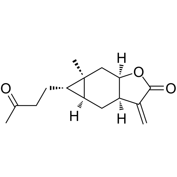
Carabrone

Based on 1 Customer Validation
Carabrone is a sesquiterpene found in the fruits of Carpesium abrotanoides and acts as a STAT3 inhibitor (Kd = 25.23 nM) with antibacterial, antitumor, and orally active properties. Carabrone covalently binds to UBE2D3 via Cys85 (Kd = 3.85 μM) and inhibits IκBα phosphorylation and NF-κB signaling. Carabrone induces ROS accumulation, loss of mitochondrial membrane potential, DNA fragmentation, and metacaspase-1-mediated apoptosis in fungi. Carabrone ameliorates autoimmune encephalomyelitis and MASH in vivo, reduces pro-inflammatory cytokines without immunotoxicity. Carabrone is used in research related to autoimmune encephalomyelitis, steatohepatitis, and fungal infections.
商品は「研究用試薬」です。人や動物の医療用・臨床診断用・食品用の製品ではありません。
研究用途以外に使用した場合、当社は一切の責任を負いかねます。
- 純度 : 99.91%
- CAS 番号: 1748-81-8
- 分子式: C15H20O3
- 分子量:248.32
-
保管条件:
4°C, protect from light
* In solvent : -80°C, 6 months; -20°C, 1 month (protect from light)

To place orders, for customer services and technical support, please contact: MedChemExpress USA
Tel: 609-228-6898 E-mail: [email protected] [email protected]


-
 生物活性
生物活性
-
化学情報
-
溶剤 & 溶解度
- プロトコル
- 純度とドキュメンテーション
- 参考文献
-
 Help & FAQs




生物活性
製品説明
IC50 & Target
[3]|
STAT3 25.23 nM (Kd) |
Cellular Effect
|
Cell Line

|
Type | Value | Description | References |
|---|---|---|---|---|
| HepG2 | IC50 |
>7.4 μM
Compound: 33
|
Cytotoxicity against human HepG2 cells assessed as reduction in cell viability after 48 hrs by MTT assay
Cytotoxicity against human HepG2 cells assessed as reduction in cell viability after 48 hrs by MTT assay
|
[PMID: 32842729] |
| Huh-7 | IC50 |
7.8 μM
Compound: 33
|
Cytotoxicity against human Huh-7 cells assessed as reduction in cell viability after 48 hrs by MTT assay
Cytotoxicity against human Huh-7 cells assessed as reduction in cell viability after 48 hrs by MTT assay
|
[PMID: 32842729] |
| Neutrophil | IC50 |
>10 μg/mL
Compound: 3
|
Inhibition of FMLP/CB-induced elastase release in human neutrophils after 5 mins by spectrophotometry
Inhibition of FMLP/CB-induced elastase release in human neutrophils after 5 mins by spectrophotometry
|
[PMID: 24997688] |
| Neutrophil | IC50 |
>10 μg/mL
Compound: 3
|
Inhibition of FMLP/CB-induced superoxide anion generation in human neutrophils by ferricytochrome c reduction method
Inhibition of FMLP/CB-induced superoxide anion generation in human neutrophils by ferricytochrome c reduction method
|
[PMID: 24997688] |
| RAW264.7 | IC50 |
6.4 μM
Compound: 3
|
Inhibition of NO production in LPS/IFN-gamma activated mouse RAW264.7 cells after 24 hrs by Griess assay
Inhibition of NO production in LPS/IFN-gamma activated mouse RAW264.7 cells after 24 hrs by Griess assay
|
[PMID: 24997688] |
| SMMC-7721 | IC50 |
>10 μM
Compound: 33
|
Cytotoxicity against human SMMC-7721 cells assessed as reduction in cell viability after 48 hrs by MTT assay
Cytotoxicity against human SMMC-7721 cells assessed as reduction in cell viability after 48 hrs by MTT assay
|
[PMID: 32842729] |
体外実験
Carabrone (0.1 μM; 30 min-6 h) induces ROS production in Gaeumannomyces graminis (Ggt) hyphae[1].
Carabrone (3.125-50 μg/mL) inhibits the activities of mitochondrial antioxidant enzymes GR, SOD, and CAT, while increasing the activity of GSH-PX and decreasing mitochondrial GSH content in Gaeumannomyces graminis (Ggt)[1].
Carabrone (0.1 μM; 2-6 h) significantly reduces the mitochondrial membrane potential of Gaeumannomyces graminis (Get) mycelia[1].
Carabrone (0.1 mM; ~6 h) undergoes a Michael addition reaction with L-cysteine (HY-Y0337) [1].
Carabrone (200 μM; 1 h) directly binds and stabilizes UBE2D3 in iBMDM whole-cell lysates[2].
Carabrone (200 μM; 1 h) increases the thermal stability of endogenous UBE2D3 in iBMDMs and THP-1 cells[2].
Carabrone (200 μM) protects endogenous UBE2D3 from proteolytic degradation in iBMDMs[2].
Carabrone directly and potently binds to recombinant human UBE2D3 with a KD of 3.85 μM[2].
Carabrone is predicted to covalently bind to the Cys85 residue of UBE2D3 via a Michael addition reaction[2].
Carabrone (50 µM; 4-6 h) directly binds to STAT3 in primary hepatocytes, as evidenced by decreased thermal stability[3].
Carabrone (90 s) directly binds to STAT3 with a KD value of 25.23 nM[3].
Carabrone (4 h) specifically binds to STAT3, as confirmed by competitive pull-down experiments[3].
Carabrone (0.1-1 µM) inhibits STAT3 activation in PO-stimulated hepatocytes[3].
Carabrone (0.1-1 µM) inhibits STAT3 nuclear translocation in PO-stimulated primary hepatocytes[3].
Carabrone inhibits STAT3 activation in PO-stimulated STAT3-knockdown L02 cells, and this inhibitory effect critically depends on Cys328[3].
Carabrone inhibits the expression of MASH-related genes in PO-stimulated STAT3-knockdown L02 cells, and this inhibitory effect is critically dependent on Cys328[3].
Carabrone (0.1 μM; 2-6 h) effectively induces apoptosis in Gaeumannomyces graminis (Ggt) and releases PS[1].
Carabrone (0.1 μM; 0-48 h) induces DNA fragmentation in Gaeumannomyces graminis (Ggt)[1].
Carabrone (0.1 μM; 0-48 h) upregulates the expression of metacaspase-1 genes Ggmet1 and Ggmet2 in Gaeumannomyces graminis (Get)[1].
Carabrone (0-100 μM; 6 h) exhibits no cytotoxicity in iBMDMs and THP-1 cells at concentrations up to 20 μM, establishing a non-cytotoxic range for subsequent experiments[2].
Carabrone (5-20 μM; 2 h) inhibits canonical NF-κB pathway activation in iBMDMs and THP-1 cells in a concentration-dependent manner[2].
Carabrone (10-20 μM) inhibits LPS-induced nuclear translocation of NF-κB p65 in iBMDMs[2].
Carabrone inhibits the NF-κB pathway in LPS (HY-D1056)-stimulated iBMDMs, and this inhibitory effect is abolished by pretreatment with β-mercaptoethanol (HY-Y0326), confirming the role of covalent cysteine binding[2].
Carabrone (5-20 μM; 2 h) inhibits LPS-induced pro-inflammatory NF-κB target gene transcription in iBMDMs in a dose-dependent manner[2].
Carabrone-mediated inhibition of the NF-κB signaling pathway in iBMDMs is specifically attenuated by Ube2d3 knockdown, indicating that its anti-inflammatory effect critically depends on the UBE2D3 protein[2].
Carabrone (0.1-1 µM) alleviates lipid accumulation in PO-treated primary hepatocytes and L02 cells[3].
Carabrone (0.1-1 µM) attenuates inflammation and fibrosis and modulates lipid metabolism gene expression in PO-treated primary hepatocytes and L02 cells[3].
Carabrone (10 mg; 6-8 h) exhibits in vitro antifungal activity against Botrytis cinerea (IC50 = 14.14 μg/mL) and Colletotrichum lagenarium (IC50 = 8.29 μg/mL) in spore germination assays[4].
Carabrone (5-100 μg/mL; 8 h) inhibits spore germination of Colletotrichum lagenarium with an EC50 of 7.10 μg/mL[5].
MedChemExpress (MCE) has not independently confirmed the accuracy of these methods. They are for reference only. Further protocols information, click here.
-
Cell Line:Gaeumannomyces graminis
-
Concentration:0.1 μM
-
Incubation Time:2, 4 and 6 h
-
Result:Exposed 3′-OH in the Get genomic DNA was broken after 6 h of incubation in the presence of carabrone acquired dUTP labeled with fluorescein (FITC) via terminal deoxynucleotidyl transferase.
Effectively induced Get apoptosis.
-
Cell Line:Gaeumannomyces graminis
-
Concentration:0.1 μM
-
Incubation Time:2, 4, 6, 12 and 24 h
-
Result:The expression levels of Ggmet1 and Ggmet2 significantly increased after 6 h.
-
Cell Line:iBMDMs and THP-1 cells
-
Concentration:0, 10, 20,40, 60, 80 and 100 μM
-
Incubation Time:6 h
-
Result:Carabrone exhibited no cytotoxicity at concentrations up to 20 μM in iBMDMs and THP-1 cells.
-
Cell Line:iBMDMs and THP-1 cells
-
Concentration:5,10 and 20 μM
-
Incubation Time:2 h
-
Result:Carabrone concentration-dependently inhibited the phosphorylation of IκBα and NF-κB p65 in iBMDMs and THP-1 cells.
-
Cell Line:iBMDMs
-
Concentration:5,10 and 20 μM
-
Incubation Time:2 h
-
Result:Carabrone dose-dependently suppressed the LPS-induced transcription of Il1b, Il6, Tnf, Nlrp3, Nos2, and Ptgs2.
-
Cell Line:iBMDMs
-
Concentration:20 μM
-
Incubation Time:2 h
-
Result:Carabrone treatment effectively suppressed the LPS-induced phosphorylation of NF-κB p65 and IκBα, and subsequent degradation of IκBα.
This suppressive effect was partially reversed in Ube2d3 siRNA-transfected cells.
Ube2d3 knockdown significantly reversed the inhibition of carabrone on Il1b, Il6, Tnf, Nlrp3, Nos2, and Ptgs2.
体内実験
Carabrone (10 mg/kg; p.o.; daily; 21 days) does not cause significant immunotoxicity in C57BL/6 mice[2].
Carabrone (1-10 mg/kg/day; p.o.; daily; 16 weeks) treatment alleviates HFHC diet-induced MASH by reducing lipid accumulation, inflammation, and fibrosis, and improving glucose metabolism[3].
Carabrone (1-10 mg/kg/day; gavage; daily; 16 weeks) significantly ameliorates HFD-induced MASH by reducing lipid accumulation, liver injury, inflammation, and fibrosis, and improving glucose sensitivity[3].
Carabrone (10 mg/kg/day; intragastric administration; daily; 8 weeks) alleviates MASH in MCD-fed mice in a STAT3-dependent manner, and its derivative CA-21 shows superior efficacy[3].
MedChemExpress (MCE) has not independently confirmed the accuracy of these methods. They are for reference only.
-
Animal Model:C57BL/6J (8-week-old female, EAE induced by MOG35-55 peptide in CFA with pertussis toxin)[2]
-
Dosage:3 mg/kg/day and 10 mg/kg/day
-
Administration:p.o.; every day; starting from day 3 post-immunization
-
Result:Significantly lowered clinical scores compared to vehicle-treated EAE controls.
Showed a dramatic reduction in inflammatory cell infiltration within the spinal cords.
Effectively preserved myelin integrity with significantly less extensive demyelinated lesions.
Significantly reduced the frequencies and absolute numbers of CNS-infiltrating immune cells, including CD4+ T cells, CD8+ T cells, B cells, monocytes/macrophages, and neutrophils.
-
Animal Model:C57BL/6J (8-week-old female, EAE induced by MOG35-55 peptide in CFA with pertussis toxin)[2]
-
Dosage:10 mg/kg
-
Administration:p.o.; daily; 21 consecutive days
-
Result:No significant differences in body weight were observed between CA-treated and vehicle control groups at days 0, 7, 14, and 21.
No significant differences in spleen size, morphology, or weight were observed at day 21.
The frequencies of B cells (B220+), total T cells (CD3+), T cell subsets (CD4+ and CD8+), and myeloid cells (Ly6G+ and Ly6C+) in the blood were unaltered.
Splenocytes showed comparable percentages of all examined immune cell subsets between CA-treated and vehicle groups.
-
Animal Model:C57BL/6J (male, 6-8 weeks old, HFHC diet-induced MASH)[3]
-
Dosage:1 and 10 mg/kg/day
-
Administration:i.g.; daily; 16 weeks
-
Result:Stabilized body weight, reduced epididymal adipose size, and decreased liver weights and the liver weight-to-body weight ratio compared to the HFHC control group.
Exhibited lower serum lipid levels, reduced hepatic lipid accumulation, decreased de novo lipogenesis, and increased β-oxidation.
Ameliorated hepatic inflammatory response and fibrosis deposition.
Markedly reduced hepatic profibrotic mediators.
Reduced liver injury markers and improved glucose tolerance and insulin tolerance, as indicated by lower blood glucose levels in OGTT and ITT assays.
-
Animal Model:C57BL/6J (male, 6-8 weeks old, HFD-induced MASH)[3]
-
Dosage:1 and 10 mg/kg/day
-
Administration:i.g.; daily; 16 weeks
-
Result:Showed significantly lower body weight compared to HFD-fed mice.
Significantly decreased the contents of liver triglycerides (TG) and total cholesterol (TC) and serum TG and TC.
Markedly inhibited the accumulation of lipid droplets in liver tissues as revealed by H&E and Oil Red O staining.
Notably decreased serum ALT and AST levels.
Exhibited lower mRNA levels of genes related to de novo lipogenesis, inflammation, and fibrosis, but higher β-oxidation compared with HFD mice.
Improved glucose sensitivity, as shown by the OGTT and ITT results.
-
Animal Model:C57BL/6J (male, 6-8 weeks old, MCD diet-induced MASH, with or without hepatic STAT3 overexpression)[3]
-
Dosage:10 mg/kg/day
-
Administration:i.g.; daily; 8 weeks
-
Result:Significantly reduced serum ALT and AST levels and liver TG and TC contents in MCD-fed mice.
Showed less steatosis and inflammatory cell infiltration in histological staining.
Decreased the expressions of inflammation (Tnfα, Mcp1, and Cxcl10) and fibrosis-related genes (Col1a1, Col3a1, and Ctgf).
Improvements were not observed in STAT3-overexpressed mice.


化学情報
-
CAS 番号 1748-81-8
-
性状 Solid
-
分子量 248.32
-
分子式 C15H20O3
-
Color White to off-white
-
SMILES
O=C1O[C@@]2([H])[C@@](C[C@]([C@@H]3CCC(C)=O)([H])[C@]3(C)C2)([H])C1=C
-
Structure Classification
-
輸送条件
Room temperature in continental US; may vary elsewhere.
-
保管条件
4°C, protect from light
* In solvent : -80°C, 6 months; -20°C, 1 month (protect from light)


溶剤 & 溶解度
体外:
DMSO : 100 mg/mL (402.71 mM; Need ultrasonic; Hygroscopic DMSO has a significant impact on the solubility of product, please use newly opened DMSO)
Please refer to the solubility information to select the appropriate solvent. Once prepared, please aliquot and store the solution to prevent product inactivation from repeated freeze-thaw cycles.
Storage method and period of stock solution: -80°C, 6 months; -20°C, 1 month (protect from light). When stored at -80°C, please use it within 6 months. When stored at -20°C, please use it within 1 month.
Please refer to the solubility information to select the appropriate solvent. Once prepared, please aliquot and store the solution to prevent product inactivation from repeated freeze-thaw cycles.
Storage method and period of stock solution: -80°C, 6 months; -20°C, 1 month (protect from light). When stored at -80°C, please use it within 6 months. When stored at -20°C, please use it within 1 month.
濃度 (開始) × 体積 (開始) = 濃度 (終了) × 体積 (終了)
体内:
Select the appropriate dissolution method based on your experimental animal and administration route.
- For the following dissolution methods, please ensure to first prepare a clear stock solution using an In Vitro approach and then sequentially add co-solvents:
- To ensure reliable experimental results, the clarified stock solution can be appropriately stored based on storage conditions. As for the working solution for In Vivo experiments, it is recommended to prepare freshly and use it on the same day.
- The percentages shown for the solvents indicate their volumetric ratio in the final prepared solution. If precipitation or phase separation occurs during preparation, heat and/or sonication can be used to aid dissolution.
Add each solvent one by one: 10% DMSO 40% PEG300 5% Tween-80 45% Saline
Solubility: ≥ 2.5 mg/mL (10.07 mM); Clear solution
This protocol yields a clear solution of ≥ 2.5 mg/mL (saturation unknown).
Taking 1 mL working solution as an example, add 100 μL DMSO stock solution (25.0 mg/mL) to 400 μL PEG300, and mix evenly; then add 50 μL Tween-80 and mix evenly; then add 450 μL Saline to adjust the volume to 1 mL.
Preparation of Saline: Dissolve 0.9 g sodium chloride in ddH₂O and dilute to 100 mL to obtain a clear Saline solution.
Add each solvent one by one: 10% DMSO 90% (20% SBE-β-CD in Saline)
Solubility: ≥ 2.5 mg/mL (10.07 mM); Clear solution
This protocol yields a clear solution of ≥ 2.5 mg/mL (saturation unknown).
Taking 1 mL working solution as an example, add 100 μL DMSO stock solution (25.0 mg/mL) to 900 μL 20% SBE-β-CD in Saline, and mix evenly.
Preparation of 20% SBE-β-CD in Saline (4°C, storage for one week): 2 g SBE-β-CD powder is dissolved in 10 mL Saline, completely dissolve until clear.
In Vivo Dissolution Calculator
Please enter the basic information of animal experiments:
-
-
-
-
Recommended: Prepare an additional quantity of animals to account for potential losses during experiments.
Please enter your animal formula composition:
-
%DMSO +
Recommended: Keep the proportion of DMSO in working solution below 2% if your animal is weak.
-
%+
-
+%Tween-80 + +
-
%Saline +
The co-solvents required include: DMSO, . All of co-solvents are available by MedChemExpress (MCE). , Tween 80. All of co-solvents are available by MedChemExpress (MCE).
Working solution concentration: 0.22 mg/mL
Method for preparing stock solution: mg drug dissolved in μL DMSO. Stock solution concentration: mg/mL. * In solvent : -80°C, 6 months; -20°C, 1 month (protect from light)
1. Take μL DMSO stock solution;
2. Add μL .
μL , mix evenly;
3. Then add μL Tween 80, mix evenly;
4. Then add μL
Please ensure that the stock solution in the first step is dissolved to a clear state, and add co-solvents in sequence. You can use ultrasonic heating (ultrasonic cleaner, recommended frequency 20-40 kHz), vortexing, etc. to assist dissolution.

プロトコル
-
Research Protocol for Infectious Diseases
Infectious-disease experiments test how pathogens interact with host barriers, innate immune receptors, inflammatory signaling, pathogen replication, and tissue injury; pattern-recognition receptors such as TLRs, RIG-I-like receptors, NOD-like receptors, and inflammasomes detect microbial molecules and activate NF-κB, interferon, and cytokine responses. The central hypothesis is that infection severity reflects the balance between pathogen burden and host response: protective inflammation restricts pathogen growth, whereas excessive or mislocalized inflammation contributes to tissue damage and disease phenotype. Unresolved questions include which host pathways are protective versus pathogenic, why some infection models fail to translate to human disease, and which combined readouts best predict clinically relevant infection outcomes.
-
Western Blot
Western blotting (WB) is a commonly used experimental method in molecular biology, biochemistry, and immunogenetics for identifying and quantifying target proteins. It combines gel electrophoresis with immunoassay, enabling researchers to analyze protein expression, post-translational modifications, and molecular weight.
-
Mitochondrial membrane-potential fluorescent assay
Mitochondrial membrane potential fluorescent assays estimate ΔΨm in living cells using lipophilic cationic dyes such as TMRM, TMRE, rhodamine 123, and JC-1, which accumulate in mitochondria according to membrane polarization; loss of signal after FCCP or CCCP treatment is interpreted as mitochondrial depolarization. TMRM/TMRE and rhodamine 123 are commonly used for semi-quantitative live-cell microscopy or flow cytometry, while JC-1 can report a shift from red aggregate fluorescence to green monomer fluorescence during depolarization; interpretation requires controls because dye concentration, quenching mode, cell type, dye efflux, and mitochondrial mass can affect fluorescence independently of ΔΨm.
-
Annexin V plus membrane-impermeant dye apoptosis staining
Annexin V-based apoptosis assays rely on the detection of phosphatidylserine (PS) externalization from the inner leaflet of the plasma membrane to the outer leaflet, an early biochemical hallmark of apoptosis. Fluorescently labeled Annexin V binds PS in a calcium-dependent manner, enabling identification of early apoptotic cells by flow cytometry or fluorescence microscopy. When combined with a membrane-impermeant DNA-binding dye (e. g. , propidium iodide), this approach allows discrimination between viable (Annexin V−/dye−), early apoptotic (Annexin V+/dye−), and late apoptotic or necrotic (Annexin V+/dye+) cell populations by assessing membrane integrity and PS exposure.
-
ROS/oxidative-stress fluorescent staining
ROS/oxidative-stress fluorescent staining uses cell-permeant fluorogenic probes that become fluorescent after oxidation inside cells or tissues; commonly used examples include DCFH-DA/DCFDA for broad cellular oxidant detection, DHE for superoxide-related signal detection, MitoSOX for mitochondrial superoxide-related signal detection, and CellROX probes for oxidative-stress-associated fluorescence readouts. The assay detects probe oxidation rather than a single ROS species unless the probe and analysis method have been chemically validated for that species. DCFH-DA enters cells, is deacetylated by intracellular esterases to DCFH, and produces fluorescent DCF after oxidation, so the readout is used as an operational measure of total cellular oxidative stress rather than a species-specific ROS measurement. DHE and MitoSOX can report superoxide-related oxidation, but red fluorescence alone can include non-specific ethidium-like oxidation products; HPLC or optimized spectral approaches are
-
Research Protocol for Inflammation-related Diseases
The NLRP3 inflammasome is a cytosolic innate immune signaling platform that integrates priming signals and danger-signal activation to promote caspase-1 activation, maturation of IL-1β and IL-18, and gasdermin D-mediated pyroptotic cell death. The core experimental logic is to determine whether inflammatory disease phenotypes are driven by increased NLRP3 expression, ASC-containing inflammasome assembly, caspase-1 cleavage, GSDMD cleavage, and extracellular release of IL-1β/IL-18 rather than by nonspecific cell injury alone. The pathway is strongly linked to inflammation-related disease phenotypes because monosodium urate crystals activate NALP3/NLRP3 inflammasome signaling in gout-like crystal inflammation, cholesterol crystals activate NLRP3 inflammasomes in atherogenesis models, and DSS-induced intestinal inflammation has been reported to involve NLRP3 inflammasome activity. However, experimental colitis studies also show context-dependent protective effects of NLRP3 inflammasome co
-
Kinase activity and phosphorylation assays
Kinase activity assays measure the ability of kinases to transfer phosphate groups from ATP to specific substrates, while phosphorylation assays detect the presence and levels of phosphorylated proteins. Common methods include radiolabeled ATP incorporation (e. g. ,), ADP release detection via bioluminescence (e. g. ,[3]), enzyme-linked immunosorbent assays (ELISA) for phospho-specific epitopes (e. g. ,[6]), and microtiter-based formats for high-throughput screening (e. g. ,[8]). The ADP-Glo assay quantifies kinase activity by measuring ADP produced during phosphorylation using a luciferase-based system. Radiometric assays involve autoradiography or scintillation counting after incorporation of 32P-labeled ATP into substrate proteins. ELISA-based approaches rely on phospho-specific antibodies to detect activated kinases in cell lysates or purified samples.
-
Apoptosis
Apoptosis, also called programmed cell death, is generally characterized by distinct morphological characteristics.
-
TUNEL staining for apoptotic DNA fragmentation
TUNEL staining detects DNA strand breaks by using terminal deoxynucleotidyl transferase to add labeled nucleotides to exposed 3′-OH DNA termini, generating either microscopic staining in fixed cells or tissue sections, or fluorescence/cytometric signal in cell suspensions. TUNEL positivity reflects DNA fragmentation but should not be interpreted alone as definitive apoptosis, because TUNEL can also label necrotic, autolytic, mechanically damaged, or DNA-repair-associated DNA breaks.
-
LPS-Induced Endotoxemia/Systemic Inflammation
Lipopolysaccharide (LPS)-induced endotoxemia is a widely used in vivo model of acute systemic inflammation in which LPS, a Gram-negative bacterial endotoxin, activates innate immune signaling primarily through TLR4, leading to rapid and transient induction of pro-inflammatory cytokines such as TNF-α, IL-6, and IL-1β in circulation and tissues. This cytokine surge is commonly used as a measurable readout of systemic inflammatory activation and immune dysregulation, and is typically assessed within hours after intraperitoneal LPS administration in mouse models of endotoxemia. The model captures key features of systemic inflammatory response syndrome, including cytokine release, immune cell activation, and downstream tissue responses, and has been used to evaluate anti-inflammatory interventions such as cytokine modulation, lipid mediators, and immune cell-targeting therapies.
-
Fluorescent plasma-membrane potential dye assay
Fluorescent plasma-membrane potential dye assays measure changes in cell membrane potential using voltage-sensitive dyes whose fluorescence changes when cells depolarize or hyperpolarize. Anionic bis-oxonol dyes such as DiBAC4(3) enter depolarized cells more readily and show increased fluorescence after intracellular binding, while hyperpolarization reduces dye accumulation and fluorescence. FMP/FLIPR membrane-potential dyes are used for faster, homogeneous microplate assays of ion-channel or receptor-mediated membrane-potential changes.
-
Mitochondrial membrane-potential and mitochondrial mass staining
Mitochondrial membrane potential staining measures the electrochemical polarization across the mitochondrial inner membrane in live cells using lipophilic cationic fluorescent probes; early rhodamine-based work showed that selective mitochondrial dye accumulation is lost when the mitochondrial transmembrane potential is dissipated. JC-1 reports mitochondrial polarization by shifting from green monomer fluorescence to red J-aggregate fluorescence as dye concentration increases within energized mitochondria; therefore, the red/green fluorescence ratio is used as a relative readout of mitochondrial membrane potential. TMRE or TMRM staining provides a single-channel relative readout because these cationic rhodamine esters accumulate in polarized mitochondria, and lower fluorescence indicates reduced mitochondrial polarization when acquisition and dye-loading conditions are controlled. Mitochondrial mass staining is commonly performed with MitoTracker Green FM or related MitoTracker dyes as
-
Apoptosis Solutions
Apoptosis is a regulated, generally non-lytic cell-death pathway that removes unwanted, damaged, infected, or abnormal cells through coordinated morphological changes, caspase activation, DNA fragmentation, and membrane remodeling. The intrinsic apoptosis pathway is controlled mainly by mitochondrial outer membrane permeabilization, BCL-2 family proteins, cytochrome c release, apoptosome formation, caspase-9 activation, and downstream executioner caspase-3/7 activation. The extrinsic apoptosis pathway is initiated by death receptors such as Fas, TNFR, and TRAIL receptors, which recruit adaptor proteins and activate caspase-8 before engaging executioner caspases or mitochondrial amplification through BID cleavage. Apoptosis is linked to many phenotypes, including cancer cell killing, tissue homeostasis, immune regulation, neurodegeneration, infection response, and treatment-induced cytotoxicity; unresolved questions include how apoptosis interacts with necroptosis, pyroptosis, ferroptos
-
Protocol for Kinase activity and phosphorylation assays
Kinase activity assays measure transfer of phosphate from ATP to a protein or peptide substrate, generating phosphorylated substrate, ADP, or incorporated radiolabeled phosphate as the readout; phosphorylation assays measure site-specific phosphorylation in cells or tissues as a proxy for kinase-pathway activation, inhibition, or substrate regulation. Phosphorylation can be detected by phospho-specific Western blot, immunoprecipitation kinase assay, phospho-immunofluorescence, phospho-flow cytometry, luminescent ADP detection, radiolabeled ATP incorporation, or reporter-based pathway assays, and these readouts can be applied to cancer cells, primary neurons, mouse tumors, organoids, inflammatory macrophages, ferroptosis studies, and mitophagy studies when the kinase target is biologically relevant.


純度とドキュメンテーション
-
データシート (303 KB)
-
SDS (251 KB)
- English - EN (251 KB)
- Français - FR (251 KB)
- Deutsch - DE (251 KB)
- Norwegian - NO (251 KB)
- Español - ES (251 KB)
- Swedish - SV (251 KB)
- Italian - IT (251 KB)
- Korean - KR (251 KB)
- Portuguese - PT (251 KB)
-
取扱説明書 (2659 KB)
参考文献
[4]. Wang H, et al. Synthesis, antifungal activities and qualitative structure activity relationship of carabrone hydrazone derivatives as potential antifungal agents. International journal of molecular sciences. 2014 Mar 11;15(3):4257-72. [Content Brief]
[5]. Feng JT, et al. Synthesis and antifungal activity of carabrone derivatives. Molecules. 2010 Sep 16;15(9):6485-92. [Content Brief]


Complete Stock Solution Preparation Table
Please refer to the solubility information to select the appropriate solvent. Once prepared, please aliquot and store the solution to prevent product inactivation from repeated freeze-thaw cycles.
Storage method and period of stock solution: -80°C, 6 months; -20°C, 1 month (protect from light). When stored at -80°C, please use it within 6 months. When stored at -20°C, please use it within 1 month.
| Optional Solvent | Concentration Solvent Mass | 1 mg | 5 mg | 10 mg | 25 mg |
|---|---|---|---|---|---|
| DMSO | 1 mM | 4.0271 mL | 20.1353 mL | 40.2706 mL | 100.6765 mL |
| 5 mM | 0.8054 mL | 4.0271 mL | 8.0541 mL | 20.1353 mL | |
| 10 mM | 0.4027 mL | 2.0135 mL | 4.0271 mL | 10.0677 mL | |
| 15 mM | 0.2685 mL | 1.3424 mL | 2.6847 mL | 6.7118 mL | |
| 20 mM | 0.2014 mL | 1.0068 mL | 2.0135 mL | 5.0338 mL | |
| 25 mM | 0.1611 mL | 0.8054 mL | 1.6108 mL | 4.0271 mL | |
| 30 mM | 0.1342 mL | 0.6712 mL | 1.3424 mL | 3.3559 mL | |
| 40 mM | 0.1007 mL | 0.5034 mL | 1.0068 mL | 2.5169 mL | |
| 50 mM | 0.0805 mL | 0.4027 mL | 0.8054 mL | 2.0135 mL | |
| 60 mM | 0.0671 mL | 0.3356 mL | 0.6712 mL | 1.6779 mL | |
| 80 mM | 0.0503 mL | 0.2517 mL | 0.5034 mL | 1.2585 mL | |
| 100 mM | 0.0403 mL | 0.2014 mL | 0.4027 mL | 1.0068 mL |


Carabrone 関連分類
HY-N5020 関連分類
 Powered by Bioz
Keywords