



MC2625
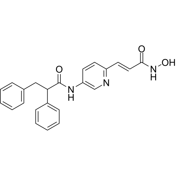
MC2625 is a potent pyridine-containing histone deacetylase (HDAC) inhibitor. MC2625 show selective HDAC3 and HDAC6 inhibition with IC50s of 80 nM and 11 nM. MC2625 increases acetyl-H3 and acetyl-tubulin levels and inhibits cancer stem cells (CSCs) growth by apoptosis induction.
For research use only. We do not sell to patients.
- CAS No.: 1776116-75-6
- Formula: C23H21N3O3
- Molecular Weight:387.43
-
Storage:
Please store the product under the recommended conditions in the Certificate of Analysis.

To place orders, for customer services and technical support, please contact: MedChemExpress USA
Tel: 609-228-6898 E-mail: [email protected] [email protected]


-
 Biological Activity
-
Chemical Information
- Protocols
- Purity & Documentation
- References
-
 Help & FAQs




Biological Activity
Description
|
HDAC3 80 nM (IC50) |
HDAC6 11 nM (IC50) |
HDAC1 1.42 μM (IC50) |
HDAC2 1.77 μM (IC50) |
HDAC4 11.7 μM (IC50) |
HDAC5 9.37 μM (IC50) |
HDAC7 8.77 μM (IC50) |
HDAC8 0.61 μM (IC50) |
HDAC9 10.6 μM (IC50) |
HDAC10 1.8 μM (IC50) |
HDAC11 10.2 μM (IC50) |
Cellular Effect
|
Cell Line

|
Type | Value | Description | References |
|---|---|---|---|---|
| A549 | IC50 |
0.22 μM
Compound: 5d; MC2625
|
Antiproliferative activity against human A549 cells incubated for 72 hrs by CellTiter-Glo luminescent cell viability assay
Antiproliferative activity against human A549 cells incubated for 72 hrs by CellTiter-Glo luminescent cell viability assay
|
[PMID: 36549114] |
| HCT-116 | IC50 |
0.07 μM
Compound: 5d; MC2625
|
Antiproliferative activity against human HCT-116 cells incubated for 72 hrs by CellTiter-Glo luminescent cell viability assay
Antiproliferative activity against human HCT-116 cells incubated for 72 hrs by CellTiter-Glo luminescent cell viability assay
|
[PMID: 36549114] |
| K562 | IC50 |
0.04 μM
Compound: 5d; MC2625
|
Antiproliferative activity against human K562 cells incubated for 72 hrs by CellTiter-Glo luminescent cell viability assay
Antiproliferative activity against human K562 cells incubated for 72 hrs by CellTiter-Glo luminescent cell viability assay
|
[PMID: 36549114] |
In Vitro
MC2625 (0.5-2 μM; 24-72 h) has potent antiproliferative effect on different sarcoma CSCs (cancer stem cells) after 72 h[1].
MC2625 (0.5-2 μM; 48 h) significantly induces apoptosis of all CSC cultures, with the exception of A204 CSCs[1].
MC2625 (0.5, 2 μM; 24 h) causes a dose-dependent increase of acetyl-histone H3[1].
MedChemExpress (MCE) has not independently confirmed the accuracy of these methods. They are for reference only.
-
Cell Line:Cancer stem cells (HOS, MG-63, RD, A204, SK-ES-1, A673)
-
Concentration:0.5, 1, 2 μM
-
Incubation Time:24, 48, 72 h
-
Result:Significantly affected MG-63, RD, and SK-ES-1 viability in 0.5 μM, while all CSCs were sensitive at 1 and 2 μM.
-
Cell Line:Cancer stem cells (HOS, MG-63, RD, A204, SK-ES-1, A673)
-
Concentration:0.5, 1, 2 μM
-
Incubation Time:48 h
-
Result:Significantly induced apoptosis of all CSC cultures, with the exception of A204 CSCs.
Generated an increase of the presence of apoptotic cells with concentrated dense granular fluorescence compared to untreated cells, especially at 1 and 2 μM.
-
Cell Line:MG-63 cancer stem cells
-
Concentration:0.5, 2 μM
-
Incubation Time:24 h
-
Result:Caused a dose-dependent increase of acetyl-histone H3.


Chemical Information
-
CAS No. 1776116-75-6
-
Molecular Weight 387.43
-
Formula C23H21N3O3
-
SMILES
O=C(NO)/C=C/C1=NC=C(C=C1)NC(C(C2=CC=CC=C2)CC3=CC=CC=C3)=O
-
Shipping
Room temperature in continental US; may vary elsewhere.
-
Storage
Please store the product under the recommended conditions in the Certificate of Analysis.


Purity & Documentation
References
[1]. Gemma Di Pompo, et al. Novel histone deacetylase inhibitors induce growth arrest, apoptosis, and differentiation in sarcoma cancer stem cells. J Med Chem. 2015 May 14;58(9):4073-9. [Content Brief]
[2]. Elisabetta Di Bello, et al. Novel pyridine-containing histone deacetylase inhibitors strongly arrest proliferation, induce apoptosis and modulate miRNAs in cancer cells. Eur J Med Chem. 2022 Dec 15;247:115022. [Content Brief]

Calculators
Concentration (start) × Volume (start) = Concentration (final) × Volume (final)


MC2625 Related Classifications
HY-152225 Related Classifications
 Powered by Bioz