



Momordin Ic
Based on 5 publication(s) in Google Scholar
Momordin Ic is an orally active triterpenoid saponin that can be isolated from Kochia scoparia. It is also a SUMO specific protease 1 (SENP1) inhibitor, SENP1/c-MYC signaling pathway inhibitor, and apoptosis inducer. Momordin Ic induces autophagy and apoptosis in liver cancer cells through the PI3K/Akt and MAPK signaling pathways mediated by reactive oxygen species. Momordin Ic has the ability to control glucose induced blood glucose elevation, inhibit gastric emptying, resist rheumatoid arthritis, reduce CCl4 (HY-Y0298) induced hepatotoxicity and anti-tumor activity.
For research use only. We do not sell to patients.
- Purity : 99.78%
- CAS No.: 96990-18-0
- Formula: C41H64O13
- Molecular Weight:764.94
-
Storage:
4°C, protect from light
* In solvent : -80°C, 6 months; -20°C, 1 month (protect from light)

To place orders, for customer services and technical support, please contact: MedChemExpress USA
Tel: 609-228-6898 E-mail: [email protected] [email protected]


-
 Biological Activity
-
Chemical Information
-
Solvent & Solubility
- Protocols
- Purity & Documentation
- References
-
 Help & FAQs


-
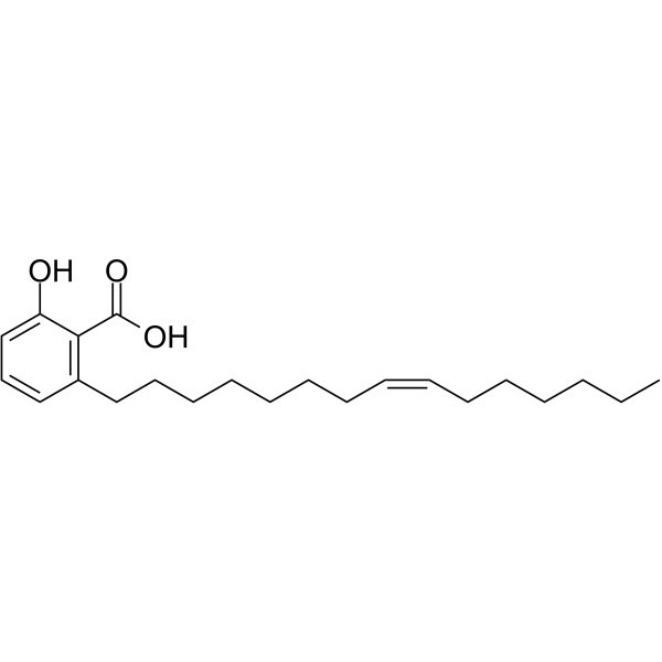
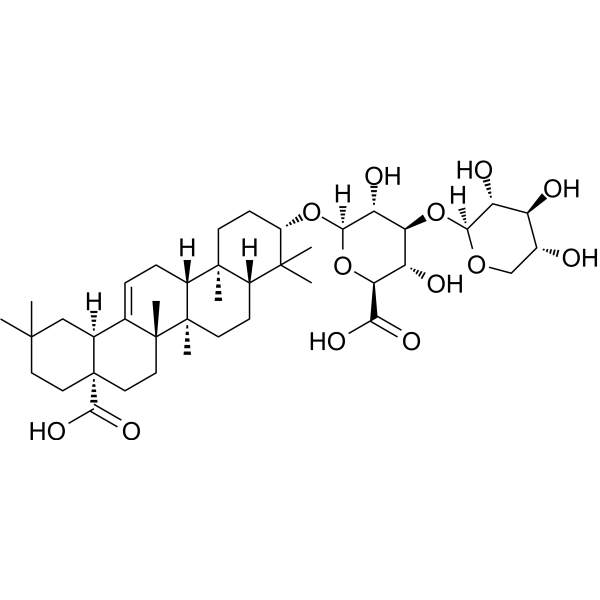
HY-N0077
Ginkgolic acidE1/E2/E3 Enzyme Inhibitor -

HY-13259
MG-132Proteasome Inhibitor -
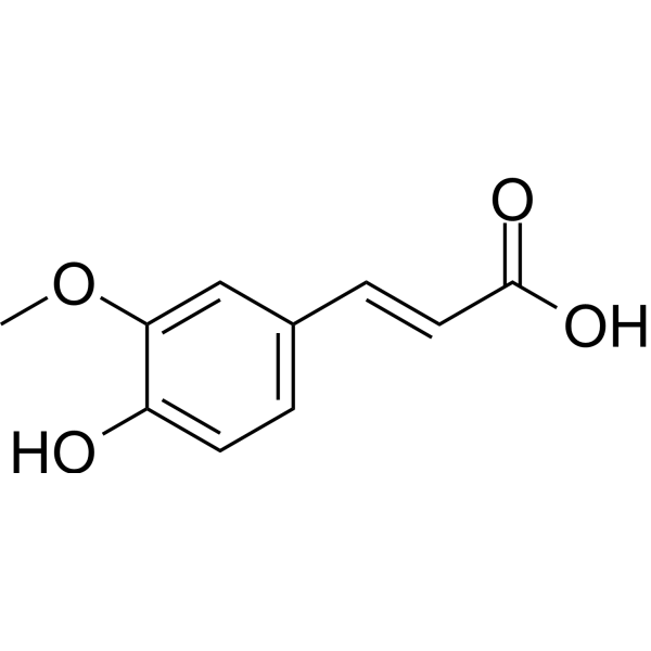
HY-N0060
Ferulic acidFGFR1 Inhibitor -

HY-Y1891
Tween 80 -
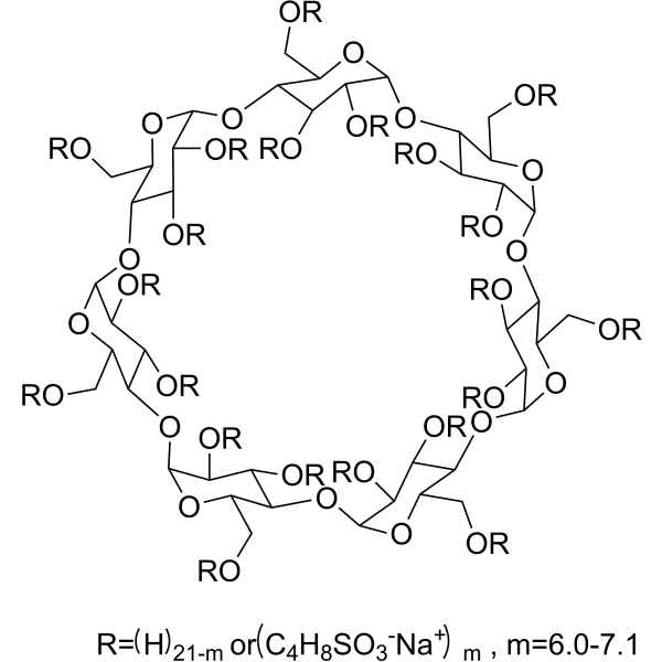
HY-17031
SBE-β-CDExcipient -
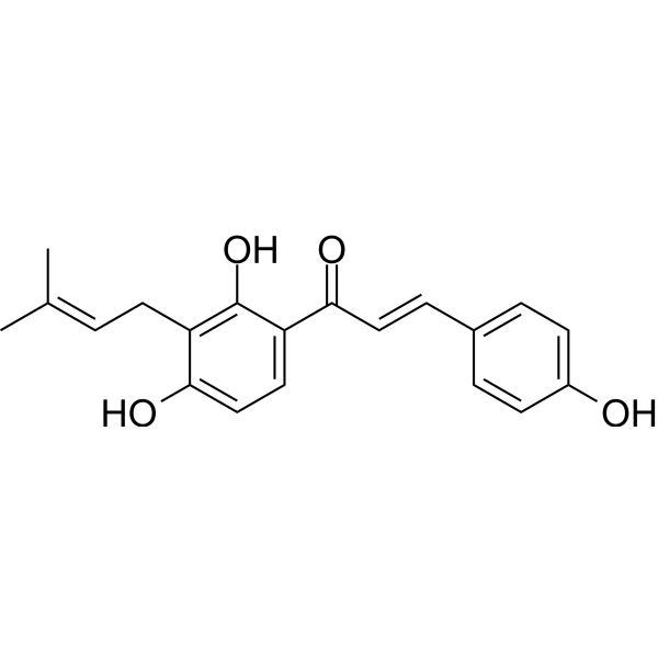
HY-13065
IsobavachalconeAkt Inhibitor -
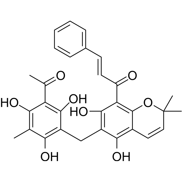
HY-18980
RottlerinPKC Inhibitor -
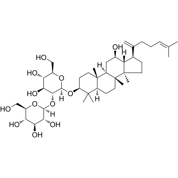
HY-N2515
Ginsenoside Rk1Apoptosis Inducer -
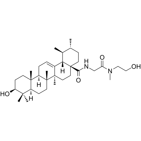
HY-134594
SENP1-IN-1SENP1 Inhibitor -

HY-N0330R
Momordin Ic (Standard)Autophagy/Apoptosis Inducer, SENP1 Inhibitor
Publications Citing Use of MedChemExpress (MCE) Momordin Ic
More Customer Validation & Images

-
WB
-
Cell Proliferation/Viability Assay
-
ELISA
-
Cell Migration/Invasion Assay
-
In Vivo Efficacy Study


Biological Activity
Description
In Vitro
Momordin Ic (0-20 μM, 48 h) relies on cholesterol and ganglioside GM1 to enhance the toxicity of recombinant protein MAP30 in breast cancer cells[2]. Momordin Ic (10 μM, 24 h) induces colon cancer cell cycle arrest and apoptosis by inhibiting the SENP1/c-MYC signaling pathway[3].
MedChemExpress (MCE) has not independently confirmed the accuracy of these methods. They are for reference only. Further protocols information, click here.
-
Cell Line:HepG2 cell; MDA-MB-231, MCF-7, HepG2, H460, A549, HeLa cells
-
Concentration:0-20 μM
-
Incubation Time:4 h; 48 h
-
Result:Inhibited growth in HepG2 cells; Enhanced the cytotoxicity of MAP30 to breast cancer cells.
-
Cell Line:HepG2 cell
-
Concentration:0-15 μM
-
Incubation Time:4 h
-
Result:Reduced the protein levels of caspase-3 and Bcl-2, and increased the protein levels of cytochrome c and Bax in the cytoplasmicsol.
-
Cell Line:HCT-8 cell, HCT-116 cells
-
Concentration:10 μM
-
Incubation Time:24 h
-
Result:Reduced the level of c-Myc protein.
In Vivo
MedChemExpress (MCE) has not independently confirmed the accuracy of these methods. They are for reference only.
-
Animal Model:Male ddY mice[4].
-
Dosage:12.5, 25, 50 mg/kg
-
Administration:Oral gavage (p.o.)
-
Result:Accelerated gastrointestinal transit in fasted mice.
-
Animal Model:Male Sprague-Dawley rat[5].
-
Dosage:10 mg/kg
-
Administration:Oral gavage (p.o.)
-
Result:Reduced the length of the lesions.
-
Animal Model:Male Sprague–Dawley rat[6].
-
Dosage:30 mg/kg
-
Administration:Oral gavage (p.o.), once a day for 14 days
-
Result:Reduced serum transaminase, lactic dehydrogenase, and γ-glutamyltransferase levels in the CCl4-treated rats.


Chemical Information
-
CAS No. 96990-18-0
-
Appearance Solid
-
Molecular Weight 764.94
-
Formula C41H64O13
-
Color White to off-white
-
SMILES
CC1(C)CC[C@@](CC[C@]2(C)C3=CC[C@@]4([H])[C@@]2(C)CC[C@]5([H])[C@]4(C)CC[C@H](O[C@]6([H])O[C@H](C(O)=O)[C@@H](O)[C@H](O[C@]7([H])OC[C@@H](O)[C@H](O)[C@H]7O)[C@H]6O)C5(C)C)(C(O)=O)[C@@]3([H])C1
-
Structure Classification
-
Initial Source
-
Shipping
Room temperature in continental US; may vary elsewhere.
-
Storage
4°C, protect from light
* In solvent : -80°C, 6 months; -20°C, 1 month (protect from light)


Publications (5)
-
Journal Impact Factor
-
Most Recent
-
Biochem Pharmacol
Momordin Ic suppresses breast cancer growth by targeting ACTL8‑dependent glutamine metabolism and PI3K/AKT/mTOR-MYC. [Abstract]2026 May:247:117761. PMID: 41621692
Momordin Ic purchased from MedChemExpress. Usage Cited in: Biochem Pharmacol. 2026 May:247:117761. [Abstract]
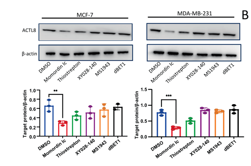
Western blot analysis showing ACTL8 protein levels in BC cells treated with DMSO, Momordin Ic, Thiostrepton, XY028-140, MS1943, or dBET1(10 μM each) for 24 h, indicating that Momordin Ic markedly reduced ACTL8 expression, The corresponding protein quantification is shown below.
Momordin Ic purchased from MedChemExpress. Usage Cited in: Biochem Pharmacol. 2026 May:247:117761. [Abstract]
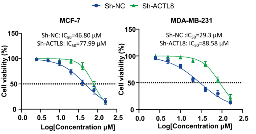
Cell viability assays revealing the IC50 values of Momordin Ic in BC cells. Dose-response curves and IC50 values of Momordin Ic in MCF-7 and MDA-MB-231 cells transfected with sh-NC or sh-ACTL8.
Momordin Ic purchased from MedChemExpress. Usage Cited in: Biochem Pharmacol. 2026 May:247:117761. [Abstract]
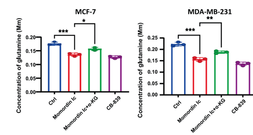
Glutamine levels were measured using an ELISA kit treated with Momordin Ic (10 μM).
Momordin Ic purchased from MedChemExpress. Usage Cited in: Biochem Pharmacol. 2026 May:247:117761. [Abstract]
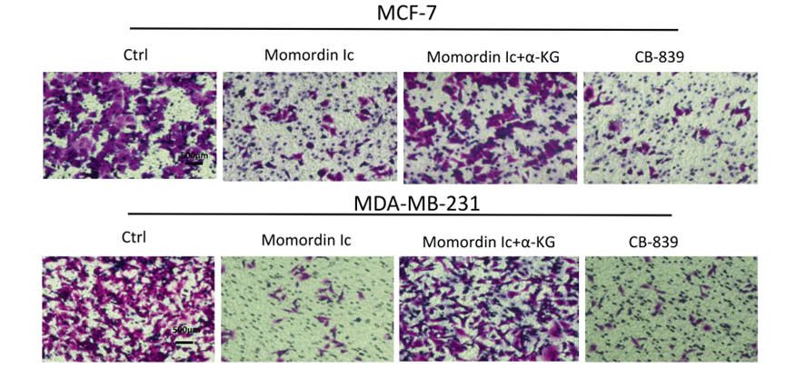
Transwell invasion assays and quantification showing that α-KG supplementation rescued the impaired invasive ability caused by Momordin Ic (10 μM).
Momordin Ic purchased from MedChemExpress. Usage Cited in: Biochem Pharmacol. 2026 May:247:117761. [Abstract]
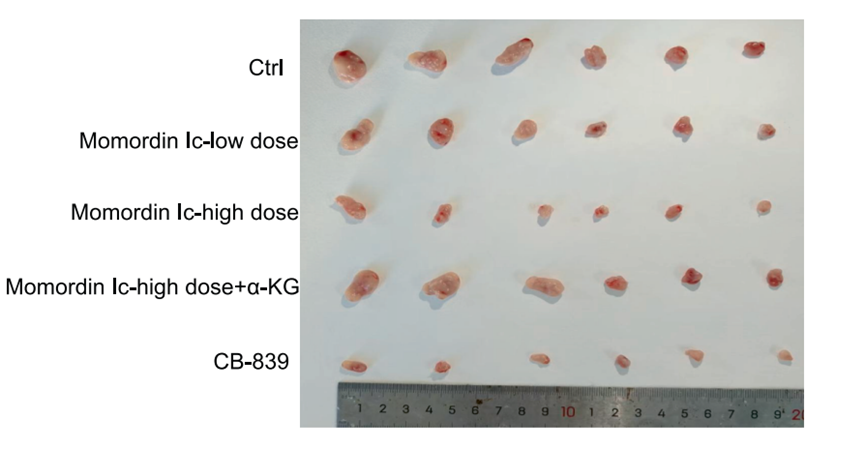
Images of subcutaneous xenograft tumors from each treatment group (Control, low-dose Momordin Ic (50 mg/kg), high-dose Momordin Ic (100 mg/kg), α-KG supplementation, and CB-839).
-
-
Int J Mol Sci
Cycloastragenol Improves Fatty Acid Metabolism Through NHR-49/FAT-7 Suppression and Potent AAK-2 Activation in Caenorhabditis elegans Obesity Model. [Abstract]2026 Jan 13;27(2):772. PMID: 41596421 -
J Pharm Pharmacol
Epidermal growth factor receptor as a potential target of momordin Ic to promote apoptosis of cholangiocarcinoma cells. [Abstract]2022 Jul 15;74(7):996-1005. PMID: 35640567 -
J Pharmacol Sci
Momordin Ic induces G0/1 phase arrest and apoptosis in colon cancer cells by suppressing SENP1/c-MYC signaling pathway. [Abstract]2021 Aug;146(4):249-258. PMID: 34049792


Solvent & Solubility
In Vitro:
DMSO : 100 mg/mL (130.73 mM; Need ultrasonic; Hygroscopic DMSO has a significant impact on the solubility of product, please use newly opened DMSO)
Please refer to the solubility information to select the appropriate solvent. Once prepared, please aliquot and store the solution to prevent product inactivation from repeated freeze-thaw cycles.
Storage method and period of stock solution: -80°C, 6 months; -20°C, 1 month (protect from light). When stored at -80°C, please use it within 6 months. When stored at -20°C, please use it within 1 month.
Please refer to the solubility information to select the appropriate solvent. Once prepared, please aliquot and store the solution to prevent product inactivation from repeated freeze-thaw cycles.
Storage method and period of stock solution: -80°C, 6 months; -20°C, 1 month (protect from light). When stored at -80°C, please use it within 6 months. When stored at -20°C, please use it within 1 month.
Concentration (start) × Volume (start) = Concentration (final) × Volume (final)
In Vivo:
Select the appropriate dissolution method based on your experimental animal and administration route.
- For the following dissolution methods, please ensure to first prepare a clear stock solution using an In Vitro approach and then sequentially add co-solvents:
- To ensure reliable experimental results, the clarified stock solution can be appropriately stored based on storage conditions. As for the working solution for In Vivo experiments, it is recommended to prepare freshly and use it on the same day.
- The percentages shown for the solvents indicate their volumetric ratio in the final prepared solution. If precipitation or phase separation occurs during preparation, heat and/or sonication can be used to aid dissolution.
Add each solvent one by one: 10% DMSO 40% PEG300 5% Tween-80 45% Saline
Solubility: ≥ 6.25 mg/mL (8.17 mM); Clear solution
This protocol yields a clear solution of ≥ 6.25 mg/mL (saturation unknown).
Taking 1 mL working solution as an example, add 100 μL DMSO stock solution (62.5 mg/mL) to 400 μL PEG300, and mix evenly; then add 50 μL Tween-80 and mix evenly; then add 450 μL Saline to adjust the volume to 1 mL.
Preparation of Saline: Dissolve 0.9 g sodium chloride in ddH₂O and dilute to 100 mL to obtain a clear Saline solution.
Add each solvent one by one: 10% DMSO 90% (20% SBE-β-CD in Saline)
Solubility: ≥ 2.5 mg/mL (3.27 mM); Clear solution
This protocol yields a clear solution of ≥ 2.5 mg/mL (saturation unknown).
Taking 1 mL working solution as an example, add 100 μL DMSO stock solution (25.0 mg/mL) to 900 μL 20% SBE-β-CD in Saline, and mix evenly.
Preparation of 20% SBE-β-CD in Saline (4°C, storage for one week): 2 g SBE-β-CD powder is dissolved in 10 mL Saline, completely dissolve until clear.
In Vivo Dissolution Calculator
Please enter the basic information of animal experiments:
-
-
-
-
Recommended: Prepare an additional quantity of animals to account for potential losses during experiments.
Please enter your animal formula composition:
-
%DMSO +
Recommended: Keep the proportion of DMSO in working solution below 2% if your animal is weak.
-
%+
-
+%Tween-80 + +
-
%Saline +
The co-solvents required include: DMSO, . All of co-solvents are available by MedChemExpress (MCE). , Tween 80. All of co-solvents are available by MedChemExpress (MCE).
Working solution concentration: 0.22 mg/mL
Method for preparing stock solution: mg drug dissolved in μL DMSO. Stock solution concentration: mg/mL. * In solvent : -80°C, 6 months; -20°C, 1 month (protect from light)
1. Take μL DMSO stock solution;
2. Add μL .
μL , mix evenly;
3. Then add μL Tween 80, mix evenly;
4. Then add μL
Please ensure that the stock solution in the first step is dissolved to a clear state, and add co-solvents in sequence. You can use ultrasonic heating (ultrasonic cleaner, recommended frequency 20-40 kHz), vortexing, etc. to assist dissolution.

Protocols
-
Collagen-Induced Arthritis
Collagen-induced arthritis (CIA) is an autoimmune murine model of rheumatoid arthritis in which immunization with type II collagen (CII) emulsified in an adjuvant induces a T cell- and autoantibody-driven inflammatory arthritis characterized by synovial hyperplasia, immune cell infiltration, and joint destruction. The model typically relies on genetically susceptible mouse strains (e. g. , DBA/1) and reproduces key features of human rheumatoid arthritis, including anti-collagen immune responses and progressive joint inflammation. Disease onset generally occurs within ~3-4 weeks after immunization, depending on antigen/adjuvant combinations and protocol variation. The immunopathology is driven by adaptive immune activation against CII, leading to systemic and local joint inflammation mediated by pro-inflammatory cytokines and effector immune cells, making CIA a standard preclinical platform for evaluating immunomodulatory and anti-arthritic interventions.
-
Apoptosis
Apoptosis, also called programmed cell death, is generally characterized by distinct morphological characteristics.
-
TUNEL staining for apoptotic DNA fragmentation
TUNEL staining detects DNA strand breaks by using terminal deoxynucleotidyl transferase to add labeled nucleotides to exposed 3′-OH DNA termini, generating either microscopic staining in fixed cells or tissue sections, or fluorescence/cytometric signal in cell suspensions. TUNEL positivity reflects DNA fragmentation but should not be interpreted alone as definitive apoptosis, because TUNEL can also label necrotic, autolytic, mechanically damaged, or DNA-repair-associated DNA breaks.
-
Annexin V plus membrane-impermeant dye apoptosis staining
Annexin V-based apoptosis assays rely on the detection of phosphatidylserine (PS) externalization from the inner leaflet of the plasma membrane to the outer leaflet, an early biochemical hallmark of apoptosis. Fluorescently labeled Annexin V binds PS in a calcium-dependent manner, enabling identification of early apoptotic cells by flow cytometry or fluorescence microscopy. When combined with a membrane-impermeant DNA-binding dye (e. g. , propidium iodide), this approach allows discrimination between viable (Annexin V−/dye−), early apoptotic (Annexin V+/dye−), and late apoptotic or necrotic (Annexin V+/dye+) cell populations by assessing membrane integrity and PS exposure.
-
ROS/oxidative-stress fluorescent staining
ROS/oxidative-stress fluorescent staining uses cell-permeant fluorogenic probes that become fluorescent after oxidation inside cells or tissues; commonly used examples include DCFH-DA/DCFDA for broad cellular oxidant detection, DHE for superoxide-related signal detection, MitoSOX for mitochondrial superoxide-related signal detection, and CellROX probes for oxidative-stress-associated fluorescence readouts. The assay detects probe oxidation rather than a single ROS species unless the probe and analysis method have been chemically validated for that species. DCFH-DA enters cells, is deacetylated by intracellular esterases to DCFH, and produces fluorescent DCF after oxidation, so the readout is used as an operational measure of total cellular oxidative stress rather than a species-specific ROS measurement. DHE and MitoSOX can report superoxide-related oxidation, but red fluorescence alone can include non-specific ethidium-like oxidation products; HPLC or optimized spectral approaches are
-
Autophagy
Autophagy is a process in which eukaryotic cells use lysosomes to degrade their own cytoplasmic proteins and damaged organelles under the regulation of autophagy related gene (Atg). Microtubule-associated proteins light chain 3 (LC3) is recognized as autophagy marker, which transfers from cytoplasmic LC3 (LC3-I) to membrane type (LC3-II). LC3-II/I ratio could be detected by Western Blot and fluorescence microscopy.
-
Apoptosis Solutions
Apoptosis is a regulated, generally non-lytic cell-death pathway that removes unwanted, damaged, infected, or abnormal cells through coordinated morphological changes, caspase activation, DNA fragmentation, and membrane remodeling. The intrinsic apoptosis pathway is controlled mainly by mitochondrial outer membrane permeabilization, BCL-2 family proteins, cytochrome c release, apoptosome formation, caspase-9 activation, and downstream executioner caspase-3/7 activation. The extrinsic apoptosis pathway is initiated by death receptors such as Fas, TNFR, and TRAIL receptors, which recruit adaptor proteins and activate caspase-8 before engaging executioner caspases or mitochondrial amplification through BID cleavage. Apoptosis is linked to many phenotypes, including cancer cell killing, tissue homeostasis, immune regulation, neurodegeneration, infection response, and treatment-induced cytotoxicity; unresolved questions include how apoptosis interacts with necroptosis, pyroptosis, ferroptos
-
Lysosome and acidic-vesicle live-cell staining
Lysosome and acidic-vesicle live-cell staining detects acidic intracellular compartments by using membrane-permeant acidotropic probes that accumulate in low-pH vesicles, including lysosomes, late endosomes, autolysosomes, and acidic phagosomes. LysoTracker staining is commonly used as an intensity-based readout of acidic lysosomal compartment abundance or enlargement, while acridine orange produces green fluorescence in less concentrated compartments and red fluorescence after concentration-dependent accumulation in acidic vesicular organelles. Loss or reduction of acridine-orange red signal can be used as a readout of lysosomal membrane permeabilization or reduced acidic-vesicle integrity. This protocol is designed for live cultured cells and can be adapted for fluorescence microscopy, high-content imaging, plate-reader readout, or flow cytometry when the selected literature supports the readout. Because these dyes report acidotropic accumulation rather than lysosome identity alone,
-
Liver Cancer Modeling
Liver cancer can be classified into primary liver cancer and secondary liver cancer. Secondary liver cancer is the metastatic liver cancer. Primary liver cancer includes hepatocellular carcinoma (HCC), intrahepatic cholangiocarcinoma (ICC) and fibrolamellar HCC, of which HCC is the most common form, accounting for approximately 90% of primary liver cancers[1]. HCC mouse models include chemical agent-induced models, transplanted tumor models, and genetic engineered models.
-
Hepatotoxicity Study
This protocol evaluates hepatotoxicity using complementary in vivo mouse APAP acute liver injury and in vitro hepatocyte-based cytotoxicity readouts. In vivo APAP injury is assessed by serum ALT/AST, liver histology, hepatic glutathione, protein adducts, necrosis, inflammation, and regeneration-related endpoints. In vitro hepatotoxicity is assessed by loss of viability, leakage of ALT/AST/LDH, oxidative-stress markers, mitochondrial function, nuclear morphology, intracellular calcium, and high-content imaging endpoints.
-
Macroautophagy Solutions
Macroautophagy is a conserved lysosome-dependent degradation pathway in which cytoplasmic material is sequestered into double-membrane autophagosomes and delivered to lysosomes for degradation and recycling. The pathway supports cellular homeostasis during nutrient limitation, organelle stress, protein-aggregate accumulation, infection, differentiation, and tissue remodeling by coupling cargo sequestration, autophagosome maturation, lysosomal fusion, and degradation of cargo-derived macromolecules. The core molecular sequence includes initiation by nutrient- and stress-regulated autophagy machinery, autophagosome nucleation, LC3/ATG8-family conjugation to autophagosomal membranes, cargo selection through receptors such as SQSTM1/p62, autophagosome-lysosome fusion, and lysosomal degradation. LC3 was identified as a mammalian homolog of yeast Atg8 that localizes to autophagosomal membranes after processing, and p62/SQSTM1 was shown to connect ubiquitinated cargo with autophagic degradati


Purity & Documentation
-
Data Sheet (283 KB)
-
SDS (394 KB)
- English - EN (394 KB)
- Français - FR (394 KB)
- Deutsch - DE (394 KB)
- Norwegian - NO (394 KB)
- Español - ES (394 KB)
- Swedish - SV (394 KB)
- Italian - IT (394 KB)
- Korean - KR (394 KB)
- Portuguese - PT (394 KB)
-
Handling Instructions (2659 KB)
References
[1]. Mi Y, et al. Momordin Ic couples apoptosis with autophagy in human hepatoblastoma cancer cells by reactive oxygen species (ROS)-mediated PI3K/Akt and MAPK signaling pathways. Free Radic Biol Med. 2016 Jan;90:230-42. [Content Brief]
[2]. Wang W, et al. Cytotoxic effects of recombinant proteins enhanced by momordin Ic are dependent on cholesterol and ganglioside GM1. Toxicon. 2023 Jun 15;229:107129. [Content Brief]
[3]. Xianjun F, et al. Momordin Ic induces G0/1 phase arrest and apoptosis in colon cancer cells by suppressing SENP1/c-MYC signaling pathway. J Pharmacol Sci. 2021 Aug;146(4):249-258. [Content Brief]
[4]. Li Y, et al. Acceleration of gastrointestinal transit by momordin Ic in mice: possible involvement of 5-hydroxytryptamine, 5-HT(2) receptors and prostaglandins. Eur J Pharmacol. 2000 Mar 24;392(1-2):71-7. [Content Brief]
[5]. Matsuda H, et al. Roles of capsaicin-sensitive sensory nerves, endogenous nitric oxide, sulfhydryls, and prostaglandins in gastroprotection by momordin Ic, an oleanolic acid oligoglycoside, on ethanol-induced gastric mucosal lesions in rats. Life Sci. 1999;65(2):PL27-32. [Content Brief]
[6]. Kim NY, et al. Momordin Ic and oleanolic acid from Kochiae Fructus reduce carbon tetrachloride-induced hepatotoxicity in rats. J Med Food. 2005 Summer;8(2):177-83. [Content Brief]


Complete Stock Solution Preparation Table
Please refer to the solubility information to select the appropriate solvent. Once prepared, please aliquot and store the solution to prevent product inactivation from repeated freeze-thaw cycles.
Storage method and period of stock solution: -80°C, 6 months; -20°C, 1 month (protect from light). When stored at -80°C, please use it within 6 months. When stored at -20°C, please use it within 1 month.
| Optional Solvent | Concentration Solvent Mass | 1 mg | 5 mg | 10 mg | 25 mg |
|---|---|---|---|---|---|
| DMSO | 1 mM | 1.3073 mL | 6.5365 mL | 13.0729 mL | 32.6823 mL |
| 5 mM | 0.2615 mL | 1.3073 mL | 2.6146 mL | 6.5365 mL | |
| 10 mM | 0.1307 mL | 0.6536 mL | 1.3073 mL | 3.2682 mL | |
| 15 mM | 0.0872 mL | 0.4358 mL | 0.8715 mL | 2.1788 mL | |
| 20 mM | 0.0654 mL | 0.3268 mL | 0.6536 mL | 1.6341 mL | |
| 25 mM | 0.0523 mL | 0.2615 mL | 0.5229 mL | 1.3073 mL | |
| 30 mM | 0.0436 mL | 0.2179 mL | 0.4358 mL | 1.0894 mL | |
| 40 mM | 0.0327 mL | 0.1634 mL | 0.3268 mL | 0.8171 mL | |
| 50 mM | 0.0261 mL | 0.1307 mL | 0.2615 mL | 0.6536 mL | |
| 60 mM | 0.0218 mL | 0.1089 mL | 0.2179 mL | 0.5447 mL | |
| 80 mM | 0.0163 mL | 0.0817 mL | 0.1634 mL | 0.4085 mL | |
| 100 mM | 0.0131 mL | 0.0654 mL | 0.1307 mL | 0.3268 mL |


Momordin Ic Related Classifications
HY-N0330 Related Classifications
 Powered by Bioz