



Moroxydine hydrochloride
Based on 1 publication(s) in Google Scholar
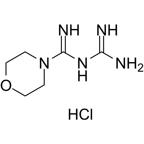
Moroxydine (ABOB) hydrochloride is a broad-spectrum agent with multi-antiviral activities against DNA and RNA viruses, including influenza virus, herpes simplex, varicella zoster, measles, mumps disease, hepatitis C virus, etc. Moroxydine hydrochloride exhibits excellent antiviral activity and shows low cytotoxicity to cells infected by dsRNA viruses (grass carp reovirus, GCRV) and large DNA viruses (giant salamander iridovirus, GSIV). Moroxydine hydrochloride blocks the GCRV-induced cytopathic effects and eliminates nucleocapsids in ctenopharyngodon idella kidney (CIK) cells to keep the normal morphological structure. Moroxydine hydrochloride significantly inhibits the apoptosis, the caspase 3 activity, Bax expression and down-regulates Bcl-2 levels.
For research use only. We do not sell to patients.
- Purity : 99.36%
- CAS No.: 3160-91-6
- Formula: C6H14ClN5O
- Molecular Weight:207.66
-
Storage:
4°C, sealed storage, away from moisture
* In solvent : -80°C, 6 months; -20°C, 1 month (sealed storage, away from moisture)

To place orders, for customer services and technical support, please contact: MedChemExpress USA
Tel: 609-228-6898 E-mail: [email protected] [email protected]


-
 Biological Activity
-
Chemical Information
-
Solvent & Solubility
- Protocols
- Purity & Documentation
- References
-
 Help & FAQs


-
HY-13318
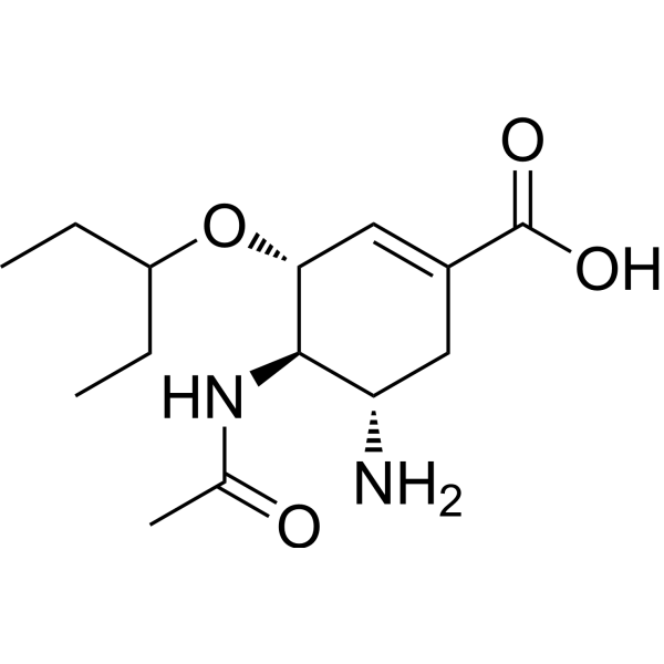
Oseltamivir acidOseltamivir phosphate Metabolite -
HY-17422
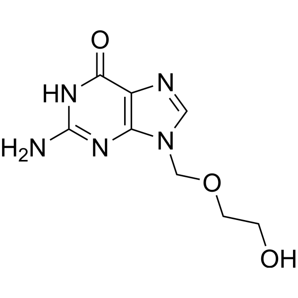
AcyclovirHSV Inhibitor -
HY-B0434
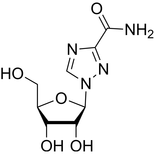
RibavirinAntiviral Agent -
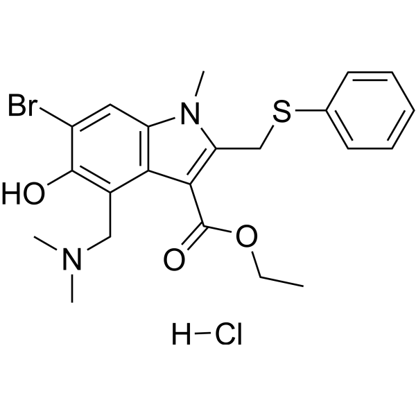
HY-14904A
Umifenovir hydrochlorideAntiviral Agent -
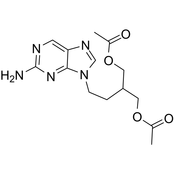
HY-17426
FamciclovirAntiviral Agent -
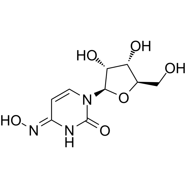
HY-125033
EIDD-1931Anti-Virus Agent -
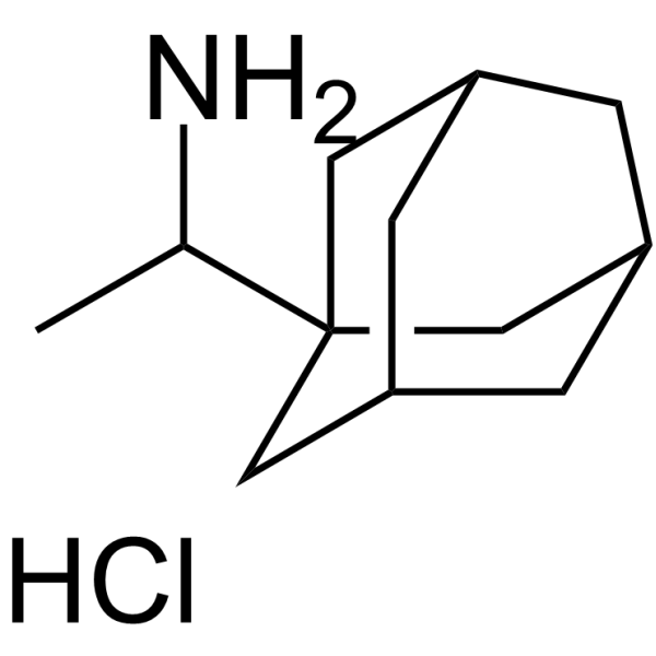
HY-B0338A
Rimantadine hydrochlorideAntiviral Agent -
HY-17015
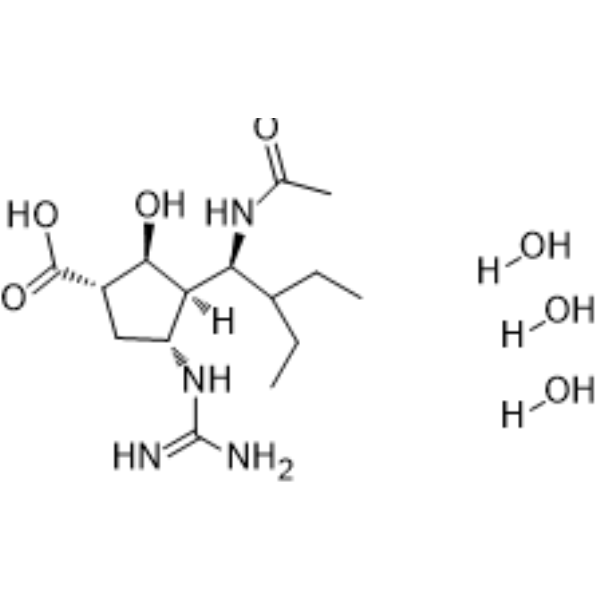
Peramivir trihydrateInfluenza Virus Inhibitor -

HY-17424R
Penciclovir (Standard)Anti-herpesvirus Agent -

HY-B0420AR
Moroxydine hydrochloride (Standard)Antiviral Agent -
HY-19743
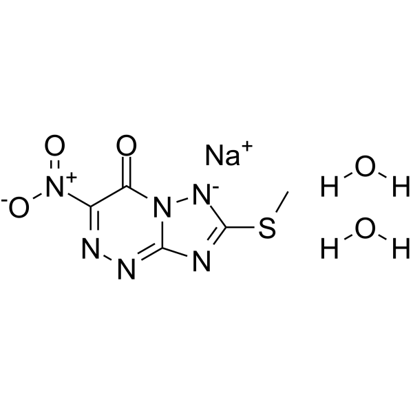
TriazavirinAntiviral Agent -
HY-13210
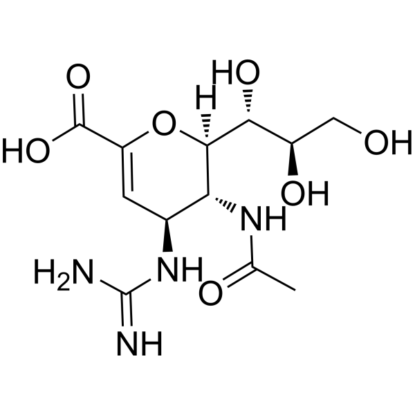
ZanamivirNeuraminidase Inhibitor
Publications Citing Use of MedChemExpress (MCE) Moroxydine hydrochloride
MoreAll Caspase Isoforms
More

Biological Activity
Description
In Vitro
Moroxydine hydrochloride (1-100 μg/mL, 24-96 h) shows protective effects against grass carp reovirus (GCRV) in Ctenopharyngodon idella kidney (CIK) cells (TC50 = 1246.0 μg/mL, SC = 218.5 μg/mL) and grass carp ovary (GCO) cells (TC50 = 117.2 μg/mL, SC = 140.9 μg/mL)[1][2].
Moroxydine hydrochloride (1-100 μg/mL, 24-96 h) shows protective effects against giant salamander iridovirus (GSIV) in epithelioma papulosum cyprinid (EPC) cells, with a TC50 of 1486.3 μg/mL and a safe concentration (SC) of 209.7 μg/mL[2].
Moroxydine hydrochloride (40 μg/mL, 48-96 h) blocks the virus-induced CPE and cell death within 96 h and maintained the normal structure and nucleocapsids[1].
Moroxydine hydrochloride (40 μg/mL, 48-96 h) significantly decreases the apoptosis in CIK cells (GCRV), GCO cells (GCRV) and EPC cells (GSIV)[1][2].
Moroxydine (ABOB) hydrochloride (12-48 h) significantly inhibits the caspase 3 activity, Bax expression and down-regulates Bcl-2 at 48 h in CIK cells[1].
Moroxydine hydrochloride (20 μg/mL, 12-72 h) shows time-dependent suppression of viral gene expression in CIK cells(GCRV), GCO cells (GCRV) and EPC cells (GSIV)[1][2].
Moroxydine hydrochloride (20 μg/mL, 72 h) arrests cell cycle in the period of G1/S and G2/M of CIK cells[3].
Moroxydine hydrochloride (3-7 days) inhibits the GCRV-induced upregulation of immune genes (IL-1β, IFN-I, TNF-α) in the kidney, liver, muscle and gill[3].
MedChemExpress (MCE) has not independently confirmed the accuracy of these methods. They are for reference only. Further protocols information, click here.
-
Cell Line:GCRV infected CIK cells
-
Concentration:1, 2.5, 6.3, 15.9, 39.8, 100 μg/mL
-
Incubation Time:24, 48, 72, 96 h
-
Result:Presented a better protective-efficiency than Ribavirin(HY-B0434).
Showed no significant difference when expose GCRV-infected CIK cells in 39.8 μg/mL compared with virus-free group at 96 h.
-
Cell Line:GCRV infected CIK cells
-
Concentration:40 μg/mL
-
Incubation Time:48 h
-
Result:Exhibited a positive inhibition in the GCRV infected cells compared with negative control.
-
Cell Line:GCRV infected CIK cells
-
Concentration:20 μg/mL
-
Incubation Time:72 h
-
Result:Arrested cell cycle in the period of G1/S and G2/M of CIK cells
-
Cell Line:GCRV infected CIK cells
-
Concentration:20 μg/mL
-
Incubation Time:12, 24, 48, 72 h
-
Result:Suppressed viral genes (vp1, vp2, vp3 and vp4) at 12 h following a slow decrease from 12 h to 72 h post medication.
Inhibited the IL-1β expression.


Chemical Information
-
CAS No. 3160-91-6
-
Appearance Solid
-
Molecular Weight 207.66
-
Formula C6H14ClN5O
-
Color White to off-white
-
SMILES
N=C(N1CCOCC1)NC(N)=N.Cl
-
Synonyms
ABOB hydrochloride
-
Shipping
Room temperature in continental US; may vary elsewhere.
-
Storage
4°C, sealed storage, away from moisture
* In solvent : -80°C, 6 months; -20°C, 1 month (sealed storage, away from moisture)


Publications (1)
-
Journal Impact Factor
-
Most Recent
-
Pharmaceuticals (Basel)
2024 Sep 19;17(9):1234. PMID: 39338396


Solvent & Solubility
In Vitro:
H2O : ≥ 100 mg/mL (481.56 mM)
DMSO : 50 mg/mL (240.78 mM; Need ultrasonic; Hygroscopic DMSO has a significant impact on the solubility of product, please use newly opened DMSO)
* "≥" means soluble, but saturation unknown.
Please refer to the solubility information to select the appropriate solvent. Once prepared, please aliquot and store the solution to prevent product inactivation from repeated freeze-thaw cycles.
Storage method and period of stock solution: -80°C, 6 months; -20°C, 1 month (sealed storage, away from moisture). When stored at -80°C, please use it within 6 months. When stored at -20°C, please use it within 1 month.
* Note: If you choose water as the stock solution, please dilute it to the working solution, then filter and sterilize it with a 0.22 μm filter before use.
Please refer to the solubility information to select the appropriate solvent. Once prepared, please aliquot and store the solution to prevent product inactivation from repeated freeze-thaw cycles.
Storage method and period of stock solution: -80°C, 6 months; -20°C, 1 month (sealed storage, away from moisture). When stored at -80°C, please use it within 6 months. When stored at -20°C, please use it within 1 month.
* Note: If you choose water as the stock solution, please dilute it to the working solution, then filter and sterilize it with a 0.22 μm filter before use.
Concentration (start) × Volume (start) = Concentration (final) × Volume (final)
In Vivo:
Select the appropriate dissolution method based on your experimental animal and administration route.
- For the following dissolution methods, please ensure to first prepare a clear stock solution using an In Vitro approach and then sequentially add co-solvents:
- To ensure reliable experimental results, the clarified stock solution can be appropriately stored based on storage conditions. As for the working solution for In Vivo experiments, it is recommended to prepare freshly and use it on the same day.
- The percentages shown for the solvents indicate their volumetric ratio in the final prepared solution. If precipitation or phase separation occurs during preparation, heat and/or sonication can be used to aid dissolution.
Add each solvent one by one: 10% DMSO 40% PEG300 5% Tween-80 45% Saline
Solubility: ≥ 2.5 mg/mL (12.04 mM); Clear solution
This protocol yields a clear solution of ≥ 2.5 mg/mL (saturation unknown).
Taking 1 mL working solution as an example, add 100 μL DMSO stock solution (25.0 mg/mL) to 400 μL PEG300, and mix evenly; then add 50 μL Tween-80 and mix evenly; then add 450 μL Saline to adjust the volume to 1 mL.
Preparation of Saline: Dissolve 0.9 g sodium chloride in ddH₂O and dilute to 100 mL to obtain a clear Saline solution.
Add each solvent one by one: 10% DMSO 90% (20% SBE-β-CD in Saline)
Solubility: ≥ 2.5 mg/mL (12.04 mM); Clear solution
This protocol yields a clear solution of ≥ 2.5 mg/mL (saturation unknown).
Taking 1 mL working solution as an example, add 100 μL DMSO stock solution (25.0 mg/mL) to 900 μL 20% SBE-β-CD in Saline, and mix evenly.
Preparation of 20% SBE-β-CD in Saline (4°C, storage for one week): 2 g SBE-β-CD powder is dissolved in 10 mL Saline, completely dissolve until clear.
For the following dissolution methods, please prepare the working solution directly:
It is recommended to prepare fresh solutions and use them promptly within a short period of time.
The percentages shown for the solvents indicate their volumetric ratio in the final prepared solution. If precipitation or phase separation occurs during preparation, heat and/or sonication can be used to aid dissolution.
Add each solvent one by one: PBS
Solubility: 100 mg/mL (481.56 mM); Clear solution; Need ultrasonic
In Vivo Dissolution Calculator
Please enter the basic information of animal experiments:
-
-
-
-
Recommended: Prepare an additional quantity of animals to account for potential losses during experiments.
Working solution concentration: 0.22 mg/mL
This product has good water solubility, please refer to the measured solubility data in water/PBS/Saline for details.

Protocols
-
Research Protocol for Infectious Diseases
Infectious-disease experiments test how pathogens interact with host barriers, innate immune receptors, inflammatory signaling, pathogen replication, and tissue injury; pattern-recognition receptors such as TLRs, RIG-I-like receptors, NOD-like receptors, and inflammasomes detect microbial molecules and activate NF-κB, interferon, and cytokine responses. The central hypothesis is that infection severity reflects the balance between pathogen burden and host response: protective inflammation restricts pathogen growth, whereas excessive or mislocalized inflammation contributes to tissue damage and disease phenotype. Unresolved questions include which host pathways are protective versus pathogenic, why some infection models fail to translate to human disease, and which combined readouts best predict clinically relevant infection outcomes.
-
RNA extraction experimental
By lysing cells, releasing RNA, and removing impurities such as proteins and DNA, high-purity RNA products are finally obtained. The commonly used traditional method is the guanidine isothiocyanate/phenol/chloroform method (Trizol), which is suitable for a variety of animal materials including animal tissues, microorganisms, cultured cells, etc., and most plant materials.
-
Apoptosis
Apoptosis, also called programmed cell death, is generally characterized by distinct morphological characteristics.
-
TUNEL staining for apoptotic DNA fragmentation
TUNEL staining detects DNA strand breaks by using terminal deoxynucleotidyl transferase to add labeled nucleotides to exposed 3′-OH DNA termini, generating either microscopic staining in fixed cells or tissue sections, or fluorescence/cytometric signal in cell suspensions. TUNEL positivity reflects DNA fragmentation but should not be interpreted alone as definitive apoptosis, because TUNEL can also label necrotic, autolytic, mechanically damaged, or DNA-repair-associated DNA breaks.
-
Cell Cytotoxicity Assay
Cytotoxicity assays are usually based on the assessment of cell membrane damage, which can also be indirectly detected by measuring cell viability. Detection methods include MTT assay, CKK-8 assay, LDH assay and ATP assay, etc.
-
Mammalian live/dead viability and cytotoxicity staining
Live/dead viability and cytotoxicity staining assays are based on the simultaneous detection of intracellular esterase activity in metabolically active (viable) cells and membrane integrity loss in non-viable cells. In commonly used dual-staining approaches, membrane-permeant fluorogenic substrates are converted by intracellular esterases into fluorescent products in live cells, while impermeant DNA-binding dyes selectively enter cells with compromised plasma membranes and label nucleic acids in dead or dying cells, enabling discrimination between viable and non-viable populations by fluorescence microscopy or flow cytometry.
-
Annexin V plus membrane-impermeant dye apoptosis staining
Annexin V-based apoptosis assays rely on the detection of phosphatidylserine (PS) externalization from the inner leaflet of the plasma membrane to the outer leaflet, an early biochemical hallmark of apoptosis. Fluorescently labeled Annexin V binds PS in a calcium-dependent manner, enabling identification of early apoptotic cells by flow cytometry or fluorescence microscopy. When combined with a membrane-impermeant DNA-binding dye (e. g. , propidium iodide), this approach allows discrimination between viable (Annexin V−/dye−), early apoptotic (Annexin V+/dye−), and late apoptotic or necrotic (Annexin V+/dye+) cell populations by assessing membrane integrity and PS exposure.
-
Apoptosis Solutions
Apoptosis is a regulated, generally non-lytic cell-death pathway that removes unwanted, damaged, infected, or abnormal cells through coordinated morphological changes, caspase activation, DNA fragmentation, and membrane remodeling. The intrinsic apoptosis pathway is controlled mainly by mitochondrial outer membrane permeabilization, BCL-2 family proteins, cytochrome c release, apoptosome formation, caspase-9 activation, and downstream executioner caspase-3/7 activation. The extrinsic apoptosis pathway is initiated by death receptors such as Fas, TNFR, and TRAIL receptors, which recruit adaptor proteins and activate caspase-8 before engaging executioner caspases or mitochondrial amplification through BID cleavage. Apoptosis is linked to many phenotypes, including cancer cell killing, tissue homeostasis, immune regulation, neurodegeneration, infection response, and treatment-induced cytotoxicity; unresolved questions include how apoptosis interacts with necroptosis, pyroptosis, ferroptos
-
Cell Viability Determination by MTT Colorimetric Assay
The following protocol uses the MTT colorimetric assay as a classic literature-established method for assessing cell viability/metabolic activity in cultured mammalian cells. MTT[3-(4,5-dimethylthiazol-2-yl)-2,5-diphenyltetrazolium bromide] is reduced by metabolically active cells to a colored formazan product; the amount of formazan is quantified spectrophotometrically and provides an indirect measure of metabolically active viable cells. Importantly, MTT reduction reflects cellular oxidoreductase/metabolic activity rather than an absolute direct count of living cells, so changes in cellular metabolism can alter the signal independently of cell number.


Purity & Documentation
-
Data Sheet (279 KB)
-
SDS (393 KB)
- English - EN (393 KB)
- Français - FR (393 KB)
- Deutsch - DE (393 KB)
- Norwegian - NO (393 KB)
- Español - ES (393 KB)
- Swedish - SV (393 KB)
- Italian - IT (393 KB)
- Korean - KR (393 KB)
- Portuguese - PT (393 KB)
-
Handling Instructions (2659 KB)
References
[1]. Yu XB, et al. Moroxydine hydrochloride inhibits grass carp reovirus replication and suppresses apoptosis in Ctenopharyngodon idella kidney cells. Antiviral Res. 2016 Jul;131:156-65. [Content Brief]
[2]. Yu XB, et al. In vitro antiviral efficacy of moroxydine hydrochloride and ribavirin against grass carp reovirus and giant salamander iridovirus. Dis Aquat Organ. 2016 Oct 27;121(3):189-199. [Content Brief]
[3]. Yu XB, et al. Effects of moroxydine hydrochloride and ribavirin on the cellular growth and immune responses by inhibition of GCRV proliferation. Res Vet Sci. 2018 Apr;117:37-44. [Content Brief]


Complete Stock Solution Preparation Table
Please refer to the solubility information to select the appropriate solvent. Once prepared, please aliquot and store the solution to prevent product inactivation from repeated freeze-thaw cycles.
Storage method and period of stock solution: -80°C, 6 months; -20°C, 1 month (sealed storage, away from moisture). When stored at -80°C, please use it within 6 months. When stored at -20°C, please use it within 1 month.
| Optional Solvent | Concentration Solvent Mass | 1 mg | 5 mg | 10 mg | 25 mg |
|---|---|---|---|---|---|
| DMSO / H2O | 1 mM | 4.8156 mL | 24.0778 mL | 48.1556 mL | 120.3891 mL |
| 5 mM | 0.9631 mL | 4.8156 mL | 9.6311 mL | 24.0778 mL | |
| 10 mM | 0.4816 mL | 2.4078 mL | 4.8156 mL | 12.0389 mL | |
| 15 mM | 0.3210 mL | 1.6052 mL | 3.2104 mL | 8.0259 mL | |
| 20 mM | 0.2408 mL | 1.2039 mL | 2.4078 mL | 6.0195 mL | |
| 25 mM | 0.1926 mL | 0.9631 mL | 1.9262 mL | 4.8156 mL | |
| 30 mM | 0.1605 mL | 0.8026 mL | 1.6052 mL | 4.0130 mL | |
| 40 mM | 0.1204 mL | 0.6019 mL | 1.2039 mL | 3.0097 mL | |
| 50 mM | 0.0963 mL | 0.4816 mL | 0.9631 mL | 2.4078 mL | |
| 60 mM | 0.0803 mL | 0.4013 mL | 0.8026 mL | 2.0065 mL | |
| 80 mM | 0.0602 mL | 0.3010 mL | 0.6019 mL | 1.5049 mL | |
| 100 mM | 0.0482 mL | 0.2408 mL | 0.4816 mL | 1.2039 mL |
* Note: If you choose water as the stock solution, please dilute it to the working solution, then filter and sterilize it with a 0.22 μm filter before use.


Moroxydine hydrochloride Related Classifications
HY-B0420A Related Classifications
 Powered by Bioz