



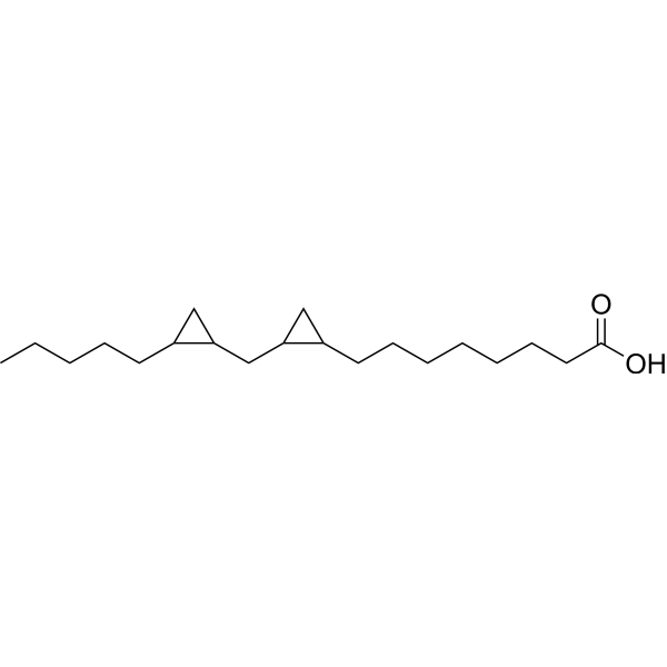
DCP-LA
Based on 9 publication(s) in Google Scholar
DCP-LA (FR236924), a linoleic acid derivative, selectively and directly activates PKCε. DCP-LA activates Ca(2+)/calmodulin-dependent protein kinase II (CaMKII) and inhibits protein phosphatase-1 (PP-1) to stimulate AMPA receptor exocytosis. DCP-LA inhibits activation of caspase-3/-9 and protects neurons at least in part from oxidative stress-induced apoptosis.
For research use only. We do not sell to patients.
- Purity : 98.0%
- CAS No.: 28399-31-7
- Formula: C20H36O2
- Molecular Weight:308.50
-
Storage:Pure form -20°C, 3 years , 4°C, 2 years ; In solvent -80°C, 6 months , -20°C, 1 month

To place orders, for customer services and technical support, please contact: MedChemExpress USA
Tel: 609-228-6898 E-mail: [email protected] [email protected]


-
 Biological Activity
-
Chemical Information
-
Solvent & Solubility
- Protocols
- Purity & Documentation
- References
-
 Help & FAQs


-
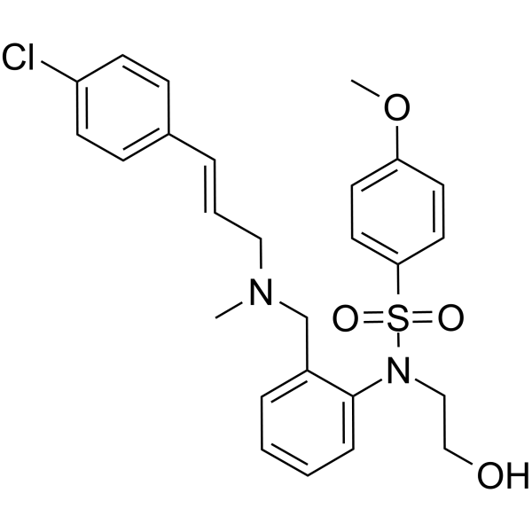
HY-15465
KN-93CaMK II Inhibitor -
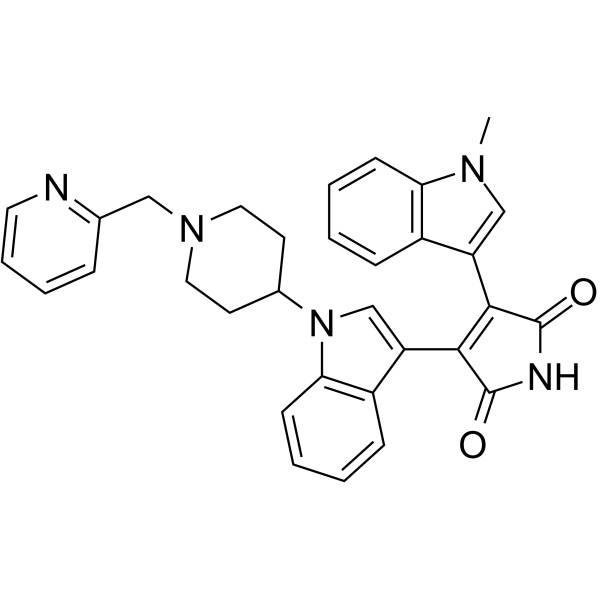
HY-10342
EnzastaurinPKCβ Inhibitor -
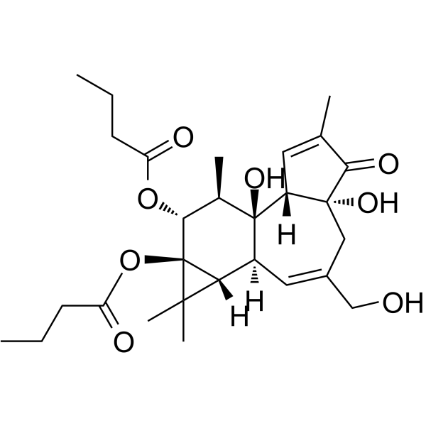
HY-18985
Phorbol 12,13-dibutyratePKC Activator -

HY-18739
Phorbol 12-myristate 13-acetatePKC Activator -
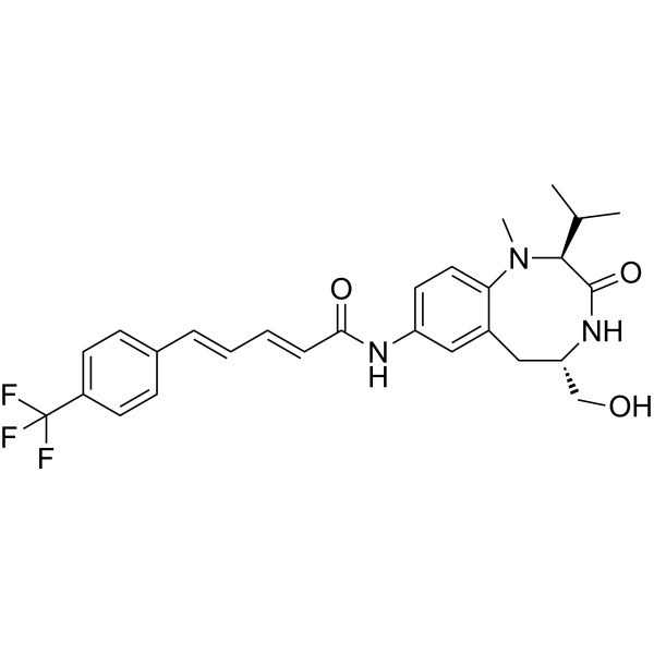
HY-12359
TPPBPKC Activator -
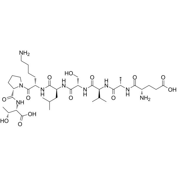
HY-P0154
Epsilon-V1-2PKCε Inhibitor -
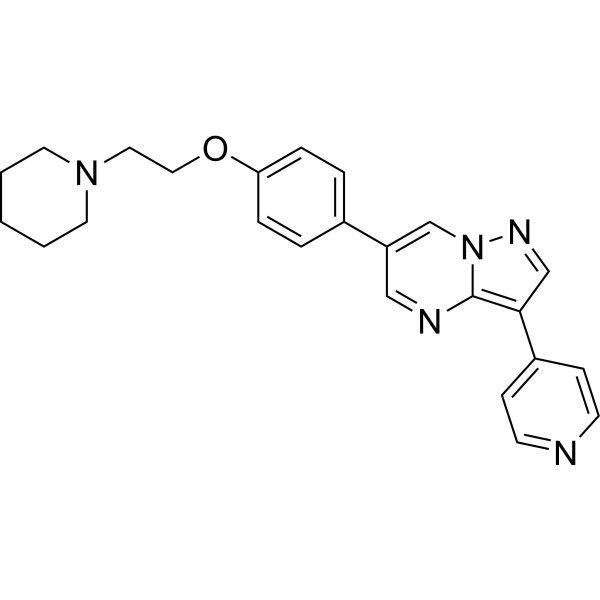
HY-13418A
DorsomorphinAMPK Inhibitor -
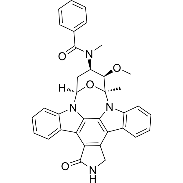
HY-10230
MidostaurinMulti-targeted Protein Kinase Inhibitor -
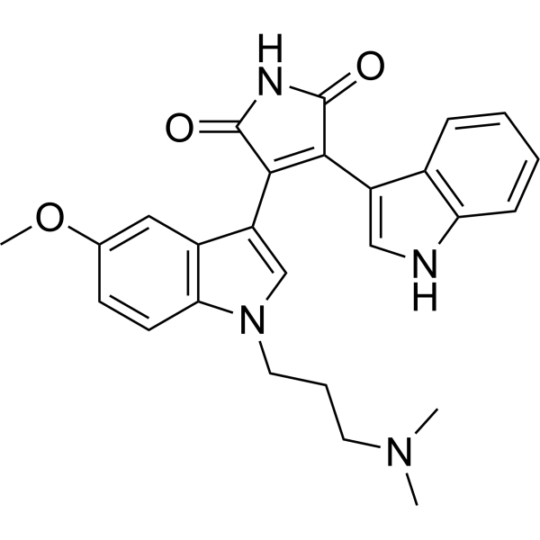
HY-13689
Go 6983PKC Inhibitor -
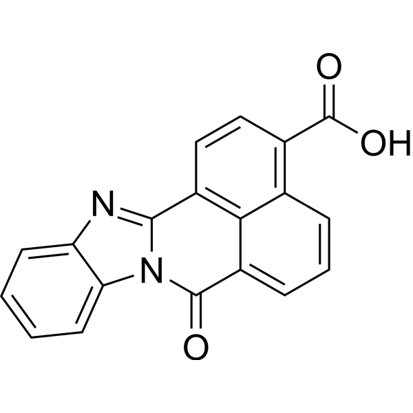
HY-19805
STO-609CaMKK Inhibitor -
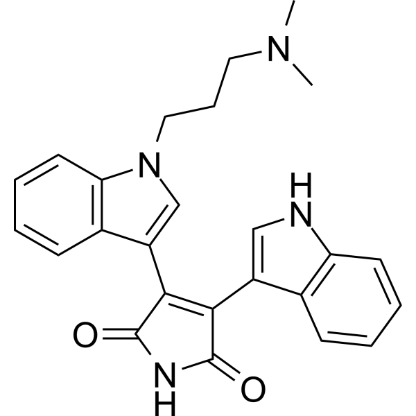
HY-13867
Bisindolylmaleimide IPKC Inhibitor -
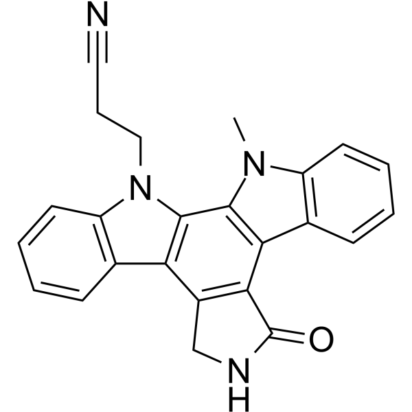
HY-10183
Go6976PKC Inhibitor
Publications Citing Use of MedChemExpress (MCE) DCP-LA
More- Cell Metab. 2023 Jan 3;35(1):101-117.e11. [Abstract]
- Cell Death Dis. 2022 Mar 28;13(3):275. [Abstract]
- J Genet Genomics. 2024 Nov;51(11):1204-1214. [Abstract]
- J Ethnopharmacol. 2025 Feb 10;338(Pt 3):119003. [Abstract]
- Cell Mol Life Sci. 2026 May 22. [Abstract]
- Biochem Pharmacol. 2025 Aug 8;242(Pt 3):117223. [Abstract]
- Transl Res. 2025 Jul:281:1-13. [Abstract]
- Int Immunopharmacol. 2026 Feb 15:171:116139. [Abstract]
- J Ovarian Res. 2024 Sep 13;17(1):185. [Abstract]
 Customer Validation & Images

-
WB

-
Cell Imaging/Staining

-
RT-PCR


Biological Activity
Description
|
PKCε |
CaMK II |
In Vitro
DCP-LA (100 nM; 15 min) activates cytosolic PKCε by binding to the phosphatidylserine binding/associating sites Arg50 and Ile89, without translocation towards the cell surface in PL-12 cells[1].
DCP-LA (10, 100, 100 nM; 10 min; 35 °C) activates CaMKII and inhibits PP-1 in rat hippocampal neurons[2].
DCP-LA (100 nM; 20 min) increases expression of the receptors on the plasma membrane, responsible for potentiate AMPA receptor responses in rat hippocampal slices[2].
DCP-LA (100 nM; 10 min) results facilitation of hippocampal synaptic transmission[2].
DCP-LA (100 nM; 24 h) prevents the Sodium nitroprusside (SNP, HY-10219) effect and rescues neurons from SNP-induced degradation[3].
DCP-LA (100 nM; 12 h) abolishes the caspase-3/9 activation induced by (SNP, HY-10219) (1 mM)[3].
MedChemExpress (MCE) has not independently confirmed the accuracy of these methods. They are for reference only. Further protocols information, click here.
-
Cell Line:Rat cerebral cortical neurons
-
Concentration:100 nM
-
Incubation Time:24 hours; accompanied with 1 mM Sodium nitroprusside (SNP)
-
Result:Prevented SNP-induced neuronal cell death.
In Vivo
MedChemExpress (MCE) has not independently confirmed the accuracy of these methods. They are for reference only.
-
Animal Model:Middle cerebral artery (MCA) occlusion in mouse model (male CB-17 mice, 5-8 weeks old)[3]
-
Dosage:1 mg/kg
-
Administration:Oral gavage; once daily for 7 days; sacrificed mice on days 28
-
Result:Significantly diminished degraded area due to cerebral infarction, with the rescued area reaching 82%.


Chemical Information
-
CAS No. 28399-31-7
-
Appearance Liquid (Density: 0.968 g/cm3)
-
Molecular Weight 308.50
-
Formula C20H36O2
-
Color Colorless to light yellow
-
SMILES
O=C(O)CCCCCCCC1C(CC2C(CCCCC)C2)C1
-
Synonyms
FR236924
-
Shipping
Room temperature in continental US; may vary elsewhere.
-
Storage
Pure form -20°C 3 years 4°C 2 years In solvent -80°C 6 months -20°C 1 month


Publications (9)
-
Journal Impact Factor
-
Most Recent
-
Cell Metab
Discovery of a potent allosteric activator of DGKQ that ameliorates obesity-induced insulin resistance via the sn-1,2-DAG-PKCε signaling axis. [Abstract]2023 Jan 3;35(1):101-117.e11. PMID: 36525963 -
Cell Death Dis
Hepatic deficiency of selenoprotein S exacerbates hepatic steatosis and insulin resistance. [Abstract]2022 Mar 28;13(3):275. PMID: 35347118 -
J Genet Genomics
Compound heterozygous mutations of NTNG2 cause intellectual disability via inhibition of the CaMKII signaling. [Abstract]2024 Nov;51(11):1204-1214. PMID: 39151821 -
J Ethnopharmacol
Buyang Huanwu Decoction restores the balance of mitochondrial dynamics after cerebral ischemia-reperfusion through calcium overload reduction by the PKCε-Nampt-Sirt5 axis. [Abstract]2025 Feb 10;338(Pt 3):119003. PMID: 39528118 -
Cell Mol Life Sci
Obesity-induced oleic acid metabolic dysregulation may exacerbate osteoarthritis through the degradation of SOX9. [Abstract]2026 May 22. PMID: 42171703
DCP-LA purchased from MedChemExpress. Usage Cited in: Cell Mol Life Sci. 2026 May 22. [Abstract]
Immunoblot detection of p-CaMK2, CaMK2, ACAN and COL2A1 protein in mouse primary chondrocytes treated with DCP-LA (0.2 µM) for 7 d.
DCP-LA purchased from MedChemExpress. Usage Cited in: Cell Mol Life Sci. 2026 May 22. [Abstract]
Alcian blue and Toluidine blue staining to examine the proteoglycan content after DCP-LA (0.2 µM) treatment for 14 d.
DCP-LA purchased from MedChemExpress. Usage Cited in: Cell Mol Life Sci. 2026 May 22. [Abstract]
Quantification of mRNA levels for ACAN, COL2A1, MMP13 and ADAMTS5 in mouse primary chondrocytes treated with DCP-LA (0.2 µM) for 3 d.
-
Biochem Pharmacol
Triptolide induces the secretion of insulin, through which it alleviates the tauopathies of Alzheimer's disease via inhibiting the phosphorylation of tau. [Abstract]2025 Aug 8;242(Pt 3):117223. PMID: 40784568 -
Transl Res
2025 Jul:281:1-13. PMID: 40409587 -
Int Immunopharmacol
Multi-omics integration identifies CACNG3 as a synaptic calcium channel regulator in Alzheimer's disease: Mechanistic insights and therapeutic implications. [Abstract]2026 Feb 15:171:116139. PMID: 41483619 -
J Ovarian Res
Long non-coding ribonucleic acid SNHG18 induced human granulosa cell apoptosis via disruption of glycolysis in ovarian aging. [Abstract]2024 Sep 13;17(1):185. PMID: 39272131


Solvent & Solubility
In Vitro:
DMSO : 100 mg/mL (324.15 mM; Need ultrasonic; Hygroscopic DMSO has a significant impact on the solubility of product, please use newly opened DMSO)
Please refer to the solubility information to select the appropriate solvent. Once prepared, please aliquot and store the solution to prevent product inactivation from repeated freeze-thaw cycles.
Storage method and period of stock solution: -80°C, 6 months; -20°C, 1 month. When stored at -80°C, please use it within 6 months. When stored at -20°C, please use it within 1 month.
Please refer to the solubility information to select the appropriate solvent. Once prepared, please aliquot and store the solution to prevent product inactivation from repeated freeze-thaw cycles.
Storage method and period of stock solution: -80°C, 6 months; -20°C, 1 month. When stored at -80°C, please use it within 6 months. When stored at -20°C, please use it within 1 month.
Concentration (start) × Volume (start) = Concentration (final) × Volume (final)
In Vivo:
Select the appropriate dissolution method based on your experimental animal and administration route.
- For the following dissolution methods, please ensure to first prepare a clear stock solution using an In Vitro approach and then sequentially add co-solvents:
- To ensure reliable experimental results, the clarified stock solution can be appropriately stored based on storage conditions. As for the working solution for In Vivo experiments, it is recommended to prepare freshly and use it on the same day.
- The percentages shown for the solvents indicate their volumetric ratio in the final prepared solution. If precipitation or phase separation occurs during preparation, heat and/or sonication can be used to aid dissolution.
Add each solvent one by one: 10% DMSO 40% PEG300 5% Tween-80 45% Saline
Solubility: ≥ 2.5 mg/mL (8.10 mM); Clear solution
This protocol yields a clear solution of ≥ 2.5 mg/mL (saturation unknown).
Taking 1 mL working solution as an example, add 100 μL DMSO stock solution (25.0 mg/mL) to 400 μL PEG300, and mix evenly; then add 50 μL Tween-80 and mix evenly; then add 450 μL Saline to adjust the volume to 1 mL.
Preparation of Saline: Dissolve 0.9 g sodium chloride in ddH₂O and dilute to 100 mL to obtain a clear Saline solution.
Add each solvent one by one: 10% DMSO 90% (20% SBE-β-CD in Saline)
Solubility: ≥ 2.5 mg/mL (8.10 mM); Clear solution
This protocol yields a clear solution of ≥ 2.5 mg/mL (saturation unknown).
Taking 1 mL working solution as an example, add 100 μL DMSO stock solution (25.0 mg/mL) to 900 μL 20% SBE-β-CD in Saline, and mix evenly.
Preparation of 20% SBE-β-CD in Saline (4°C, storage for one week): 2 g SBE-β-CD powder is dissolved in 10 mL Saline, completely dissolve until clear.
In Vivo Dissolution Calculator
Please enter the basic information of animal experiments:
-
-
-
-
Recommended: Prepare an additional quantity of animals to account for potential losses during experiments.
Please enter your animal formula composition:
-
%DMSO +
Recommended: Keep the proportion of DMSO in working solution below 2% if your animal is weak.
-
%+
-
+%Tween-80 + +
-
%Saline +
The co-solvents required include: DMSO, . All of co-solvents are available by MedChemExpress (MCE). , Tween 80. All of co-solvents are available by MedChemExpress (MCE).
Working solution concentration: 0.22 mg/mL
Method for preparing stock solution: mg drug dissolved in μL DMSO. Stock solution concentration: mg/mL.
1. Take μL DMSO stock solution;
2. Add μL .
μL , mix evenly;
3. Then add μL Tween 80, mix evenly;
4. Then add μL
Please ensure that the stock solution in the first step is dissolved to a clear state, and add co-solvents in sequence. You can use ultrasonic heating (ultrasonic cleaner, recommended frequency 20-40 kHz), vortexing, etc. to assist dissolution.

Protocols
-
Apoptosis
Apoptosis, also called programmed cell death, is generally characterized by distinct morphological characteristics.
-
TUNEL staining for apoptotic DNA fragmentation
TUNEL staining detects DNA strand breaks by using terminal deoxynucleotidyl transferase to add labeled nucleotides to exposed 3′-OH DNA termini, generating either microscopic staining in fixed cells or tissue sections, or fluorescence/cytometric signal in cell suspensions. TUNEL positivity reflects DNA fragmentation but should not be interpreted alone as definitive apoptosis, because TUNEL can also label necrotic, autolytic, mechanically damaged, or DNA-repair-associated DNA breaks.
-
Annexin V plus membrane-impermeant dye apoptosis staining
Annexin V-based apoptosis assays rely on the detection of phosphatidylserine (PS) externalization from the inner leaflet of the plasma membrane to the outer leaflet, an early biochemical hallmark of apoptosis. Fluorescently labeled Annexin V binds PS in a calcium-dependent manner, enabling identification of early apoptotic cells by flow cytometry or fluorescence microscopy. When combined with a membrane-impermeant DNA-binding dye (e. g. , propidium iodide), this approach allows discrimination between viable (Annexin V−/dye−), early apoptotic (Annexin V+/dye−), and late apoptotic or necrotic (Annexin V+/dye+) cell populations by assessing membrane integrity and PS exposure.
-
ROS/oxidative-stress fluorescent staining
ROS/oxidative-stress fluorescent staining uses cell-permeant fluorogenic probes that become fluorescent after oxidation inside cells or tissues; commonly used examples include DCFH-DA/DCFDA for broad cellular oxidant detection, DHE for superoxide-related signal detection, MitoSOX for mitochondrial superoxide-related signal detection, and CellROX probes for oxidative-stress-associated fluorescence readouts. The assay detects probe oxidation rather than a single ROS species unless the probe and analysis method have been chemically validated for that species. DCFH-DA enters cells, is deacetylated by intracellular esterases to DCFH, and produces fluorescent DCF after oxidation, so the readout is used as an operational measure of total cellular oxidative stress rather than a species-specific ROS measurement. DHE and MitoSOX can report superoxide-related oxidation, but red fluorescence alone can include non-specific ethidium-like oxidation products; HPLC or optimized spectral approaches are
-
Apoptosis Solutions
Apoptosis is a regulated, generally non-lytic cell-death pathway that removes unwanted, damaged, infected, or abnormal cells through coordinated morphological changes, caspase activation, DNA fragmentation, and membrane remodeling. The intrinsic apoptosis pathway is controlled mainly by mitochondrial outer membrane permeabilization, BCL-2 family proteins, cytochrome c release, apoptosome formation, caspase-9 activation, and downstream executioner caspase-3/7 activation. The extrinsic apoptosis pathway is initiated by death receptors such as Fas, TNFR, and TRAIL receptors, which recruit adaptor proteins and activate caspase-8 before engaging executioner caspases or mitochondrial amplification through BID cleavage. Apoptosis is linked to many phenotypes, including cancer cell killing, tissue homeostasis, immune regulation, neurodegeneration, infection response, and treatment-induced cytotoxicity; unresolved questions include how apoptosis interacts with necroptosis, pyroptosis, ferroptos


Purity & Documentation
-
Data Sheet (272 KB)
-
SDS (393 KB)
- English - EN (393 KB)
- Français - FR (393 KB)
- Deutsch - DE (393 KB)
- Norwegian - NO (393 KB)
- Español - ES (393 KB)
- Swedish - SV (393 KB)
- Italian - IT (393 KB)
- Korean - KR (393 KB)
- Portuguese - PT (393 KB)
-
Handling Instructions (2659 KB)
References
[1]. Kanno T, et al. DCP-LA Activates Cytosolic PKCε by Interacting with the Phosphatidylserine Binding/Associating Sites Arg50 and Ile89 in the C2-Like Domain. Cell Physiol Biochem. 2015;37(1):193-200. [Content Brief]
[2]. Kanno T, et al. DCP-LA stimulates AMPA receptor exocytosis through CaMKII activation due to PP-1 inhibition. J Cell Physiol. 2009 Oct;221(1):183-8. [Content Brief]
[3]. Yaguchi T, et al. Linoleic acid derivative DCP-LA protects neurons from oxidative stress-induced apoptosis by inhibiting caspase-3/-9 activation. Neurochem Res. 2010 May;35(5):712-7. [Content Brief]


Complete Stock Solution Preparation Table
Please refer to the solubility information to select the appropriate solvent. Once prepared, please aliquot and store the solution to prevent product inactivation from repeated freeze-thaw cycles.
Storage method and period of stock solution: -80°C, 6 months; -20°C, 1 month. When stored at -80°C, please use it within 6 months. When stored at -20°C, please use it within 1 month.
| Optional Solvent | Concentration Solvent Mass | 1 mg | 5 mg | 10 mg | 25 mg |
|---|---|---|---|---|---|
| DMSO | 1 mM | 3.2415 mL | 16.2075 mL | 32.4149 mL | 81.0373 mL |
| 5 mM | 0.6483 mL | 3.2415 mL | 6.4830 mL | 16.2075 mL | |
| 10 mM | 0.3241 mL | 1.6207 mL | 3.2415 mL | 8.1037 mL | |
| 15 mM | 0.2161 mL | 1.0805 mL | 2.1610 mL | 5.4025 mL | |
| 20 mM | 0.1621 mL | 0.8104 mL | 1.6207 mL | 4.0519 mL | |
| 25 mM | 0.1297 mL | 0.6483 mL | 1.2966 mL | 3.2415 mL | |
| 30 mM | 0.1080 mL | 0.5402 mL | 1.0805 mL | 2.7012 mL | |
| 40 mM | 0.0810 mL | 0.4052 mL | 0.8104 mL | 2.0259 mL | |
| 50 mM | 0.0648 mL | 0.3241 mL | 0.6483 mL | 1.6207 mL | |
| 60 mM | 0.0540 mL | 0.2701 mL | 0.5402 mL | 1.3506 mL | |
| 80 mM | 0.0405 mL | 0.2026 mL | 0.4052 mL | 1.0130 mL | |
| 100 mM | 0.0324 mL | 0.1621 mL | 0.3241 mL | 0.8104 mL |


DCP-LA Related Classifications
HY-108599 Related Classifications
 Powered by Bioz