
Quinocetone
Based on 1 publication(s) in Google Scholar
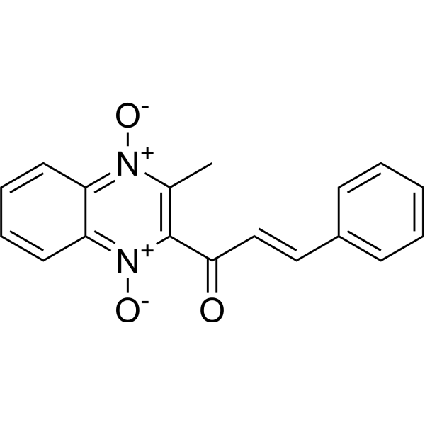
Quinocetone is an orally active animal feed additive used to increase the meat production of livestock and poultry. Quinocetone exhibits antibacterial activity against a variety of pathogenic microorganisms. Quinocetone exhibits tissue-specific (liver, lymphocyte) toxicity. Quinocetone induces autophagy in cells through the ATF6/DAPK1 pathway. Quinocetone activates the NF-κB and iNOS pathways, leading to cell apoptosis, hepatocyte vacuolar degeneration and fibrosis. Quinocetone can inhibit Nrf2/HO-1 and induce the generation of reactive oxygen species (ROS), leading to oxidative stress and DNA damage.
商品は「研究用試薬」です。人や動物の医療用・臨床診断用・食品用の製品ではありません。
研究用途以外に使用した場合、当社は一切の責任を負いかねます。
- 純度 : 98.25%
- CAS 番号: 81810-66-4
- 分子式: C18H14N2O3
- 分子量:306.32
-
保管条件:Powder -20°C, 3 years , 4°C, 2 years ; In solvent -80°C, 6 months , -20°C, 1 month
MedChemExpress(MCE)の使用を引用している文献 Quinocetone
MoreAntibiotic アイソフォーム固有の製品をすべて表示
More
生物活性
製品説明
|
NF-κB |
iNOS |
DAPK1 |
HO-1 |
Cellular Effect
|
Cell Line
|
Type | Value | Description | References |
|---|---|---|---|---|
| CCRF-CEM | IC50 |
5.92 μM
Compound: 3a
|
Growth inhibition of human CEM cells after 3 days
|
[PMID: 19427790] |
| HL-60 | CC50 |
4.1 μM
Compound: 3a
|
Cytotoxicity against human HL60 cells after 48 hrs
|
[PMID: 19427790] |
| HSC-2 | CC50 |
8.4 μM
Compound: 3a
|
Cytotoxicity against human HSC2 cells after 48 hrs
|
[PMID: 19427790] |
| HSC-3 | CC50 |
9.7 μM
Compound: 3a
|
Cytotoxicity against human HSC3 cells after 48 hrs
|
[PMID: 19427790] |
| HSC-4 | CC50 |
9.6 μM
Compound: 3a
|
Cytotoxicity against human HSC4 cells after 48 hrs
|
[PMID: 19427790] |
| MOLT-4 | IC50 |
6.76 μM
Compound: 3a
|
Growth inhibition of human Molt4/C8 cells after 3 days
|
[PMID: 19427790] |
体外実験
Quinocetone is active against Microsporum canis, Mycoplasma gallisepticum and Mycoplasma hyopneumoniae with MICs of 8, 8 and 16 μg/mL but shows no significant inhibitory effect on various animal viruses including bursal disease virus, porcine reproductive and respiratory syndrome virus, porcine parvovirus and classical swine fever virus[1].
Quinocetone (0-10 μg/mL, 0-24 h) triggers ER stress-induced autophagy via ATF6/DAPK1-modulated mAtg9a trafficking in HepG2 cells[2].
Quinocetone (0-20 μg/mL, 6-24 h) inhibits in human peripheral lymphocytes growth, triggers ROS increase and induced DNA damage[3].
MedChemExpress (MCE) has not independently confirmed the accuracy of these methods. They are for reference only. Further protocols information, click here.
-
Cell Line:HepG2 cells
-
Concentration:0, 1.25, 2.5, 5, 7.5, 10 μg/mL
-
Incubation Time:0, 1.5, 3, 6, 9, 12, 24 h
-
Result:Significantly increased the LC3-II/LC3-I ratio.
Dose-dependently upregulated BiP and CHOP expression and increased ATF6 cleavage.
Significantly upregulated DAPK1 expression and MRLC phosphorylation.
-
Cell Line:HepG2 cells
-
Concentration:0, 1.25, 2.5, 5, 7.5, 10 μg/mL
-
Incubation Time:0, 1.5, 3, 6, 9, 12, 24 h
-
Result:Increased BiP, HerpUD and sec24D transcription.
Increased DAPK1 mRNA.
体内実験
MedChemExpress (MCE) has not independently confirmed the accuracy of these methods. They are for reference only.
-
Animal Model:Hepatocyte damage assay established in adult male SD rats[4]
-
Dosage:50, 800 and 2400 mg/kg
-
Administration:Intragastrical administration (p.o.), once daily for 13 weeks
-
Result:Kept all rats survived and no obviously significant changes were observed in mortality, illness and clinical signs.
Induced liver damage and liver was likely to be the target organ at high dose.
Aggravated ROS accumulation in liver and aggravated DNA damage at high dose.
Aggravated inflammation and apoptosis of hepatocyte at high dose.
Inhibited aggravated inflammation and apoptosis of hepatocyte at high dose.
化学情報
-
CAS 番号 81810-66-4
-
性状 Solid
-
分子量 306.32
-
分子式 C18H14N2O3
-
Color White to yellow
-
SMILES
O=C(C1=C(C)[N+]([O-])=C2C=CC=CC2=[N+]1[O-])/C=C/C3=CC=CC=C3
-
輸送条件
Room temperature in continental US; may vary elsewhere.
-
保管条件
Powder -20°C 3 years 4°C 2 years In solvent -80°C 6 months -20°C 1 month

Publications (1)
-
Journal Impact Factor
-
Most Recent
-
Chin Herb Med
Integrated network pharmacology, transcriptomics and experimental validation to explore mechanisms of Wenyang Jiedu Granule on IAV-induced pneumonia. [Abstract]2026 Feb 11;18(2):241-261. PMID: 41971577
溶剤 & 溶解度
体外:
DMSO : 100 mg/mL (326.46 mM; Need ultrasonic; Hygroscopic DMSO has a significant impact on the solubility of product, please use newly opened DMSO)
Please refer to the solubility information to select the appropriate solvent. Once prepared, please aliquot and store the solution to prevent product inactivation from repeated freeze-thaw cycles.
Storage method and period of stock solution: -80°C, 6 months; -20°C, 1 month. When stored at -80°C, please use it within 6 months. When stored at -20°C, please use it within 1 month.
Please refer to the solubility information to select the appropriate solvent. Once prepared, please aliquot and store the solution to prevent product inactivation from repeated freeze-thaw cycles.
Storage method and period of stock solution: -80°C, 6 months; -20°C, 1 month. When stored at -80°C, please use it within 6 months. When stored at -20°C, please use it within 1 month.
濃度 (開始) × 体積 (開始) = 濃度 (終了) × 体積 (終了)
プロトコル
-
Research Protocol for Infectious Diseases
Infectious-disease experiments test how pathogens interact with host barriers, innate immune receptors, inflammatory signaling, pathogen replication, and tissue injury; pattern-recognition receptors such as TLRs, RIG-I-like receptors, NOD-like receptors, and inflammasomes detect microbial molecules and activate NF-κB, interferon, and cytokine responses. The central hypothesis is that infection severity reflects the balance between pathogen burden and host response: protective inflammation restricts pathogen growth, whereas excessive or mislocalized inflammation contributes to tissue damage and disease phenotype. Unresolved questions include which host pathways are protective versus pathogenic, why some infection models fail to translate to human disease, and which combined readouts best predict clinically relevant infection outcomes.
-
Collagen: Sirius Red Staining
Sirius Red or picrosirius red staining is a histochemical method for visualizing collagen-rich extracellular matrix in tissue sections, and collagen fibers are detected as red-stained structures under bright-field microscopy with enhanced birefringence under polarized light. Picrosirius red is useful for assessing total collagen organization, distribution, and fibrosis burden, but polarized color should not be interpreted as a definitive collagen type I versus type III readout because color is affected by fiber orientation, thickness, and packing.
-
Annexin V plus membrane-impermeant dye apoptosis staining
Annexin V-based apoptosis assays rely on the detection of phosphatidylserine (PS) externalization from the inner leaflet of the plasma membrane to the outer leaflet, an early biochemical hallmark of apoptosis. Fluorescently labeled Annexin V binds PS in a calcium-dependent manner, enabling identification of early apoptotic cells by flow cytometry or fluorescence microscopy. When combined with a membrane-impermeant DNA-binding dye (e. g. , propidium iodide), this approach allows discrimination between viable (Annexin V−/dye−), early apoptotic (Annexin V+/dye−), and late apoptotic or necrotic (Annexin V+/dye+) cell populations by assessing membrane integrity and PS exposure.
-
ROS/oxidative-stress fluorescent staining
ROS/oxidative-stress fluorescent staining uses cell-permeant fluorogenic probes that become fluorescent after oxidation inside cells or tissues; commonly used examples include DCFH-DA/DCFDA for broad cellular oxidant detection, DHE for superoxide-related signal detection, MitoSOX for mitochondrial superoxide-related signal detection, and CellROX probes for oxidative-stress-associated fluorescence readouts. The assay detects probe oxidation rather than a single ROS species unless the probe and analysis method have been chemically validated for that species. DCFH-DA enters cells, is deacetylated by intracellular esterases to DCFH, and produces fluorescent DCF after oxidation, so the readout is used as an operational measure of total cellular oxidative stress rather than a species-specific ROS measurement. DHE and MitoSOX can report superoxide-related oxidation, but red fluorescence alone can include non-specific ethidium-like oxidation products; HPLC or optimized spectral approaches are
-
Connective Tissue: Masson's Trichrome/Collagen Trichrome Staining
Masson’s Trichrome (collagen/trichrome staining) is a histological technique that differentially stains tissue compartments using sequential acidic dyes to distinguish collagen from muscle and cytoplasmic components based on dye affinity and tissue permeability differences, enabling visualization of fibrosis and connective tissue architecture in histological sections. The classical formulation typically uses Weigert's iron hematoxylin for nuclear staining, Biebrich scarlet-acid fuchsin for cytoplasm and muscle, and aniline blue (or light green variants) for collagen, producing a characteristic blue/green collagen signal contrasted against red cytoplasm and dark nuclei. The staining principle relies on selective displacement of smaller dye molecules by larger anionic dyes in collagen-rich regions under controlled acidified conditions, which enhances collagen-specific dye retention. This property makes the method widely used for fibrosis assessment in organs such as heart, liver, lung, a
-
Research Protocol for Inflammation-related Diseases
The NLRP3 inflammasome is a cytosolic innate immune signaling platform that integrates priming signals and danger-signal activation to promote caspase-1 activation, maturation of IL-1β and IL-18, and gasdermin D-mediated pyroptotic cell death. The core experimental logic is to determine whether inflammatory disease phenotypes are driven by increased NLRP3 expression, ASC-containing inflammasome assembly, caspase-1 cleavage, GSDMD cleavage, and extracellular release of IL-1β/IL-18 rather than by nonspecific cell injury alone. The pathway is strongly linked to inflammation-related disease phenotypes because monosodium urate crystals activate NALP3/NLRP3 inflammasome signaling in gout-like crystal inflammation, cholesterol crystals activate NLRP3 inflammasomes in atherogenesis models, and DSS-induced intestinal inflammation has been reported to involve NLRP3 inflammasome activity. However, experimental colitis studies also show context-dependent protective effects of NLRP3 inflammasome co
-
Bacterial live/dead nucleic-acid viability staining
The LIVE/DEAD bacterial viability staining method is based on differential permeability of nucleic-acid-binding fluorescent dyes, most commonly SYTO 9 and propidium iodide (PI), which enables discrimination of bacterial populations with intact versus compromised cytoplasmic membranes. SYTO 9 penetrates both intact and damaged bacterial membranes and binds nucleic acids to produce green fluorescence, whereas propidium iodide penetrates only cells with compromised membranes and fluoresces red while also reducing SYTO 9 signal through competitive binding and fluorescence interactions. The resulting fluorescence pattern is interpreted as a proxy for membrane integrity, which is widely used as an indicator of bacterial viability in microscopy, flow cytometry, and spectroscopic platforms. However, mechanistic studies show that SYTO 9 and PI interactions involve displacement and fluorescence resonance energy transfer effects, which can influence signal interpretation depending on dye ratios a
-
Fibrosis/Collagen Morphometry
Fibrosis and collagen morphometry is based on the quantitative visualization of fibrillar collagen deposition in tissue sections using histochemical stains such as Sirius Red (Picrosirius Red) or Masson's trichrome, followed by image-based or polarization-enhanced analysis to estimate collagen proportional area as a surrogate of extracellular matrix accumulation during fibrotic remodeling. Sirius Red combined with polarized light microscopy enhances detection of collagen fibers due to birefringence properties, enabling more specific visualization of collagen type I and III fibrils compared to conventional bright-field histology, while whole-section or region-restricted digital morphometry reduces field-selection bias in fibrosis assessment. Alternative quantitative approaches include second harmonic generation (SHG) and two-photon excited fluorescence microscopy, which enable label-free detection of fibrillar collagen and have been validated against histological staining and biochemica
-
Apoptosis
Apoptosis, also called programmed cell death, is generally characterized by distinct morphological characteristics.
-
TUNEL staining for apoptotic DNA fragmentation
TUNEL staining detects DNA strand breaks by using terminal deoxynucleotidyl transferase to add labeled nucleotides to exposed 3′-OH DNA termini, generating either microscopic staining in fixed cells or tissue sections, or fluorescence/cytometric signal in cell suspensions. TUNEL positivity reflects DNA fragmentation but should not be interpreted alone as definitive apoptosis, because TUNEL can also label necrotic, autolytic, mechanically damaged, or DNA-repair-associated DNA breaks.
-
Autophagy
Autophagy is a process in which eukaryotic cells use lysosomes to degrade their own cytoplasmic proteins and damaged organelles under the regulation of autophagy related gene (Atg). Microtubule-associated proteins light chain 3 (LC3) is recognized as autophagy marker, which transfers from cytoplasmic LC3 (LC3-I) to membrane type (LC3-II). LC3-II/I ratio could be detected by Western Blot and fluorescence microscopy.
-
Apoptosis Solutions
Apoptosis is a regulated, generally non-lytic cell-death pathway that removes unwanted, damaged, infected, or abnormal cells through coordinated morphological changes, caspase activation, DNA fragmentation, and membrane remodeling. The intrinsic apoptosis pathway is controlled mainly by mitochondrial outer membrane permeabilization, BCL-2 family proteins, cytochrome c release, apoptosome formation, caspase-9 activation, and downstream executioner caspase-3/7 activation. The extrinsic apoptosis pathway is initiated by death receptors such as Fas, TNFR, and TRAIL receptors, which recruit adaptor proteins and activate caspase-8 before engaging executioner caspases or mitochondrial amplification through BID cleavage. Apoptosis is linked to many phenotypes, including cancer cell killing, tissue homeostasis, immune regulation, neurodegeneration, infection response, and treatment-induced cytotoxicity; unresolved questions include how apoptosis interacts with necroptosis, pyroptosis, ferroptos
-
Lysosome and acidic-vesicle live-cell staining
Lysosome and acidic-vesicle live-cell staining detects acidic intracellular compartments by using membrane-permeant acidotropic probes that accumulate in low-pH vesicles, including lysosomes, late endosomes, autolysosomes, and acidic phagosomes. LysoTracker staining is commonly used as an intensity-based readout of acidic lysosomal compartment abundance or enlargement, while acridine orange produces green fluorescence in less concentrated compartments and red fluorescence after concentration-dependent accumulation in acidic vesicular organelles. Loss or reduction of acridine-orange red signal can be used as a readout of lysosomal membrane permeabilization or reduced acidic-vesicle integrity. This protocol is designed for live cultured cells and can be adapted for fluorescence microscopy, high-content imaging, plate-reader readout, or flow cytometry when the selected literature supports the readout. Because these dyes report acidotropic accumulation rather than lysosome identity alone,
-
Macroautophagy Solutions
Macroautophagy is a conserved lysosome-dependent degradation pathway in which cytoplasmic material is sequestered into double-membrane autophagosomes and delivered to lysosomes for degradation and recycling. The pathway supports cellular homeostasis during nutrient limitation, organelle stress, protein-aggregate accumulation, infection, differentiation, and tissue remodeling by coupling cargo sequestration, autophagosome maturation, lysosomal fusion, and degradation of cargo-derived macromolecules. The core molecular sequence includes initiation by nutrient- and stress-regulated autophagy machinery, autophagosome nucleation, LC3/ATG8-family conjugation to autophagosomal membranes, cargo selection through receptors such as SQSTM1/p62, autophagosome-lysosome fusion, and lysosomal degradation. LC3 was identified as a mammalian homolog of yeast Atg8 that localizes to autophagosomal membranes after processing, and p62/SQSTM1 was shown to connect ubiquitinated cargo with autophagic degradati
-
Genotoxicity/Mutagenicity Study
The bacterial reverse mutation assay detects point mutations that restore amino-acid prototrophy in auxotrophic Salmonella typhimurium or Escherichia coli tester strains; after exposure to a test article, mutagenic activity is read out as an increased number of revertant colonies on minimal agar compared with the vehicle control. The assay uses tester strains with different mutation targets so that base-substitution and frameshift mutagens can be detected, and testing is performed with and without exogenous mammalian metabolic activation because some chemicals require biotransformation to become mutagenic.
純度とドキュメンテーション
-
データシート (279 KB)
-
SDS (418 KB)
- English - EN (418 KB)
- Français - FR (418 KB)
- Deutsch - DE (418 KB)
- Norwegian - NO (418 KB)
- Español - ES (418 KB)
- Swedish - SV (418 KB)
- Italian - IT (418 KB)
- Korean - KR (418 KB)
- Portuguese - PT (418 KB)
-
取扱説明書 (2659 KB)
参考文献
[1]. Zhao Y, et al. In vitro antimicrobial activities of animal-used quinoxaline 1,4-di-N-oxides against mycobacteria, mycoplasma and fungi. BMC Vet Res. 2016 Sep 6;12(1):186. [Content Brief]
[2]. Zhou Y, et al. Quinocetone triggered ER stress-induced autophagy via ATF6/DAPK1-modulated mAtg9a trafficking. Cell Biol Toxicol. 2016 Apr;32(2):141-52. [Content Brief]
[3]. Yang W, et al. Quinocetone triggers oxidative stress and induces cytotoxicity and genotoxicity in human peripheral lymphocytes of both genders. J Sci Food Agric. 2013 Apr;93(6):1317-25. [Content Brief]
[4]. Yu M, et al. Quinocetone-induced Nrf2/HO-1 pathway suppression aggravates hepatocyte damage of Sprague-Dawley rats. Food Chem Toxicol. 2014 Jul;69:210-9. doi: 10.1016/j.fct.2014.04.026. Epub 2014 Apr 30. PMID: 24795230. [Content Brief]
Complete Stock Solution Preparation Table
Please refer to the solubility information to select the appropriate solvent. Once prepared, please aliquot and store the solution to prevent product inactivation from repeated freeze-thaw cycles.
Storage method and period of stock solution: -80°C, 6 months; -20°C, 1 month. When stored at -80°C, please use it within 6 months. When stored at -20°C, please use it within 1 month.
| Optional Solvent | Concentration Solvent Mass | 1 mg | 5 mg | 10 mg | 25 mg |
|---|---|---|---|---|---|
| DMSO | 1 mM | 3.2646 mL | 16.3228 mL | 32.6456 mL | 81.6140 mL |
| 5 mM | 0.6529 mL | 3.2646 mL | 6.5291 mL | 16.3228 mL | |
| 10 mM | 0.3265 mL | 1.6323 mL | 3.2646 mL | 8.1614 mL | |
| 15 mM | 0.2176 mL | 1.0882 mL | 2.1764 mL | 5.4409 mL | |
| 20 mM | 0.1632 mL | 0.8161 mL | 1.6323 mL | 4.0807 mL | |
| 25 mM | 0.1306 mL | 0.6529 mL | 1.3058 mL | 3.2646 mL | |
| 30 mM | 0.1088 mL | 0.5441 mL | 1.0882 mL | 2.7205 mL | |
| 40 mM | 0.0816 mL | 0.4081 mL | 0.8161 mL | 2.0403 mL | |
| 50 mM | 0.0653 mL | 0.3265 mL | 0.6529 mL | 1.6323 mL | |
| 60 mM | 0.0544 mL | 0.2720 mL | 0.5441 mL | 1.3602 mL | |
| 80 mM | 0.0408 mL | 0.2040 mL | 0.4081 mL | 1.0202 mL | |
| 100 mM | 0.0326 mL | 0.1632 mL | 0.3265 mL | 0.8161 mL |

