



Rapamycin-d3
Based on 1 publication(s) in Google Scholar
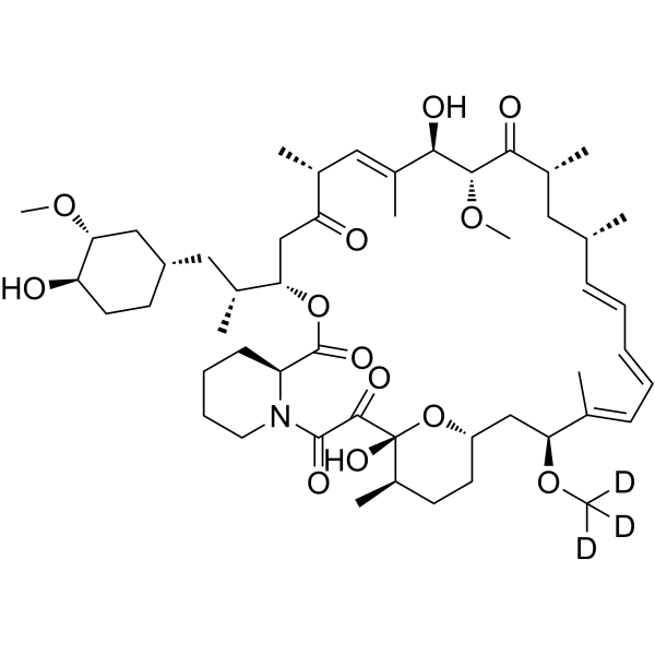
Rapamycin-d3 (Sirolimus-d3; AY-22989-d3; NSC 226080-d3) is the deuterated-labeled Rapamycin (HY-10219). Rapamycin (Sirolimus; AY 22989) is a potent and specific blood-brain barrier-transmissible mTOR inhibitor with an IC50 of 0.1 nM in HEK293 cells. Rapamycin is a molecular glue that binds FKBP12 and mTOR proteins together, thereby inhibiting mTOR kinase activity. Rapamycin binds to FKBP12 and specifically acts as an allosteric inhibitor of mTORC1. Rapamycin is an autophagy activator, an immunosuppressant.
For research use only. We do not sell to patients.
- Purity : 95.92%
- CAS No.: 392711-19-2
- Formula: C51H76D3NO13
- Molecular Weight:917.19
-
Storage:Powder -20°C, 3 years ; In solvent -80°C, 6 months , -20°C, 1 month

To place orders, for customer services and technical support, please contact: MedChemExpress USA
Tel: 609-228-6898 E-mail: [email protected] [email protected]


-
 Biological Activity
-
Chemical Information
- Protocols
- Purity & Documentation
- References
-
 Help & FAQs


-
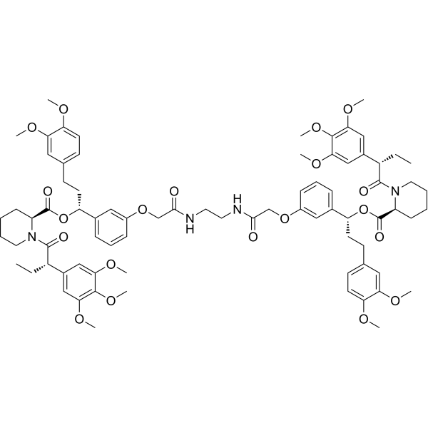
HY-16046
RimiducidDimerization Inducer -

HY-10219
RapamycinmTOR Inhibitor -
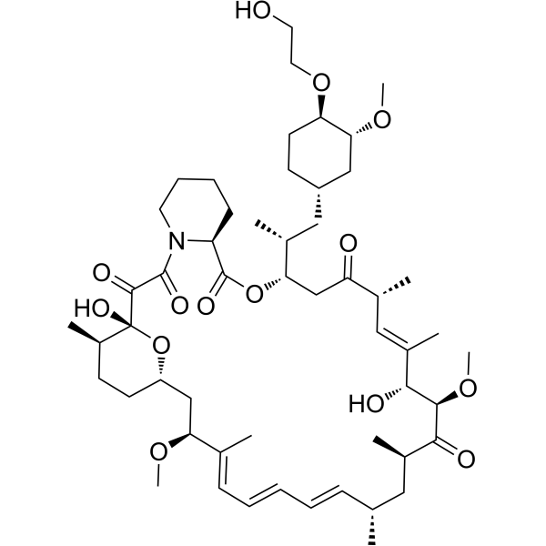
HY-10218
EverolimusmTORC Inhibitor -
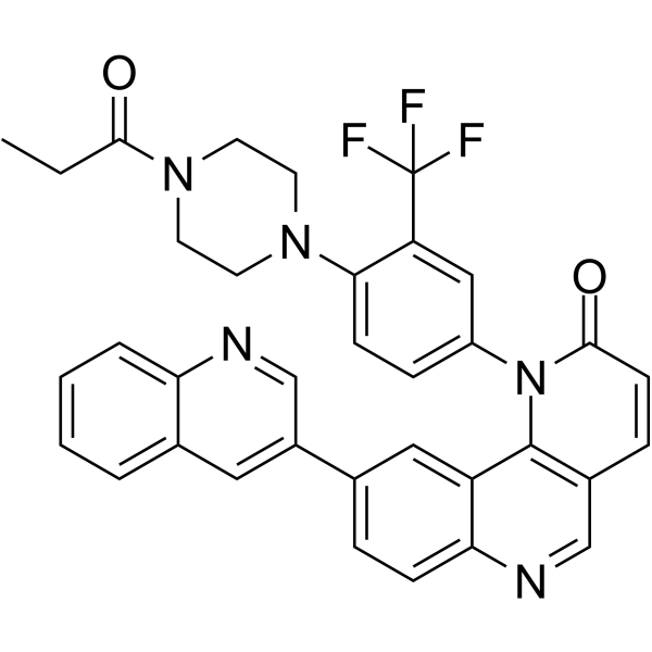
HY-13003
Torin 1mTOR Inhibitor -
HY-10422
AZD-8055mTOR Inhibitor -

HY-10219G
Rapamycin (GMP)mTOR Inhibitor -

HY-10219R
Rapamycin (Standard)Reference Standard -
HY-50908
RidaforolimusmTOR Inhibitor -
HY-10423
OSI-027mTOR Inhibitor -
HY-10297
OmipalisibPI3K/mTOR Inhibitor -
HY-10218R
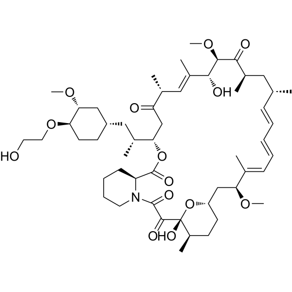
Everolimus (Standard)mTORC Inhibitor -
HY-12034
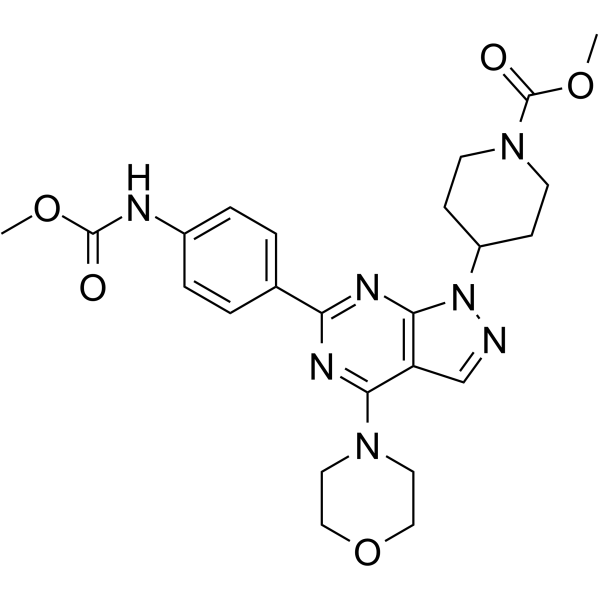
WYE-354mTOR inhibitor
Publications Citing Use of MedChemExpress (MCE) Rapamycin-d3
MoreAll Endogenous Metabolite Isoforms
MoreAll Antibiotic Isoforms
More

Biological Activity
Description
IC50 & Target
|
mTOR 0.1 nM (IC50, in HEK293 cells ) |
Microbial Metabolite |
FKBP12 |
Autophagy |
In Vitro
Stable heavy isotopes of hydrogen, carbon, and other elements have been incorporated into drug molecules, largely as tracers for quantitation during the drug development process. Deuteration has gained attention because of its potential to affect the pharmacokinetic and metabolic profiles of drugs.
MedChemExpress (MCE) has not independently confirmed the accuracy of these methods. They are for reference only. Further protocols information, click here.
-
Cell Line:Lung cancer cell lines A549, SPC-A-1, 95D and NCI-H446
-
Concentration:12.5 nM, 25 nM, 50 nM, 100 nM
-
Incubation Time:24 hours
-
Result:Treatment exerted modest inhibitory effect on lung cancer cell proliferation in a dose-dependent manner in all cell lines.
-
Cell Line:95D cells
-
Concentration:10 nM and 20 nM
-
Incubation Time:24 hours
-
Result:Combination treatment with RP-56976 decreased phosphorylation of ERK.
In Vivo
-
Animal Model:24 male nu/nu mice aged 4-5 week old (15-20 g)[4]
-
Dosage:2.0 mg/kg
-
Administration:Intraperitoneal injection; every other day; 28 days
-
Result:Had a moderate inhibitory effect in monotherapy group. The combination with Metformin exerted a significantly increased inhibition of tumor growth.
Application
1. This compound can be used as a tracer
2. This compound can be used as an internal standard for quantitative analysis by NMR, GC-MS, or LC-MS.


Chemical Information
-
CAS No. 392711-19-2
-
Unlabeled CAS 53123-88-9
-
Appearance Solid
-
Molecular Weight 917.19
-
Formula C51H76D3NO13
-
Color White to off-white
-
SMILES
O=C([C@@]1(O[C@@H](CC[C@H]1C)C[C@@H](/C(C)=C/C=C/C=C/[C@H](C2)C)OC([2H])([2H])[2H])O)C(N3[C@@H](CCCC3)C(O[C@@H](CC([C@@H](/C=C([C@H]([C@H](C([C@@H]2C)=O)OC)O)\C)C)=O)[C@@H](C[C@@H]4CC[C@H]([C@@H](C4)OC)O)C)=O)=O
-
Synonyms
Sirolimus-d3; AY-22989-d3; NSC 226080-d3
-
Shipping
Room temperature in continental US; may vary elsewhere.
-
Storage
Powder -20°C 3 years In solvent -80°C 6 months -20°C 1 month


Publications (1)
-
Journal Impact Factor
-
Most Recent
-
Ecotoxicol Environ Saf
ATF4-mediated different mode of interaction between autophagy and mTOR determines cell fate dependent on the level of ER stress induced by Cr(VI). [Abstract]2024 Aug:281:116639. PMID: 38964069

Protocols
-
Kinase activity and phosphorylation assays
Kinase activity assays measure the ability of kinases to transfer phosphate groups from ATP to specific substrates, while phosphorylation assays detect the presence and levels of phosphorylated proteins. Common methods include radiolabeled ATP incorporation (e. g. ,), ADP release detection via bioluminescence (e. g. ,[3]), enzyme-linked immunosorbent assays (ELISA) for phospho-specific epitopes (e. g. ,[6]), and microtiter-based formats for high-throughput screening (e. g. ,[8]). The ADP-Glo assay quantifies kinase activity by measuring ADP produced during phosphorylation using a luciferase-based system. Radiometric assays involve autoradiography or scintillation counting after incorporation of 32P-labeled ATP into substrate proteins. ELISA-based approaches rely on phospho-specific antibodies to detect activated kinases in cell lysates or purified samples.
-
Cell Cytotoxicity Assay
Cytotoxicity assays are usually based on the assessment of cell membrane damage, which can also be indirectly detected by measuring cell viability. Detection methods include MTT assay, CKK-8 assay, LDH assay and ATP assay, etc.
-
Autophagy
Autophagy is a process in which eukaryotic cells use lysosomes to degrade their own cytoplasmic proteins and damaged organelles under the regulation of autophagy related gene (Atg). Microtubule-associated proteins light chain 3 (LC3) is recognized as autophagy marker, which transfers from cytoplasmic LC3 (LC3-I) to membrane type (LC3-II). LC3-II/I ratio could be detected by Western Blot and fluorescence microscopy.
-
Protocol for Kinase activity and phosphorylation assays
Kinase activity assays measure transfer of phosphate from ATP to a protein or peptide substrate, generating phosphorylated substrate, ADP, or incorporated radiolabeled phosphate as the readout; phosphorylation assays measure site-specific phosphorylation in cells or tissues as a proxy for kinase-pathway activation, inhibition, or substrate regulation. Phosphorylation can be detected by phospho-specific Western blot, immunoprecipitation kinase assay, phospho-immunofluorescence, phospho-flow cytometry, luminescent ADP detection, radiolabeled ATP incorporation, or reporter-based pathway assays, and these readouts can be applied to cancer cells, primary neurons, mouse tumors, organoids, inflammatory macrophages, ferroptosis studies, and mitophagy studies when the kinase target is biologically relevant.
-
Lysosome and acidic-vesicle live-cell staining
Lysosome and acidic-vesicle live-cell staining detects acidic intracellular compartments by using membrane-permeant acidotropic probes that accumulate in low-pH vesicles, including lysosomes, late endosomes, autolysosomes, and acidic phagosomes. LysoTracker staining is commonly used as an intensity-based readout of acidic lysosomal compartment abundance or enlargement, while acridine orange produces green fluorescence in less concentrated compartments and red fluorescence after concentration-dependent accumulation in acidic vesicular organelles. Loss or reduction of acridine-orange red signal can be used as a readout of lysosomal membrane permeabilization or reduced acidic-vesicle integrity. This protocol is designed for live cultured cells and can be adapted for fluorescence microscopy, high-content imaging, plate-reader readout, or flow cytometry when the selected literature supports the readout. Because these dyes report acidotropic accumulation rather than lysosome identity alone,
-
Macroautophagy Solutions
Macroautophagy is a conserved lysosome-dependent degradation pathway in which cytoplasmic material is sequestered into double-membrane autophagosomes and delivered to lysosomes for degradation and recycling. The pathway supports cellular homeostasis during nutrient limitation, organelle stress, protein-aggregate accumulation, infection, differentiation, and tissue remodeling by coupling cargo sequestration, autophagosome maturation, lysosomal fusion, and degradation of cargo-derived macromolecules. The core molecular sequence includes initiation by nutrient- and stress-regulated autophagy machinery, autophagosome nucleation, LC3/ATG8-family conjugation to autophagosomal membranes, cargo selection through receptors such as SQSTM1/p62, autophagosome-lysosome fusion, and lysosomal degradation. LC3 was identified as a mammalian homolog of yeast Atg8 that localizes to autophagosomal membranes after processing, and p62/SQSTM1 was shown to connect ubiquitinated cargo with autophagic degradati


Purity & Documentation
-
Data Sheet (281 KB)
-
SDS (604 KB)
- English - EN (604 KB)
- Français - FR (604 KB)
- Deutsch - DE (604 KB)
- Norwegian - NO (604 KB)
- Español - ES (604 KB)
- Swedish - SV (604 KB)
- Italian - IT (604 KB)
- Korean - KR (604 KB)
- Portuguese - PT (604 KB)
-
Handling Instructions (2659 KB)
References
[1]. Edwards SR, et al. The rapamycin-binding domain of the protein kinase mammalian target of rapamycin is a destabilizing domain. J Biol Chem, 2007, 282(18), 13395-13401. [Content Brief]
[2]. Rangaraju S, et al. Rapamycin activates autophagy and improves myelination in explant cultures from neuropathicmice. J Neurosci. 2010 Aug 25;30(34):11388-97. [Content Brief]
[3]. Niu H, et al. Rapamycin potentiates cytotoxicity by RP-56976 possibly through downregulation of Survivin in lung cancer cells. J Exp Clin Cancer Res. 2011 Mar 10;30:28. [Content Brief]
[4]. Zhang JW, et al. Metformin synergizes with rapamycin to inhibit the growth of pancreatic cancer in vitro and in vivo. Oncol Lett. 2018 Feb;15(2):1811-1816. [Content Brief]
[5]. Svensson JE, et al. Evaluating the effect of rapamycin treatment in Alzheimer's disease and aging using in vivo imaging: the ERAP phase IIa clinical study protocol. BMC Neurol. 2024 Apr 4;24(1):111. [Content Brief]
[6]. Soini L, et al. Molecular glues to stabilise protein-protein interactions. Curr Opin Chem Biol. 2022 Aug;69:102169. [Content Brief]

Calculators
Concentration (start) × Volume (start) = Concentration (final) × Volume (final)


Rapamycin-d3 Related Classifications
HY-10219S Related Classifications
 Powered by Bioz