



S1g-10

Based on 1 Customer Validation
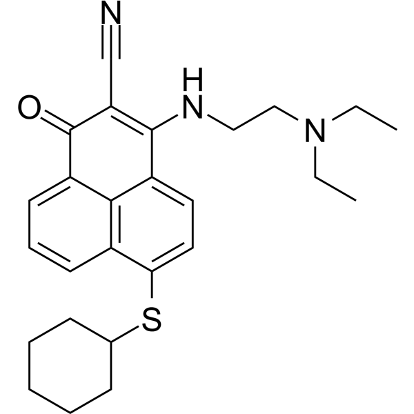
S1g-10 is an Hsp70-Bim protein-protein interaction inhibitor and an Hsp70 ATPase inhibitor. S1g-10 binds the Hsp70 nucleotide-binding domain at the Bim interface, inhibits Bim-stimulated ATPase activity, and selectively disrupts Hsp70-Bim without affecting Hsp70-Bag3, thereby inhibiting mitophagy (Autophagy), inducing apoptosis (Apoptosis), and reducing the survival and self-renewal of CML leukemia stem cells. S1g-10 can be used for research on chronic myeloid leukemia and breast cancer.
For research use only. We do not sell to patients.
- Purity : 99.08%
- CAS No.: 3032432-71-3
- Formula: C26H31N3OS
- Molecular Weight:433.61
-
Storage:Powder -20°C, 3 years , 4°C, 2 years ; In solvent -80°C, 6 months , -20°C, 1 month

To place orders, for customer services and technical support, please contact: MedChemExpress USA
Tel: 609-228-6898 E-mail: [email protected] [email protected]


-
 Biological Activity
-
Chemical Information
-
Solvent & Solubility
- Protocols
- Purity & Documentation
- References
-
 Help & FAQs




Biological Activity
Description
IC50 & Target
[4]|
HSP70 688 nM (Kd) |
Cellular Effect
|
Cell Line

|
Type | Value | Description | References |
|---|---|---|---|---|
| BV-173 | EC50 |
0.62 μM
|
Apoptosis induction against human CML BV173 cells assessed by Annexin V-FITC staining.
Apoptosis induction against human CML BV173 cells assessed by Annexin V-FITC staining.
|
38011535 |
| K562 | EC50 |
1.3 μM
|
Apoptosis induction against human CML K562 cells assessed by Annexin V-FITC staining.
Apoptosis induction against human CML K562 cells assessed by Annexin V-FITC staining.
|
38011535 |
| K562 | EC50 |
0.34 μM
|
Apoptosis induction against human TKI-resistant CML K562-R3 cells assessed by Annexin V-FITC staining.
Apoptosis induction against human TKI-resistant CML K562-R3 cells assessed by Annexin V-FITC staining.
|
38011535 |
| MDA-MB-231 | IC50 |
14.9 μM
|
Cytotoxicity against human MDA-MB-231 breast cancer cells assessed as viability reduction after 48 hrs incubation by CCK-8 assay.
Cytotoxicity against human MDA-MB-231 breast cancer cells assessed as viability reduction after 48 hrs incubation by CCK-8 assay.
|
40938081 |
| BT-549 | IC50 |
16.7 μM
|
Cytotoxicity against human BT549 breast cancer cells assessed as viability reduction after 48 hrs incubation by CCK-8 assay.
Cytotoxicity against human BT549 breast cancer cells assessed as viability reduction after 48 hrs incubation by CCK-8 assay.
|
40938081 |
| MCF7 | IC50 |
27.0 μM
|
Cytotoxicity against human MCF7 breast cancer cells assessed as viability reduction after 48 hrs incubation by CCK-8 assay.
Cytotoxicity against human MCF7 breast cancer cells assessed as viability reduction after 48 hrs incubation by CCK-8 assay.
|
40938081 |
| SK-BR-3 | IC50 |
24.2 μM
|
Cytotoxicity against human SKBR3 breast cancer cells assessed as viability reduction after 48 hrs incubation by CCK-8 assay.
Cytotoxicity against human SKBR3 breast cancer cells assessed as viability reduction after 48 hrs incubation by CCK-8 assay.
|
40938081 |
| CAMA-1 | IC50 |
19.0 μM
|
Cytotoxicity against human CAMA1 breast cancer cells assessed as viability reduction after 48 hrs incubation by CCK-8 assay.
Cytotoxicity against human CAMA1 breast cancer cells assessed as viability reduction after 48 hrs incubation by CCK-8 assay.
|
40938081 |
| T47D | IC50 |
22.3 μM
|
Cytotoxicity against human T47D breast cancer cells assessed as viability reduction after 48 hrs incubation by CCK-8 assay.
Cytotoxicity against human T47D breast cancer cells assessed as viability reduction after 48 hrs incubation by CCK-8 assay.
|
40938081 |
| MDA-MB-468 | IC50 |
16.0 μM
|
Cytotoxicity against human MDA-MB-468 breast cancer cells assessed as viability reduction after 48 hrs incubation by CCK-8 assay.
Cytotoxicity against human MDA-MB-468 breast cancer cells assessed as viability reduction after 48 hrs incubation by CCK-8 assay.
|
40938081 |
| MDA-MB-157 | IC50 |
15.4 μM
|
Cytotoxicity against human MDA-MB-157 breast cancer cells assessed as viability reduction after 48 hrs incubation by CCK-8 assay.
Cytotoxicity against human MDA-MB-157 breast cancer cells assessed as viability reduction after 48 hrs incubation by CCK-8 assay.
|
40938081 |
| HEK-293T | IC50 |
> 30 μM
|
Cytotoxicity against human embryonic kidney HEK-293T cells assessed as viability reduction after 48 hrs incubation by CCK-8 assay.
Cytotoxicity against human embryonic kidney HEK-293T cells assessed as viability reduction after 48 hrs incubation by CCK-8 assay.
|
40938081 |
| BV-173 | EC50 |
0.66 μM
|
Induction of apoptosis in human CML BV173 cells after incubation with S1g-10 for 48 hrs assessed by Annexin V-FITC flow cytometry.
Induction of apoptosis in human CML BV173 cells after incubation with S1g-10 for 48 hrs assessed by Annexin V-FITC flow cytometry.
|
39012838 |
| K562 | EC50 |
1.20 μM
|
Induction of apoptosis in human CML K562 cells after incubation with S1g-10 for 48 hrs assessed by Annexin V-FITC flow cytometry.
Induction of apoptosis in human CML K562 cells after incubation with S1g-10 for 48 hrs assessed by Annexin V-FITC flow cytometry.
|
39012838 |
| K562 | EC50 |
0.37 μM
|
Induction of apoptosis in human CML TKI-resistant K562-R3 cells after incubation with S1g-10 for 48 hrs assessed by Annexin V-FITC flow cytometry.
Induction of apoptosis in human CML TKI-resistant K562-R3 cells after incubation with S1g-10 for 48 hrs assessed by Annexin V-FITC flow cytometry.
|
39012838 |
In Vitro
S1g-10 (1 μM; week 1 of a 6-week culture; colonies counted after 14 days) reduces LTC-IC colony formation by 50%-70% in CML#1 CD34+CD38− cells without affecting healthy donor CD34+CD38− cells[1].
S1g-10 (1 μM; 6-24 h) does not change the levels of AKT, eIF4E, or Raf-1 in CML#1 CD34+ cells, but decreases these proteins in CD34− differentiated CML cells[1].
S1g-10 (1 μM; 24 h) upregulates hematopoietic lineage/differentiation markers, including CD11b, CD48, and CD71, in CML#3 CD34+ cells[1].
S1g-10 (1 μM; 3 days) reduces the absolute number of CD34+CD38− LSCs in CML#2-5, while no additional effect is observed in cells exposed to BafA1[1].
S1g-10 perturbs Hsp70−Bim signaling in K562 cells and reduces the expression/phosphorylation of AKT and EIF4e[2].
S1g-10 (48 h) inhibits the viability of MDA-MB-231, BT549, MCF7, SKBR3, CAMA1, T47D, MDA-MB-468, and MDA-MB-157 breast cancer cells with IC50 values of 14.9, 16.7, 27.0, 24.2, 19.0, 22.3, 16.0, and 15.4 µM, respectively, whereas the IC50 in HEK-293T cells is > 30 µM[3].
S1g-10 (3 μM; 12 hours) selectively disrupts the Hsp70-Bim PPI in K562 cells without affecting Hsp70-Bag3[4].
S1g-10 prevents SB-2-mediated BRD4 degradation in U-87 MG cells by competing with Hsp70 and disrupting the BRD4-Hsp70TAC-Hsp70 ternary complex[5].
S1g-10 blocks SP-2-induced PD-L1 degradation in MDA-MB-231 cells by competing with Hsp70 and preventing the formation of the PD-L1-Hsp70TAC-Hsp70 ternary complex[5].
S1g-10 (2 µM; 12 hours) does not inhibit Bcr-Abl Y412 phosphorylation in K562R cells[6].
S1g-10 (10 µM; 12 hours) does not disrupt the Hsp70-Bcr-Abl interaction in K562R cells[6].
S1g-10 (50 µM) inhibits the Bcr-Abl DBD-enhanced Hsp70 ATPase activity in K562R shBcr-Abl and HL-60 cell lysates[6].
S1g-10 (2 µM; 12 hours) disrupts Hsp70-Bim, Hsp70-AKT, and Hsp70-eIF4E interactions and reduces AKT/eIF4E levels in K562R shBcr-Abl/DBD cells[6].
S1g-10 (2 µM; 12 h) prevents the restoration of Bcr-Abl DBD-dependent nuclear export of MYC and survivin in K562R cells[6].
S1g-10 (48 h) selectively induces apoptosis in CML cells, including TKI-resistant K562-R3, with an EC50 of 0.37 μM, without affecting HEK293 and BaF3 cells[4].
S1g-10 (2 µM) restores apoptosis in K562R/K562 cells expressing Bcr-Abl DBD[6].
S1g-10 (1 μM; 12 h) inhibits autophagy more strongly in primary CML CD34+ stem/progenitor cells than in CD34− differentiated CML cells[1].
S1g-10 (1 μM; 24 h) increases CD11b and E2F2 in CML#3 CD34+ cells, which depends on the inhibition of mitophagy[1].
S1g-10 (1 μM; 12 h) inhibits mitophagy in Bcr-Abl-expressing BaF3 and K562 cells, but not in Bcr-Abl-negative or Bcr-Abl-knockdown cells[1].
S1g-10 (1 μM) reduces the colocalization of LC3 with mitochondria in CML LSCs[1].
S1g-10 (1 μM; 24 h) decreases the G0 phase proportion and increases the S-G2-M phase in CML LSCs, indicating loss of quiescence[1].
S1g-10 induces apoptosis in BV173, K562, and K562-R3 CML cells with EC50 values of 0.34-1.3 μM, and does not affect HEK293 and BaF3 cells[2].
S1g-10-induced apoptosis in K562 cells depends on Hsp70 and Bim, supporting the inhibition of Hsp70−Bim PPI targeting[2].
S1g-10 (0-100 µM; 30 minutes) inhibits the single-turnover ATPase activity of recombinant GRP75, GRP78, and HSP70, with IC50 values in the low micromolar range[3].
S1g-10 (1-5 µM; 6 hours) selectively modulates cytosolic HSP70-Bim signaling in HeLa cells and downregulates AKT/eIF4E at 1 µM, while not affecting GRP75- or GRP78-mediated pathways at 5 µM[3].
S1g-10 (1 μM; 12 h) increases mitochondrial proteins 2-3 fold in CML#2 CD34+ cells and selectively does not affect ER proteins[1].
S1g-10 (50 µM; 30 minutes) exhibits lower subcellular accumulation in isolated mitochondria and ER than the organelle-targeted derivatives 5 and 8[3].
S1g-10 (1 μM; 12 h) increases mitochondrial content by approximately 7-fold in CML#2 CD34+CD38− LSCs and by approximately 1.5-fold in differentiated CML cells, and does not affect healthy HSCs[1].
S1g-10 (5 µM; 30 minutes) shows no obvious colocalization with Mito-Tracker Red or ER-Tracker Red in HeLa cells[3].
S1g-10 (1 μM; 12 h) increases mitochondrial ROS in primary CML#1-5 CD34+ cells[1].
S1g-10 (compound 21) disrupts the Hsp70−Bim PPI in vitro, Ki = 0.25 μM[2].
S1g-10 selectively inhibits Bim-stimulated Hsp70 ATPase activity, IC50 = 15.8 μM[2].
S1g-10 binds the Hsc70 NBD/Hsp70 interface and significantly perturbs Asp225, sharing the binding interface with S1g-2[2].
S1g-10 (1-5 μM) selectively disrupts the Hsp70−Bim PPI in K562 cells without affecting Hsp70−Bag3[2].
S1g-10 (10 min) inhibits the Hsp70-Bim PPI in FPA with an IC50 of 0.80 μM[4].
S1g-10 (0.15 mM) directly binds Hsp70 in ITC with a Kd of 688 nM[4].
The water solubility of S1g-10 (10 mg dissolved in 1 mL water; 24 hours) is 7.19 μg/mL[4].
S1g-10 (50 µM) attenuates Bim-stimulated Hsp70 ATPase activity[6].
S1g-10 (50 µM) eliminates Hsp70/Bim-dependent Rhodanese (HY-P2864) aggregation inhibition[6].
MedChemExpress (MCE) has not independently confirmed the accuracy of these methods. They are for reference only.
-
Cell Line:BaF3, Bcr-Abl-transfected BaF3, K562, and Bcr-Abl shRNA-transfected K562 cells
-
Concentration:1 μM
-
Incubation Time:12 h
-
Result:Led to significant inhibition of mitophagy in Bcr-Abl-transformed BaF3 and K562 cells, but not in BaF3 and Bcr-Abl-knockdown K562 cells, as evidenced by accumulations of COX IV and NDUFB8, LC3 conversion, and changes in p62.
In mitochondrial fractions, decreased LC3-II and p62 levels in Bcr-Abl-expressing cells but not in Bcr-Abl-non-expressing cells.
Decreased 561 nm/488 nm mKeima fluorescent ratios in K562 cells.
-
Cell Line:HeLa cells
-
Concentration:1 µM; 5 µM
-
Incubation Time:6 h
-
Result:Significantly disrupted the HSP70-Bim PPI and downregulated HSP70 clients AKT and eIF4E at 1 µM.
Did not affect the GRP75-p53 interaction and did not increase eIF2α phosphorylation, ATF4, or CHOP expression at 5 µM.
-
Cell Line:human CML cell lines K562, BV173, TKI-resistant K562-R3, and human normal HEK293 and BaF3 cells
-
Concentration:0.66 μM (BV173 EC50); 1.20 μM (K562 EC50); 0.37 μM (K562-R3 EC50); 15 μM (HEK293/BaF3)
-
Incubation Time:48 h
-
Result:Induced apoptosis in CML cell lines with EC50 values of 0.66 μM in BV173, 1.20 μM in K562, and 0.37 μM in K562-R3; in HEK293 and BaF3 cells, the apoptotic rate was < 10% at 15 μM.
-
Cell Line:K562R CML cells
-
Concentration:2 µM
-
Incubation Time:12 h
-
Result:Did not reduce Y412-phosphorylated Bcr-Abl; relative level was 1.1 (DMSO control 1.0).
-
Cell Line:K562R cells
-
Concentration:2 µM
-
Incubation Time:12 h
-
Result:Abolished the Bcr-Abl DBD-dependent restoration of cytoplasmic-to-nuclear ratios of MYC and survivin at 2 µM.
Parmacokinetics
| Species | Dose | Route | Cmax | Tmax | T1/2 | CL | AUC0-t |
|---|---|---|---|---|---|---|---|
| Rat[2] | 3 mg/kg | i.p. | 2.3 μg/mL | 1.0 h | 5.2 h | 4.1 mL/min/kg | 729100 min·ng/mL |
In Vivo
S1g-10 (2.0 mg/kg; i.p.; once daily; 14 days) reduces tumor growth in K562-R1 Imatinib (HY-15463)-resistant CML xenografts in BALB/c nu/nu mice, with T/C = 33.51%[2].
S1g-10 (2 mg/kg; i.p.; once daily; 5 days) inhibits mitophagy in human CML LSCs within the bone marrow in a short-term human CD34+ CML cell xenograft model[1].
MedChemExpress (MCE) has not independently confirmed the accuracy of these methods. They are for reference only.
-
Animal Model:NYG (NOD-Prkdcem1 Il2rgem1; female; 7-8 weeks old; sublethally irradiated (2.25 Gy))[1]
-
Dosage:2 mg/kg
-
Administration:i.p.; once daily; 5 days
-
Result:Inhibited mitophagy in bone marrow-located human CML LSCs, shown by decreased p62/TOMM20 co-localization.
Mitochondria/nuclear ratio was elevated in human CD45+CD34+CD38− cells from mice compared with vehicle control mice.
-
Animal Model:NYG (NOD-Prkdcem1 Il2rgem1; female; 7-8 weeks old; sublethally irradiated (2.25 Gy))[1]
-
Dosage:2 mg/kg
-
Administration:i.p.; once daily; 14 days
-
Result:Quantitative flow cytometry of sorted human CD45+ cells from bone marrow showed that CD45+CD34+CD38− CML cells were nearly completely eradicated in 3 of 5 mice.
In the remaining two mice, 65% and 66% of CD45+CD34+CD38− CML cells in bone marrow were eliminated.
Reduced the number of LSCs by more than 80% after two weeks of injection.
-
Animal Model:BALB/c nu/nu (male, 6 weeks old)[2]
-
Dosage:2.0 mg/kg
-
Administration:i.p.; once daily; 14 days
-
Result:A reduction in tumor growth and decreased tumor weight were observed compared with vehicle-injected controls (T/C = 33.51%).
Disruption of the Hsp70−Bim PPI and destabilization of oncogenic clients AKT and EIF4e were detected in excised tumors.
- Annexin V plus membrane-impermeant dye apoptosis staining
- Immunoprecipitation
- Protocol for Bimolecular Fluorescence Complementation (BiFC) Assay
- Breast Cancer Modeling
- Detection of Apoptosis
- TUNEL staining for apoptotic DNA fragmentation
- Pull-down
- Autophagy
- Apoptosis Solutions
- Lysosome and acidic-vesicle live-cell staining
- Mitophagy Solutions
- Macroautophagy Solutions
- hPSC maintenance and expansion
- Co-Immunoprecipitation
- Protocol for Yeast Two-Hybrid (Y2H) Assay


Chemical Information
-
CAS No. 3032432-71-3
-
Appearance Solid
-
Molecular Weight 433.61
-
Formula C26H31N3OS
-
Color Light yellow to yellow
-
SMILES
O=C1C(C#N)=C(C2=CC=C(C3=C2C1=CC=C3)SC4CCCCC4)NCCN(CC)CC
-
Shipping
Room temperature in continental US; may vary elsewhere.
-
Storage
Powder -20°C 3 years 4°C 2 years In solvent -80°C 6 months -20°C 1 month


Solvent & Solubility
In Vitro:
DMSO : 25 mg/mL (57.66 mM; Need ultrasonic; Hygroscopic DMSO has a significant impact on the solubility of product, please use newly opened DMSO)
Please refer to the solubility information to select the appropriate solvent. Once prepared, please aliquot and store the solution to prevent product inactivation from repeated freeze-thaw cycles.
Storage method and period of stock solution: -80°C, 6 months; -20°C, 1 month. When stored at -80°C, please use it within 6 months. When stored at -20°C, please use it within 1 month.
Concentration (start) × Volume (start) = Concentration (final) × Volume (final)
In Vivo:
Select the appropriate dissolution method based on your experimental animal and administration route.
- For the following dissolution methods, please ensure to first prepare a clear stock solution using an In Vitro approach and then sequentially add co-solvents:
- To ensure reliable experimental results, the clarified stock solution can be appropriately stored based on storage conditions. As for the working solution for In Vivo experiments, it is recommended to prepare freshly and use it on the same day.
- The percentages shown for the solvents indicate their volumetric ratio in the final prepared solution. If precipitation or phase separation occurs during preparation, heat and/or sonication can be used to aid dissolution.
Add each solvent one by one: 10% DMSO 40% PEG300 5% Tween-80 45% Saline
Solubility: ≥ 1 mg/mL (2.31 mM); Clear solution
This protocol yields a clear solution of ≥ 1 mg/mL (saturation unknown).
Taking 1 mL working solution as an example, add 100 μL DMSO stock solution (10.0 mg/mL) to 400 μL PEG300, and mix evenly; then add 50 μL Tween-80 and mix evenly; then add 450 μL Saline to adjust the volume to 1 mL.
Preparation of Saline: Dissolve 0.9 g sodium chloride in ddH₂O and dilute to 100 mL to obtain a clear Saline solution.
Add each solvent one by one: 10% DMSO 90% (20% SBE-β-CD in Saline)
Solubility: ≥ 1 mg/mL (2.31 mM); Clear solution
This protocol yields a clear solution of ≥ 1 mg/mL (saturation unknown).
Taking 1 mL working solution as an example, add 100 μL DMSO stock solution (10.0 mg/mL) to 900 μL 20% SBE-β-CD in Saline, and mix evenly.
Preparation of 20% SBE-β-CD in Saline (4°C, storage for one week): 2 g SBE-β-CD powder is dissolved in 10 mL Saline, completely dissolve until clear.
In Vivo Dissolution Calculator
Please enter the basic information of animal experiments:
-
-
-
-
Recommended: Prepare an additional quantity of animals to account for potential losses during experiments.
Please enter your animal formula composition:
-
%DMSO +
Recommended: Keep the proportion of DMSO in working solution below 2% if your animal is weak.
-
%+
-
+%Tween-80 + +
-
%Saline +
The co-solvents required include: DMSO, . All of co-solvents are available by MedChemExpress (MCE). , Tween 80. All of co-solvents are available by MedChemExpress (MCE).
Working solution concentration: 0.22 mg/mL
Method for preparing stock solution: mg drug dissolved in μL DMSO. Stock solution concentration: mg/mL.
1. Take μL DMSO stock solution;
2. Add μL .
μL , mix evenly;
3. Then add μL Tween 80, mix evenly;
4. Then add μL
Please ensure that the stock solution in the first step is dissolved to a clear state, and add co-solvents in sequence. You can use ultrasonic heating (ultrasonic cleaner, recommended frequency 20-40 kHz), vortexing, etc. to assist dissolution.


Purity & Documentation
-
Data Sheet (296 KB)
-
SDS (251 KB)
- English - EN (251 KB)
- Français - FR (251 KB)
- Deutsch - DE (251 KB)
- Norwegian - NO (251 KB)
- Español - ES (251 KB)
- Swedish - SV (251 KB)
- Italian - IT (251 KB)
- Korean - KR (251 KB)
- Portuguese - PT (251 KB)
-
Handling Instructions (2659 KB)
References
[2]. Wang Z, et al. Exploiting the "Hot-Spots" of Hsp70Bim ProteinProtein Interaction to Optimize the 1-Oxo-1-phenalene-2,3-dicarbonitrile Analogues as Specific Hsp70Bim Inhibitors. Journal of medicinal chemistry. 2023 Dec 14;66(23):16377-16387. [Content Brief]
[3]. Jiang M, et al. Revealing GRP75-Mediated Mitochondrial Stability That Promotes Breast Cancer Survival through Subcellular Anchoring of 70 kDa Heat Shock Protein Inhibitors. Journal of medicinal chemistry. 2025 Sep 25;68(18):19169-19183. [Content Brief]
[4]. Jiang M, et al. Discovery of Biphenyl Derivatives to Target Hsp70-Bim Protein-Protein Interaction in Chronic Myeloid Leukemia by Scaffold Hopping Strategy. Journal of medicinal chemistry. 2024 Jul 25;67(14):12068-12084. [Content Brief]
[5]. Wang Z, et al. Hsp70-Targeting Chimeras Enable Dual Proteasomal and Lysosomal Degradation of Intracellular and Extracellular Proteins. Journal of medicinal chemistry. 2026 Apr 09;69(7):8417-8432. [Content Brief]
[6]. Zhang H, et al. A kinase-independent Bcr-Abl function mediating an Hsp70-Bim protein-protein interaction in chronic myeloid leukemia. International journal of biological macromolecules. 2025 May;310(Pt 3):143249. [Content Brief]


Complete Stock Solution Preparation Table
Please refer to the solubility information to select the appropriate solvent. Once prepared, please aliquot and store the solution to prevent product inactivation from repeated freeze-thaw cycles.
Storage method and period of stock solution: -80°C, 6 months; -20°C, 1 month. When stored at -80°C, please use it within 6 months. When stored at -20°C, please use it within 1 month.
| Optional Solvent | Concentration Solvent Mass | 1 mg | 5 mg | 10 mg | 25 mg |
|---|---|---|---|---|---|
| DMSO | 1 mM | 2.3062 mL | 11.5311 mL | 23.0622 mL | 57.6555 mL |
| 5 mM | 0.4612 mL | 2.3062 mL | 4.6124 mL | 11.5311 mL | |
| 10 mM | 0.2306 mL | 1.1531 mL | 2.3062 mL | 5.7655 mL | |
| 15 mM | 0.1537 mL | 0.7687 mL | 1.5375 mL | 3.8437 mL | |
| 20 mM | 0.1153 mL | 0.5766 mL | 1.1531 mL | 2.8828 mL | |
| 25 mM | 0.0922 mL | 0.4612 mL | 0.9225 mL | 2.3062 mL | |
| 30 mM | 0.0769 mL | 0.3844 mL | 0.7687 mL | 1.9218 mL | |
| 40 mM | 0.0577 mL | 0.2883 mL | 0.5766 mL | 1.4414 mL | |
| 50 mM | 0.0461 mL | 0.2306 mL | 0.4612 mL | 1.1531 mL |


S1g-10 Related Classifications
HY-162008 Related Classifications
 Powered by Bioz