



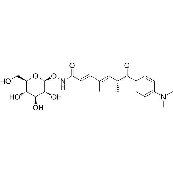
Trichostatin C
Trichostatin C is an inhibitor for histone deacetylase (HDAC), induces apoptosis and arrests cell cycle at G2/M phase, and exhibits anticancer activity against lung cancer and urothelial bladder cancer. Trichostatin C induces differentation of Friend leukemic cells. Trichostatin C exhibits antifungal activity.
For research use only. We do not sell to patients.
- CAS No.: 68676-88-0
- Formula: C23H32N2O8
- Molecular Weight:464.51
-
Storage:
Please store the product under the recommended conditions in the Certificate of Analysis.

To place orders, for customer services and technical support, please contact: MedChemExpress USA
Tel: 609-228-6898 E-mail: [email protected] [email protected]


-
 Biological Activity
-
Chemical Information
- Protocols
- Purity & Documentation
- References
-
 Help & FAQs


-
HY-100871
WT-161HDAC6 Inhibitor -
HY-10221
VorinostatHDAC Inhibitor -
HY-12163
EntinostatHDAC Class I Inhibitor -
HY-13322
PracinostatHDAC/MBLAC2 Inhibitor -

HY-100871R
WT-161 (Standard)HDAC6 Inhibitor -

HY-10221R
Vorinostat (Standard)HDAC Inhibitor -
HY-13265
AR-42Pan HDAC Inhibitor -
HY-19348
Pimelic Diphenylamide 106HDAC Inhibitor -
HY-18976
UF010HDAC Inhibitor -
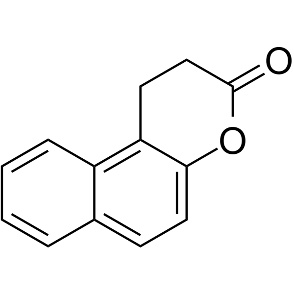
HY-100585
SplitomicinSir2p Inhibitor -
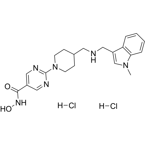
HY-15433A
Quisinostat dihydrochloridePan-HDAC Inhibitor -
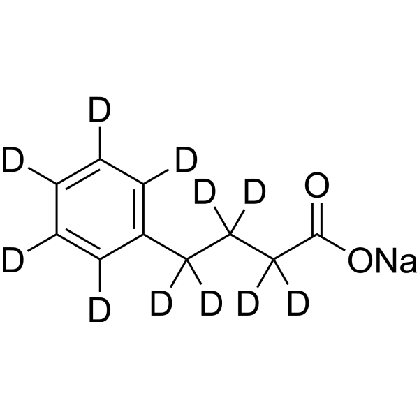
HY-15654S
Phenylbutyrate-d11 sodiumStable Isotope


Biological Activity
Description
IC50 & Target
|
HDAC1 |
HDAC6 |
Cellular Effect
|
Cell Line

|
Type | Value | Description | References |
|---|---|---|---|---|
| A2058 | IC50 |
37.3 μM
Compound: 3
|
Cytotoxicity against human A2058 cells assessed as reduction in cell viability
Cytotoxicity against human A2058 cells assessed as reduction in cell viability
|
[PMID: 32005414] |
| A549 | IC50 |
25.01 μM
Compound: 3
|
Cytotoxicity against human A549 cells assessed as reduction in cell viability
Cytotoxicity against human A549 cells assessed as reduction in cell viability
|
[PMID: 32005414] |
| HEK293 | IC50 |
>100 μM
Compound: 10
|
Cytotoxicity against human HEK293 cells after 96 hrs by MTT assay
Cytotoxicity against human HEK293 cells after 96 hrs by MTT assay
|
[PMID: 25556102] |
| HeLa | IC50 |
>200 μM
Compound: 10
|
Cytotoxicity against human HeLa cells after 96 hrs by MTT assay
Cytotoxicity against human HeLa cells after 96 hrs by MTT assay
|
[PMID: 25556102] |
| HepG2 | EC50 |
46.02 μM
Compound: 10
|
Up-regulation of transcriptional activity of human CLA-1 promoter region -1055 to -62bp transfected in HepG2 cells by luciferase reporter gene assay
Up-regulation of transcriptional activity of human CLA-1 promoter region -1055 to -62bp transfected in HepG2 cells by luciferase reporter gene assay
|
[PMID: 25556102] |
| HepG2 | IC50 |
>200 μM
Compound: 10
|
Cytotoxicity against human HepG2 cells after 96 hrs by MTT assay
Cytotoxicity against human HepG2 cells after 96 hrs by MTT assay
|
[PMID: 25556102] |
| HepG2 | IC50 |
17.82 μM
Compound: 3
|
Cytotoxicity against human HepG2 cells assessed as reduction in cell viability
Cytotoxicity against human HepG2 cells assessed as reduction in cell viability
|
[PMID: 32005414] |
| L02 | IC50 |
>200 μM
Compound: 10
|
Cytotoxicity against human LO2 cells after 96 hrs by MTT assay
Cytotoxicity against human LO2 cells after 96 hrs by MTT assay
|
[PMID: 25556102] |
| MCF7 | IC50 |
18.87 μM
Compound: 3
|
Cytotoxicity against human MCF7 cells assessed as reduction in cell viability
Cytotoxicity against human MCF7 cells assessed as reduction in cell viability
|
[PMID: 32005414] |
| MIA PaCa-2 | IC50 |
37.88 μM
Compound: 3
|
Cytotoxicity against human MIAPaCa2 cells assessed as reduction in cell viability
Cytotoxicity against human MIAPaCa2 cells assessed as reduction in cell viability
|
[PMID: 32005414] |
| SW480 | IC50 |
72 μM
Compound: 5
|
Cytotoxicity against human SW480 cells assessed as reduction in cell viability incubated for 72 hrs by EZ-Cytox colorimetric assay
Cytotoxicity against human SW480 cells assessed as reduction in cell viability incubated for 72 hrs by EZ-Cytox colorimetric assay
|
[PMID: 34455777] |
| U2OS | IC50 |
>200 μM
Compound: 10
|
Cytotoxicity against human U2OS cells after 96 hrs by MTT assay
Cytotoxicity against human U2OS cells after 96 hrs by MTT assay
|
[PMID: 25556102] |
| U2OS | IC50 |
94.9 μM
Compound: 5
|
Cytotoxicity against human U2OS cells assessed as reduction in cell viability incubated for 72 hrs by EZ-Cytox colorimetric assay
Cytotoxicity against human U2OS cells assessed as reduction in cell viability incubated for 72 hrs by EZ-Cytox colorimetric assay
|
[PMID: 34455777] |
In Vitro
Trichostatin C (0-100 μM) exhibits inhibitory activity against HDAC1 and HDAC6, inhibits proliferations of cancer cells A549, J82 and SK-BR-3, with IC50s of 6.24, 4.16 and 0.6 μM, respectively[1].
Trichostatin C (0-10 μM) induces apoptosis in cells J82 through activation of caspase 3 and caspase 7[1].
MedChemExpress (MCE) has not independently confirmed the accuracy of these methods. They are for reference only.
-
Cell Line:A549, J82 and SK-BR-3
-
Concentration:0-100 μM
-
Incubation Time:72 h
-
Result:Inhibits proliferations of A549, J82 and SK-BR-3.
-
Cell Line:J82
-
Concentration:20-80 μM
-
Incubation Time:24 h
-
Result:Induced acetylation of α-Tubulin and Histone 3 (markers of HDAC1 and HDAC6 inhibition).
-
Cell Line:J-82
-
Concentration:0.1-10 μM
-
Incubation Time:48 h
-
Result:Activated 23.1, 61.7 and 62.3% caspase 3/7 at 0.1 μM, 1 μM and 10 μM.


Chemical Information
-
CAS No. 68676-88-0
-
Molecular Weight 464.51
-
Formula C23H32N2O8
-
SMILES
O=C(NO[C@H]1[C@@H]([C@H]([C@@H]([C@@H](CO)O1)O)O)O)/C=C/C(C)=C/[C@@H](C)C(C2=CC=C(N(C)C)C=C2)=O
-
Structure Classification
-
Initial Source
Streptomyces sp. RK98-A74
-
Shipping
Room temperature in continental US; may vary elsewhere.
-
Storage
Please store the product under the recommended conditions in the Certificate of Analysis.


Purity & Documentation
References

Calculators
Concentration (start) × Volume (start) = Concentration (final) × Volume (final)


Trichostatin C Related Classifications
HY-126566 Related Classifications
 Powered by Bioz