



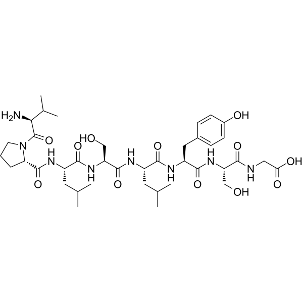
VPLSLYSG
VPLSLYSG is an octapeptide that can be degraded by matrix metalloproteinase-9 (MMP-9), MMP-1 and MMP-2. VPLSLYSG has potential applications in MMP substrates.
For research use only. We do not sell to patients.
- CAS No.: 425632-67-3
- Formula: C39H62N8O12
- Molecular Weight:834.96
-
Storage:
Please store the product under the recommended conditions in the Certificate of Analysis.

To place orders, for customer services and technical support, please contact: MedChemExpress USA
Tel: 609-228-6898 E-mail: [email protected] [email protected]


-
 Biological Activity
-
Chemical Information
- Protocols
- Purity & Documentation
- References
-
 Help & FAQs




Biological Activity
Description


Chemical Information
-
CAS No. 425632-67-3
-
Molecular Weight 834.96
-
Formula C39H62N8O12
-
Sequence
Val-Pro-Leu-Ser-Leu-Tyr-Ser-Gly
-
Sequence Shortening
VPLSLYSG
-
Shipping
Room temperature in continental US; may vary elsewhere.
-
Storage
Please store the product under the recommended conditions in the Certificate of Analysis.

Protocols
-
Mitochondrial membrane-potential fluorescent assay
Mitochondrial membrane potential fluorescent assays estimate ΔΨm in living cells using lipophilic cationic dyes such as TMRM, TMRE, rhodamine 123, and JC-1, which accumulate in mitochondria according to membrane polarization; loss of signal after FCCP or CCCP treatment is interpreted as mitochondrial depolarization. TMRM/TMRE and rhodamine 123 are commonly used for semi-quantitative live-cell microscopy or flow cytometry, while JC-1 can report a shift from red aggregate fluorescence to green monomer fluorescence during depolarization; interpretation requires controls because dye concentration, quenching mode, cell type, dye efflux, and mitochondrial mass can affect fluorescence independently of ΔΨm.
-
Mitochondrial membrane-potential and mitochondrial mass staining
Mitochondrial membrane potential staining measures the electrochemical polarization across the mitochondrial inner membrane in live cells using lipophilic cationic fluorescent probes; early rhodamine-based work showed that selective mitochondrial dye accumulation is lost when the mitochondrial transmembrane potential is dissipated. JC-1 reports mitochondrial polarization by shifting from green monomer fluorescence to red J-aggregate fluorescence as dye concentration increases within energized mitochondria; therefore, the red/green fluorescence ratio is used as a relative readout of mitochondrial membrane potential. TMRE or TMRM staining provides a single-channel relative readout because these cationic rhodamine esters accumulate in polarized mitochondria, and lower fluorescence indicates reduced mitochondrial polarization when acquisition and dye-loading conditions are controlled. Mitochondrial mass staining is commonly performed with MitoTracker Green FM or related MitoTracker dyes as


Purity & Documentation
References
[1]. Suzuki H, et al. Accurate targeting of activated macrophages based on synergistic activation of functional molecules uptake by scavenger receptor and matrix metalloproteinase. ACS Chem Biol. 2008 Aug 15;3(8):471-9. [Content Brief]
[2]. Patterson J, et al. Enhanced proteolytic degradation of molecularly engineered PEG hydrogels in response to MMP-1 and MMP-2. Biomaterials. 2010 Oct;31(30):7836-45. [Content Brief]
[3]. Wieduwild R, et al. Assembling and decorating hyaluronan hydrogels with twin protein superglues to mimic cell-cell interactions. Biomaterials. 2018 Oct;180:253-264. [Content Brief]

Calculators
Concentration (start) × Volume (start) = Concentration (final) × Volume (final)


VPLSLYSG Related Classifications
HY-P4574 Related Classifications
 Powered by Bioz