



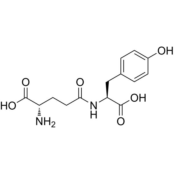
γ-Glu-Tyr
Based on 1 publication(s) in Google Scholar
γ-Glu-Tyr is an orally active gut microbiota-derived metabolite and a competitive DPP-IV inhibitor with an IC50 of 6.77 mM against hDPP-IV. γ-Glu-Tyr weakly activates hCaSR. γ-Glu-Tyr decreases colonic pro-inflammatory cytokine (IL-6, IL-1β, TNF-α) levels. γ-Glu-Tyr activates the cAMP/PKA signaling pathway and inhibits NF-κB phosphorylation, thereby reducing inflammatory responses. γ-Glu-Tyr can be used for research on type 2 diabetes and ulcerative colitis.
For research use only. We do not sell to patients.
- Purity : 99.37%
- CAS No.: 7432-23-7
- Formula: C14H18N2O6
- Molecular Weight:310.30
-
Storage:
Sealed storage, away from moisture and light.
Powder -80°C, 2 years , -20°C, 1 year* In solvent : -80°C, 6 months; -20°C, 1 month (sealed storage, away from moisture and light)

To place orders, for customer services and technical support, please contact: MedChemExpress USA
Tel: 609-228-6898 E-mail: [email protected] [email protected]


-
 Biological Activity
-
Chemical Information
-
Solvent & Solubility
- Protocols
- Purity & Documentation
- References
-
 Help & FAQs


-
HY-101056
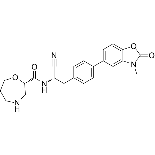
BrensocatibDPP1 Inhibitor -
HY-13233A
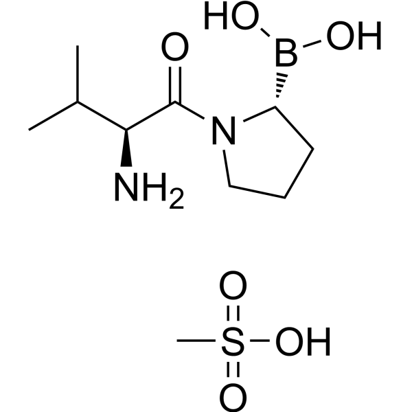
Talabostat mesylateDipeptidyl Peptidase Inhibitor -
HY-10285
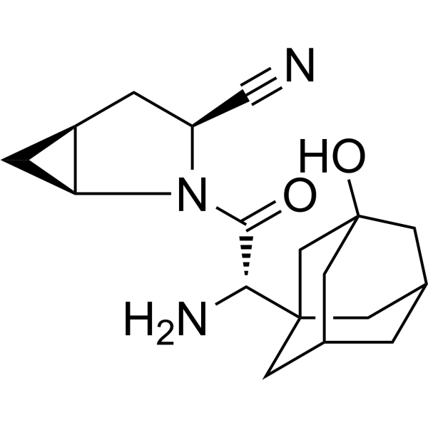
SaxagliptinDPP-4 Inhibitor -
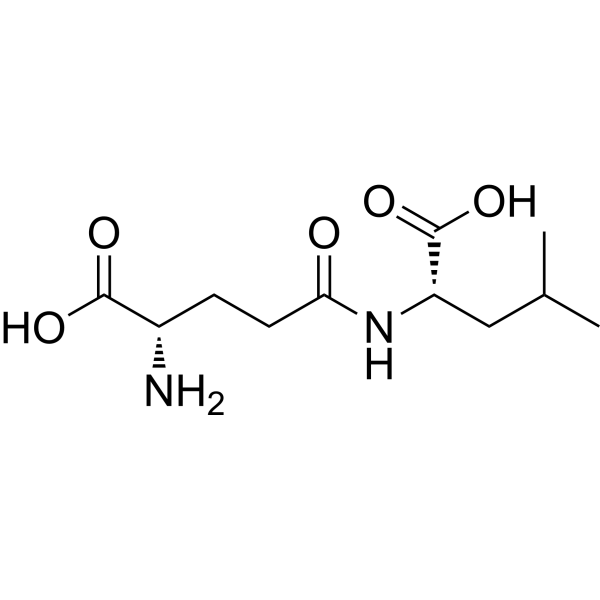
HY-P4632
H-γ-Glu-Leu-OHDipeptide -
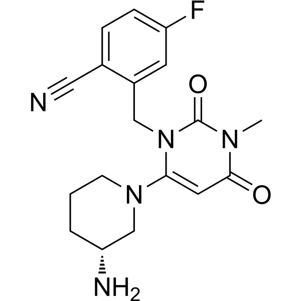
HY-15408
TrelagliptinDPP-4 Inhibitor -
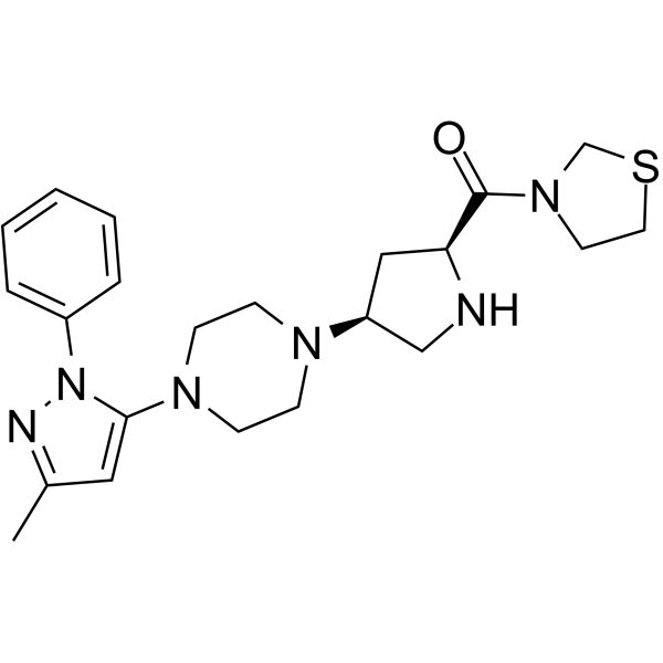
HY-14806
TeneligliptinDPP-4 Inhibitor -
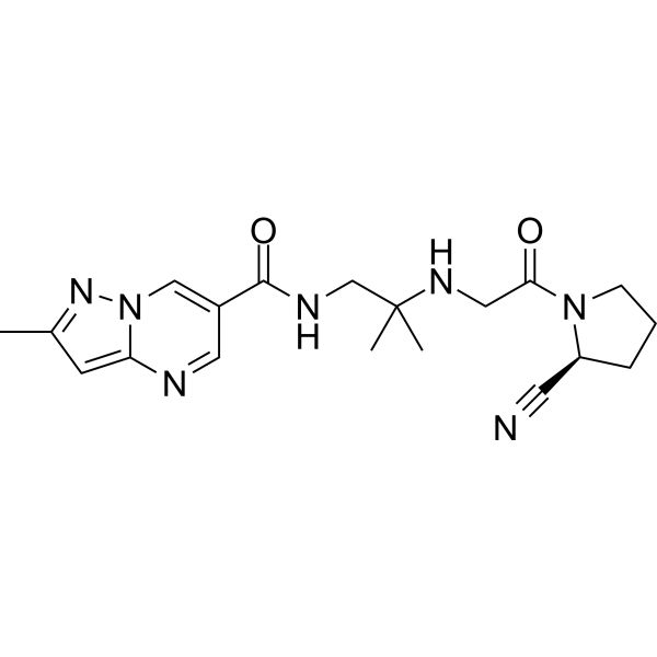
HY-14877
AnagliptinDPP-4 Inhibitor -
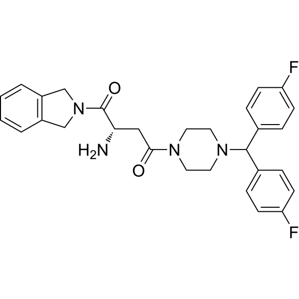
HY-116304
1G244DPP8/9 Inhibitor -
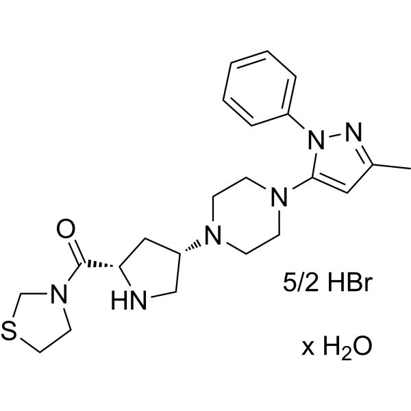
HY-14806B
Teneligliptin hydrobromide hydrateDPP-4 Inhibitor -
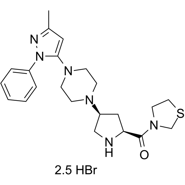
HY-14806A
Teneligliptin hydrobromideDipeptidyl Peptidase Inhibitor -
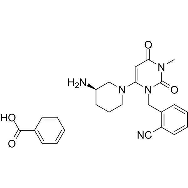
HY-A0023
Alogliptin BenzoateDPP-4 Inhibitor -
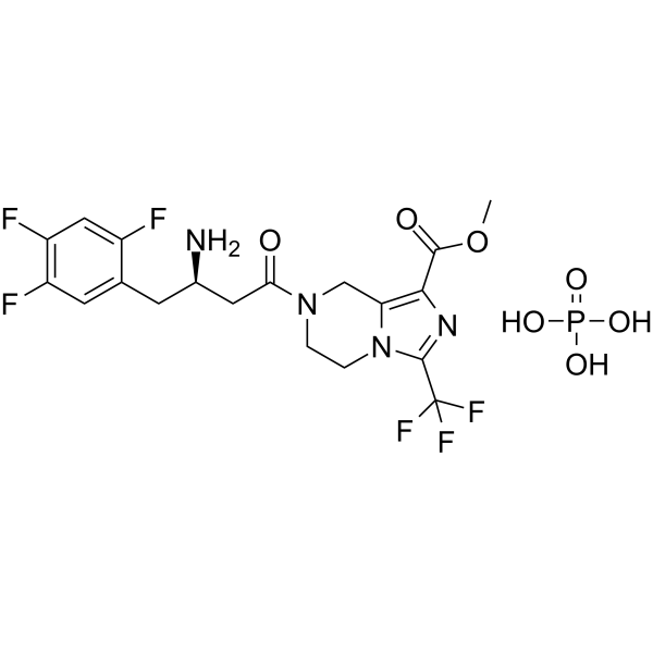
HY-112668
Retagliptin phosphateDPP-4 Inhibitor
Publications Citing Use of MedChemExpress (MCE) γ-Glu-Tyr
MoreAll Endogenous Metabolite Isoforms
More

Biological Activity
Description
|
DPP-4 6.774 mM (IC50) |
Microbial Metabolite |
IL-6 |
IL-1β |
Cellular Effect
|
Cell Line

|
Type | Value | Description | References |
|---|---|---|---|---|
| HEK-293T | EC50 |
508 μM
|
Activation of hCaSR in HEK293T cells cotransfected with hCaSR and hGα15 assessed as increase in intracellular Ca2+ levels measured by Fluo-8 AM fluorescence.
Activation of hCaSR in HEK293T cells cotransfected with hCaSR and hGα15 assessed as increase in intracellular Ca2+ levels measured by Fluo-8 AM fluorescence.
|
32202205 |
In Vitro
γ-Glu-Tyr (0.0125-2.5 mg/mL; 30 min) acts as a competitive inhibitor of human DPP-IV with an IC50 of 6.774 mM[1].
γ-Glu-Tyr weakly activates hCaSR in HEK293T cells with an EC50 of 508 μM, confirming that the γ-glutamyl moiety in the N-terminus and Tyr residue in the C-terminus contribute to its potency[2].
γ-Glu-Tyr (0-75 μM; 24 h) is non-cytotoxic up to 75 μM and optimally reduces IL-1β release at 50 μM in LPS-stimulated RAW264.7 cells[3].
γ-Glu-Tyr (50 μM; 24 h) inhibits IL-1β release in LPS-stimulated RAW264.7 cells by activating the cAMP/PKA/NF-κB pathway[3].
MedChemExpress (MCE) has not independently confirmed the accuracy of these methods. They are for reference only. Further protocols information, click here.
-
Cell Line:RAW264.7
-
Concentration:0-75 μM
-
Incubation Time:24 h
-
Result:Demonstrated non-cytotoxicity up to 75 μM.
Showed optimal reduction of IL-1β levels at 50 μM compared with the 0 μM control.
-
Cell Line:RAW264.7
-
Concentration:50 μM
-
Incubation Time:24 h
-
Result:Increased p-PKA expression (0.72 vs.
LPS: 0.16).
Decreased p-NF-κB expression (0.21 vs.
LPS: 0.95).
In Vivo
γ-Glu-Tyr (50 mg/kg; i.g.; daily; 10 days) protects mice against DSS-induced ulcerative colitis by activating the cAMP/PKA/NF-κB pathway[3].
MedChemExpress (MCE) has not independently confirmed the accuracy of these methods. They are for reference only.
-
Animal Model:C57BL/6 (male, 6 weeks old, 20-24 g, DSS-induced)[3]
-
Dosage:50 mg/kg
-
Administration:i.g.; daily; 10 days
-
Result:Significantly improved body weight.
Decreased DAI scores.
Increased colon length.
Mitigated colonic histopathological damage and inflammation.
Decreased colonic levels of IL-6, IL-1β, and TNF-α.
Decreased levels of γ-linolenic acid.
Increased cAMP levels and p-PKA expression (0.81 vs.
DSS: 0.15).
Decreased p-NF-κB levels (0.19 vs.
DSS: 0.76).
-
Animal Model:C57BL/6 (male, 6 weeks old, 20-24 g, antibiotic cocktail pretreatment followed by DSS-induced)[3]
-
Dosage:50 mg/kg (combined with 80 mg/kg AA)
-
Administration:i.g.; daily; 10 days
-
Result:Significantly enhanced the beneficial effects of AA on DSS-induced body weight loss, colon length shortening, and colonic pathological changes and inflammation.
Decreased levels of γ-linolenic acid in the colon.
Reversed the negative regulatory effects of DSS on the cAMP/PKA/NF-κB pathway by increasing cAMP and p-PKA levels and decreasing p-NF-κB levels.
-
Animal Model:C57BL/6 (male, 6 weeks old, 20-24 g, DSS-induced)[3]
-
Dosage:50 mg/kg (γ-Glu-Tyr); 10 mg/kg (H-89); 1 mg/kg (NF-κB activator 2)
-
Administration:i.g. (daily, 10 days); s.c. (daily, 10 days); i.p. (daily, 10 days)
-
Result:Increased cAMP levels and p-PKA expression.
Decreased p-NF-κB expression.
Co-treatment with H-89 reversed the γ-Glu-Tyr-induced increase in p-PKA (0.24 vs. γ-Glu-Tyr: 0.85) and p-NF-κB (0.66 vs. γ-Glu-Tyr: 0.12).
NF-κB activator 2 reversed the suppression of p-NF-κB expression (0.81 vs. γ-Glu-Tyr: 0.12).
Attenuated protective effects against DSS-induced reductions in body weight, increases in DAI scores, decreases in colon length, and improvements in colonic pathological damage and inflammation following co-treatment with either H-89 or NF-κB activator 2.


Chemical Information
-
CAS No. 7432-23-7
-
Appearance Solid
-
Molecular Weight 310.30
-
Formula C14H18N2O6
-
Color White to off-white
-
Sequence
{γ-Glu}-Tyr
-
Sequence Shortening
{γ-Glu}-Y
-
Shipping
Room temperature in continental US; may vary elsewhere.
-
Storage
Sealed storage, away from moisture and light
Powder -80°C 2 years -20°C 1 year * In solvent : -80°C, 6 months; -20°C, 1 month (sealed storage, away from moisture and light)


Publications (1)
-
Journal Impact Factor
-
Most Recent


Solvent & Solubility
In Vitro:
DMSO : 100 mg/mL (322.27 mM; Need ultrasonic; Hygroscopic DMSO has a significant impact on the solubility of product, please use newly opened DMSO)
H2O : 12.5 mg/mL (40.28 mM; Need ultrasonic)
Please refer to the solubility information to select the appropriate solvent. Once prepared, please aliquot and store the solution to prevent product inactivation from repeated freeze-thaw cycles.
Storage method and period of stock solution: -80°C, 6 months; -20°C, 1 month (sealed storage, away from moisture and light). When stored at -80°C, please use it within 6 months. When stored at -20°C, please use it within 1 month.
* Note: If you choose water as the stock solution, please dilute it to the working solution, then filter and sterilize it with a 0.22 μm filter before use.
Please refer to the solubility information to select the appropriate solvent. Once prepared, please aliquot and store the solution to prevent product inactivation from repeated freeze-thaw cycles.
Storage method and period of stock solution: -80°C, 6 months; -20°C, 1 month (sealed storage, away from moisture and light). When stored at -80°C, please use it within 6 months. When stored at -20°C, please use it within 1 month.
* Note: If you choose water as the stock solution, please dilute it to the working solution, then filter and sterilize it with a 0.22 μm filter before use.
Concentration (start) × Volume (start) = Concentration (final) × Volume (final)
In Vivo:
Select the appropriate dissolution method based on your experimental animal and administration route.
- For the following dissolution methods, please ensure to first prepare a clear stock solution using an In Vitro approach and then sequentially add co-solvents:
- To ensure reliable experimental results, the clarified stock solution can be appropriately stored based on storage conditions. As for the working solution for In Vivo experiments, it is recommended to prepare freshly and use it on the same day.
- The percentages shown for the solvents indicate their volumetric ratio in the final prepared solution. If precipitation or phase separation occurs during preparation, heat and/or sonication can be used to aid dissolution.
Add each solvent one by one: 10% DMSO 40% PEG300 5% Tween-80 45% Saline
Solubility: ≥ 2.5 mg/mL (8.06 mM); Clear solution
This protocol yields a clear solution of ≥ 2.5 mg/mL (saturation unknown).
Taking 1 mL working solution as an example, add 100 μL DMSO stock solution (25.0 mg/mL) to 400 μL PEG300, and mix evenly; then add 50 μL Tween-80 and mix evenly; then add 450 μL Saline to adjust the volume to 1 mL.
Preparation of Saline: Dissolve 0.9 g sodium chloride in ddH₂O and dilute to 100 mL to obtain a clear Saline solution.
Add each solvent one by one: 10% DMSO 90% (20% SBE-β-CD in Saline)
Solubility: ≥ 2.5 mg/mL (8.06 mM); Clear solution
This protocol yields a clear solution of ≥ 2.5 mg/mL (saturation unknown).
Taking 1 mL working solution as an example, add 100 μL DMSO stock solution (25.0 mg/mL) to 900 μL 20% SBE-β-CD in Saline, and mix evenly.
Preparation of 20% SBE-β-CD in Saline (4°C, storage for one week): 2 g SBE-β-CD powder is dissolved in 10 mL Saline, completely dissolve until clear.
In Vivo Dissolution Calculator
Please enter the basic information of animal experiments:
-
-
-
-
Recommended: Prepare an additional quantity of animals to account for potential losses during experiments.
Please enter your animal formula composition:
-
%DMSO +
Recommended: Keep the proportion of DMSO in working solution below 2% if your animal is weak.
-
%+
-
+%Tween-80 + +
-
%Saline +
The co-solvents required include: DMSO, . All of co-solvents are available by MedChemExpress (MCE). , Tween 80. All of co-solvents are available by MedChemExpress (MCE).
Working solution concentration: 0.22 mg/mL
Method for preparing stock solution: mg drug dissolved in μL DMSO. Stock solution concentration: mg/mL. * In solvent : -80°C, 6 months; -20°C, 1 month (sealed storage, away from moisture and light)
1. Take μL DMSO stock solution;
2. Add μL .
μL , mix evenly;
3. Then add μL Tween 80, mix evenly;
4. Then add μL
Please ensure that the stock solution in the first step is dissolved to a clear state, and add co-solvents in sequence. You can use ultrasonic heating (ultrasonic cleaner, recommended frequency 20-40 kHz), vortexing, etc. to assist dissolution.

Protocols
-
Kinase activity and phosphorylation assays
Kinase activity assays measure the ability of kinases to transfer phosphate groups from ATP to specific substrates, while phosphorylation assays detect the presence and levels of phosphorylated proteins. Common methods include radiolabeled ATP incorporation (e. g. ,), ADP release detection via bioluminescence (e. g. ,[3]), enzyme-linked immunosorbent assays (ELISA) for phospho-specific epitopes (e. g. ,[6]), and microtiter-based formats for high-throughput screening (e. g. ,[8]). The ADP-Glo assay quantifies kinase activity by measuring ADP produced during phosphorylation using a luciferase-based system. Radiometric assays involve autoradiography or scintillation counting after incorporation of 32P-labeled ATP into substrate proteins. ELISA-based approaches rely on phospho-specific antibodies to detect activated kinases in cell lysates or purified samples.
-
Western Blot
Western blotting (WB) is a commonly used experimental method in molecular biology, biochemistry, and immunogenetics for identifying and quantifying target proteins. It combines gel electrophoresis with immunoassay, enabling researchers to analyze protein expression, post-translational modifications, and molecular weight.
-
Cell Cytotoxicity Assay
Cytotoxicity assays are usually based on the assessment of cell membrane damage, which can also be indirectly detected by measuring cell viability. Detection methods include MTT assay, CKK-8 assay, LDH assay and ATP assay, etc.
-
DSS-Induced Colitis
Dextran sulfate sodium (DSS)-induced colitis is generated by administering DSS in mouse drinking water, producing epithelial injury, barrier disruption, weight loss, diarrhea, fecal blood, colon shortening, histologic mucosal damage, and inflammatory mediator changes; the model is mainly used to study acute or chronic intestinal inflammation resembling selected features of ulcerative colitis. DSS injury is interpreted through clinical and tissue readouts rather than a single molecular endpoint: daily body weight, stool consistency, and bleeding are combined into a disease activity index, while colon length, histology, cytokines, myeloperoxidase activity, intestinal permeability, and tight-junction markers provide complementary measures of inflammation and barrier damage.
-
TNBS-Induced Colitis
TNBS-induced colitis is produced by intrarectal delivery of 2,4,6-trinitrobenzene sulfonic acid in ethanol, where ethanol disrupts the mucosal barrier and TNBS haptenates colonic proteins, generating immune-mediated colonic inflammation with weight loss, diarrhea, ulceration, transmural injury, inflammatory-cell infiltration, and cytokine responses. The model is used as an experimental intestinal inflammation model with Crohn’s disease–like features, especially when Th1-type responses, IL-12–dependent inflammation, chronic relapsing inflammation, or fibrosis-related endpoints are studied.
-
Research Protocol for Microbiome Analysis
Microbiome analysis characterizes microbial communities in biological or environmental samples by measuring community composition, diversity, taxonomic structure, functional potential, and associations with host or environmental phenotypes. 16S rRNA gene amplicon sequencing is commonly used for bacterial and archaeal taxonomic profiling, while shotgun metagenomics provides higher taxonomic resolution and direct functional information, including microbial genes, pathways, viruses, fungi, and antimicrobial-resistance genes when sequencing depth and host-DNA contamination are adequately controlled. Microbiome results are strongly affected by sample collection, storage, DNA extraction, contamination, sequencing method, reference database, and bioinformatic pipeline; therefore, standardized protocols, negative controls, mock communities, and transparent analysis workflows are required. Unresolved issues include low-biomass contamination, compositional-data bias, inconsistent species-level c
-
Research Protocol for Inflammation-related Diseases
The NLRP3 inflammasome is a cytosolic innate immune signaling platform that integrates priming signals and danger-signal activation to promote caspase-1 activation, maturation of IL-1β and IL-18, and gasdermin D-mediated pyroptotic cell death. The core experimental logic is to determine whether inflammatory disease phenotypes are driven by increased NLRP3 expression, ASC-containing inflammasome assembly, caspase-1 cleavage, GSDMD cleavage, and extracellular release of IL-1β/IL-18 rather than by nonspecific cell injury alone. The pathway is strongly linked to inflammation-related disease phenotypes because monosodium urate crystals activate NALP3/NLRP3 inflammasome signaling in gout-like crystal inflammation, cholesterol crystals activate NLRP3 inflammasomes in atherogenesis models, and DSS-induced intestinal inflammation has been reported to involve NLRP3 inflammasome activity. However, experimental colitis studies also show context-dependent protective effects of NLRP3 inflammasome co
-
Protocol for Kinase activity and phosphorylation assays
Kinase activity assays measure transfer of phosphate from ATP to a protein or peptide substrate, generating phosphorylated substrate, ADP, or incorporated radiolabeled phosphate as the readout; phosphorylation assays measure site-specific phosphorylation in cells or tissues as a proxy for kinase-pathway activation, inhibition, or substrate regulation. Phosphorylation can be detected by phospho-specific Western blot, immunoprecipitation kinase assay, phospho-immunofluorescence, phospho-flow cytometry, luminescent ADP detection, radiolabeled ATP incorporation, or reporter-based pathway assays, and these readouts can be applied to cancer cells, primary neurons, mouse tumors, organoids, inflammatory macrophages, ferroptosis studies, and mitophagy studies when the kinase target is biologically relevant.
-
Research Protocol for Metabolic Diseases
AMP-activated protein kinase, AMPK, is a conserved cellular energy sensor that responds to reduced cellular energy status and coordinates metabolism by increasing ATP-generating catabolic pathways while suppressing ATP-consuming anabolic processes. In metabolic disease research, the AMPK pathway is experimentally relevant because it regulates hepatic lipid synthesis, fatty acid oxidation, glucose production, skeletal-muscle glucose disposal, mTORC1-linked biosynthesis, autophagy, mitochondrial homeostasis, and whole-body energy balance. The central pathway logic is that energy stress, metformin, exercise-like stimulation, or direct AMPK activators increase AMPKα Thr172 phosphorylation and downstream substrate phosphorylation, including ACC and RAPTOR. Phosphorylation of ACC suppresses lipogenesis and supports fatty acid oxidation, whereas phosphorylation of RAPTOR suppresses mTORC1 signaling and links cellular energy status to growth and protein synthesis control. The pathway is linked
-
Pyroptosis Solutions
Pyroptosis is a lytic inflammatory cell-death pathway executed by gasdermin pores, most classically through inflammasome-mediated activation of caspase-1, cleavage of gasdermin D, membrane pore formation, LDH release, and secretion of IL-1β and IL-18. The canonical pathway is commonly modeled by priming cells with an inflammatory signal such as LPS to induce pro-IL-1β and inflammasome components, followed by an activation signal such as ATP or nigericin to activate NLRP3, ASC speck formation, caspase-1 cleavage, GSDMD cleavage, cytokine release, and pyroptotic membrane rupture. The non-canonical pathway is triggered when cytosolic LPS activates mouse caspase-11 or human caspase-4/5, leading to GSDMD cleavage and pyroptosis, and this can secondarily activate NLRP3-dependent IL-1β release. Pyroptosis is linked to inflammatory injury, infection, cancer, liver disease, ocular disease, placental inflammation, and other disease phenotypes, but unresolved questions include which gasdermin fam


Purity & Documentation
-
Data Sheet (292 KB)
-
SDS (252 KB)
- English - EN (252 KB)
- Français - FR (252 KB)
- Deutsch - DE (252 KB)
- Norwegian - NO (252 KB)
- Español - ES (252 KB)
- Swedish - SV (252 KB)
- Italian - IT (252 KB)
- Korean - KR (252 KB)
- Portuguese - PT (252 KB)
-
Handling Instructions (2659 KB)
References


Complete Stock Solution Preparation Table
Please refer to the solubility information to select the appropriate solvent. Once prepared, please aliquot and store the solution to prevent product inactivation from repeated freeze-thaw cycles.
Storage method and period of stock solution: -80°C, 6 months; -20°C, 1 month (sealed storage, away from moisture and light). When stored at -80°C, please use it within 6 months. When stored at -20°C, please use it within 1 month.
| Optional Solvent | Concentration Solvent Mass | 1 mg | 5 mg | 10 mg | 25 mg |
|---|---|---|---|---|---|
| H2O / DMSO | 1 mM | 3.2227 mL | 16.1134 mL | 32.2269 mL | 80.5672 mL |
| 5 mM | 0.6445 mL | 3.2227 mL | 6.4454 mL | 16.1134 mL | |
| 10 mM | 0.3223 mL | 1.6113 mL | 3.2227 mL | 8.0567 mL | |
| 15 mM | 0.2148 mL | 1.0742 mL | 2.1485 mL | 5.3711 mL | |
| 20 mM | 0.1611 mL | 0.8057 mL | 1.6113 mL | 4.0284 mL | |
| 25 mM | 0.1289 mL | 0.6445 mL | 1.2891 mL | 3.2227 mL | |
| 30 mM | 0.1074 mL | 0.5371 mL | 1.0742 mL | 2.6856 mL | |
| 40 mM | 0.0806 mL | 0.4028 mL | 0.8057 mL | 2.0142 mL | |
| DMSO | 50 mM | 0.0645 mL | 0.3223 mL | 0.6445 mL | 1.6113 mL |
| 60 mM | 0.0537 mL | 0.2686 mL | 0.5371 mL | 1.3428 mL | |
| 80 mM | 0.0403 mL | 0.2014 mL | 0.4028 mL | 1.0071 mL | |
| 100 mM | 0.0322 mL | 0.1611 mL | 0.3223 mL | 0.8057 mL |
* Note: If you choose water as the stock solution, please dilute it to the working solution, then filter and sterilize it with a 0.22 μm filter before use.


γ-Glu-Tyr Related Classifications
HY-P4633 Related Classifications
 Powered by Bioz
Keywords
- γ-Glu-Tyr
- 7432-23-7
- Dipeptidyl Peptidase
- Endogenous Metabolite
- CaSR
- Interleukin Related
- TNF Receptor
- PKA
- NF-κB
- competitive inhibitor
- human calcium-sensing receptor
- kokumi
- cAMP/PKA signaling pathway
- ulcerative colitis
- gut microbiota-derived metabolite
- dipeptidyl peptidase-IV
- orthosteric agonist
- positive allosteric modulator
- Inhibitor
- inhibitor
- inhibit