



2,5-Hexanedione-d10

Based on 1 Customer Validation
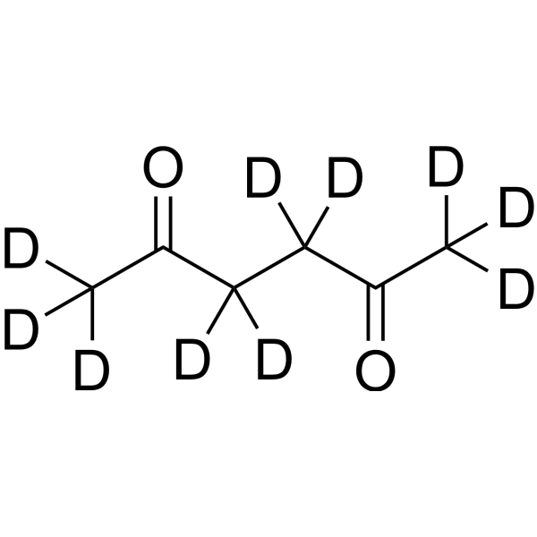
2,5-Hexanedione-d10 (2,5-HD-d10) is the deuterium labeled 2,5-Hexanedione. Hexane-2,5-dione (2,5-HD) is an orally active, CNS-penetrant cytotoxic agent. Hexane-2,5-dione reduces BCL-2 and β-catenin/TCF transcriptional activity, increases BAX and active caspase-3 expression, and promotes apoptosis. Hexane-2,5-dione causes an accumulation of neurofilaments within axons in rats. Hexane-2,5-dione can be used for the research of neurodegenerative diseases.
For research use only. We do not sell to patients.
- Purity : 99.43%
- CAS No.: 97135-07-4
- Formula: C6D10O2
- Molecular Weight:124.20
-
Storage:Pure form -20°C, 3 years , 4°C, 2 years ; In solvent -80°C, 6 months , -20°C, 1 month

To place orders, for customer services and technical support, please contact: MedChemExpress USA
Tel: 609-228-6898 E-mail: [email protected] [email protected]


-
 Biological Activity
-
Chemical Information
- Protocols
- Purity & Documentation
- References
-
 Help & FAQs


All Caspase Isoforms
More

Biological Activity
Description
In Vitro
Stable heavy isotopes of hydrogen, carbon, and other elements have been incorporated into drug molecules, largely as tracers for quantitation during the drug development process. Deuteration has gained attention because of its potential to affect the pharmacokinetic and metabolic profiles of drugs[1].
MedChemExpress (MCE) has not independently confirmed the accuracy of these methods. They are for reference only.
Application
1. This compound can be used as a tracer
2. This compound can be used as an internal standard for quantitative analysis by NMR, GC-MS, or LC-MS.


Chemical Information
-
CAS No. 97135-07-4
-
Unlabeled CAS 110-13-4
-
Appearance Oil
-
Molecular Weight 124.20
-
Formula C6D10O2
-
Color Colorless to light yellow
-
SMILES
[2H]C([2H])([2H])C(C([2H])([2H])C([2H])([2H])C(C([2H])([2H])[2H])=O)=O
-
Synonyms
2,5-HD-d10
-
Shipping
Room temperature in continental US; may vary elsewhere.
-
Storage
Pure form -20°C 3 years 4°C 2 years In solvent -80°C 6 months -20°C 1 month


Purity & Documentation
-
Data Sheet (275 KB)
-
SDS (558 KB)
- English - EN (558 KB)
- Français - FR (558 KB)
- Deutsch - DE (558 KB)
- Norwegian - NO (558 KB)
- Español - ES (558 KB)
- Swedish - SV (558 KB)
- Italian - IT (558 KB)
- Korean - KR (558 KB)
- Portuguese - PT (558 KB)
-
Handling Instructions (2659 KB)
References
[1]. Russak EM, et al. Impact of Deuterium Substitution on the Pharmacokinetics of Pharmaceuticals. Ann Pharmacother. 2019 Feb;53(2):211-216. [Content Brief]
[2]. Sun Y, et al. 2,5-Hexanedione induces human ovarian granulosa cell apoptosis through BCL-2, BAX, and CASPASE-3 signaling pathways. Arch Toxicol. 2012 Feb;86(2):205-15. [Content Brief]
[3]. DeCaprio AP, et al. Comparative neurotoxicity and pyrrole-forming potential of 2,5-hexanedione and perdeuterio-2,5-hexanedione in the rat. Toxicol Appl Pharmacol. 1988 Jan;92(1):75-85. [Content Brief]
[4]. Boekelheide K, et al. 2,5-hexanedione-induced testicular injury. Annu Rev Pharmacol Toxicol. 2003;43:125-47. [Content Brief]
[5]. Planas AM, et al. Uncoupling of cerebral glucose supply and utilization after hexane-2,5-dione intoxication in the rat. J Neurochem. 1987 Mar;48(3):816-23. [Content Brief]
[6]. Xu X, et al. The Wnt/β-catenin pathway is involved in 2,5-hexanedione-induced ovarian granulosa cell cycle arrest. Ecotoxicol Environ Saf. 2023 Dec;268:115720. [Content Brief]
[7]. Pereira ME, et al. 2,5-Hexanedione inhibits rat brain acetylcholinesterase activity in vitro. Toxicol Lett. 2004 Feb 2;146(3):269-74. [Content Brief]

Calculators
Concentration (start) × Volume (start) = Concentration (final) × Volume (final)


2,5-Hexanedione-d10 Related Classifications
HY-Y0315S Related Classifications
 Powered by Bioz