



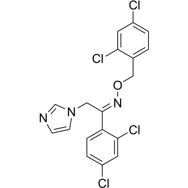
Oxiconazole
Based on 2 publication(s) in Google Scholar
Oxiconazole (Ro 13-8996) is a broad spectrum anti-fungal agent which can inhibit the growth of Candida, Aspergillus and Trichophyton. Oxiconazole is also a highly efficacious activator of CYP3A4 transactivation, which could be antagonized by Rifampicin (HY-B0272) in a competitive manner. Oxiconazole exhibits inhibitory effect against colorectal cancer (CRC) via peroxiredoxin-2 (PRDX2)-mediated autophagy arrest.
연구목적의 판매만을 진행합니다. 환자를 대상으로 한 판매는 하지 않습니다.
- CAS No.: 64211-45-6
- 화학식: C18H13Cl4N3O
- 분자량:429.13
-
보관:
Please store the product under the recommended conditions in the Certificate of Analysis.

To place orders, for customer services and technical support, please contact: MedChemExpress USA
Tel: 609-228-6898 E-mail: [email protected] [email protected]


-
 Biological Activity
-
Chemical Information
- Protocol
- 순도&문서
- References
-
 Help & FAQs


Publications Citing Use of MedChemExpress (MCE) Oxiconazole
MoreAll Antibiotic Isoforms
More

Biological Activity
제품 설명
IC50 & Target
[2]|
CYP3A4 |
Cellular Effect
|
Cell Line

|
Type | Value | Description | References |
|---|---|---|---|---|
| BV-2 | EC50 |
13.5 μM
Compound: Oxiconazole
|
Inhibition of LPS induced NO production in mouse BV-2 cells by Griess reagent based analysis
Inhibition of LPS induced NO production in mouse BV-2 cells by Griess reagent based analysis
|
[PMID: 34875520] |
In Vitro
Oxiconazole (24 h; 0-40 μM) inhibits CRC cell growth[3].
Oxiconazole has antifungal activity against Candida, Aspergillus and Trichophyton[1].
Antifungal Activities of Oxiconazole[1].
| Candidaalbicans | Candidaglabrata | Candidaparapsilosis | Aspergillusfumigatus | Aspergillusflavus | Trichophytonmentagrophytes | Trichophytonrubrum | |
| Oxiconazole | 0.03 μg/mL | 0.01 μg/mL | 0.008 μg/mL | 2 μg/mL | 2 μg/mL | 2 μg/mL | 2 μg/mL |
MedChemExpress (MCE) has not independently confirmed the accuracy of these methods. They are for reference only.
-
Cell Line:HCT116, SW480, RKO, DLD-1, SW620, LoVo and NCM460
-
Concentration:0-40 μM
-
Incubation Time:24 h
-
Result:Exhibited inhibitory activity against HCT116, SW480, RKO, DLD-1, SW620, LoVo and NCM460 with IC50s of 25.86 μM, 27.34 μM, 21.01 μM, 25.56 μM, 21.75 μM, 24.87 μM and 126.4 μM.
In Vivo
MedChemExpress (MCE) has not independently confirmed the accuracy of these methods. They are for reference only.
-
Animal Model:BALB/c nude mice (injected subcutaneously with HCT116 cells (1×10⁷/mouse)[3]
-
Dosage:50 mg/kg/day
-
Administration:IP; for 12 days
-
Result:Significantly restrained CRC cell growth and showed no obvious side effects.


Chemical Information
-
CAS No. 64211-45-6
-
분자량 429.13
-
화학식 C18H13Cl4N3O
-
SMILES
ClC1=CC=C(/C(CN2C=CN=C2)=N/OCC3=CC=C(Cl)C=C3Cl)C(Cl)=C1
-
Synonyms
Ro 13-8996 free base
-
선적
Room temperature in continental US; may vary elsewhere.
-
보관
Please store the product under the recommended conditions in the Certificate of Analysis.


Publications (2)
-
Journal Impact Factor
-
Most Recent
-
Int J Biol Sci
2022 May 21;18(9):3747-3761. PMID: 35813474 -


순도&문서
References
[1]. Rossello A, et al. Synthesis, antifungal activity, and molecular modeling studies of new inverted oxime ethers of oxiconazole. J Med Chem. 2002 Oct 24;45(22):4903-12. [Content Brief]
[2]. Svecova L, et al. Azole antimycotics differentially affect rifampicin-induced pregnane X receptor-mediated CYP3A4 gene expression. Drug Metab Dispos. 2008 Feb;36(2):339-48. [Content Brief]
[3]. Shi J, et al. Repurposing Oxiconazole against Colorectal Cancer via PRDX2-mediated Autophagy Arrest. Int J Biol Sci. 2022 May 21;18(9):3747-3761. [Content Brief]

Calculators
Concentration (start) × Volume (start) = Concentration (final) × Volume (final)


Oxiconazole Related Classifications
HY-B1324A Related Classifications
 Powered by Bioz