



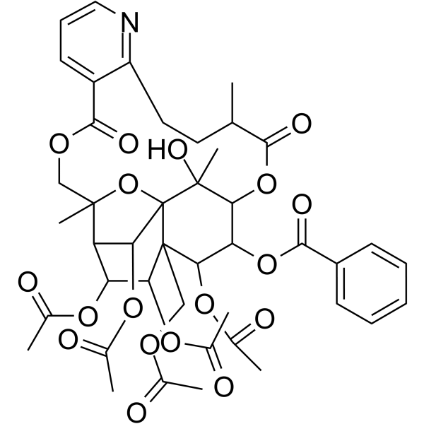
Wilforine
Based on 1 publication(s) in Google Scholar
Wilforine is an orally active JAK-STAT pathway inhibitor with immunomodulatory effects and the ability to inhibit osteoclast fusion. Wilforine disrupts lipid raft integrity, reprograms cholesterol and glycosphingolipid metabolic pathways, regulates NF-κB and the complement system, and modulates the expression of various interleukins. Wilforine also inhibits the Wnt11/β-catenin signaling pathway and suppresses the proliferation of fibroblast-like synoviocytes. Wilforine can serve as a quality and pharmacokinetic marker for Tripterygium glycoside tablets, and can be applied to research on related diseases such as rheumatoid arthritis, inflammatory osteolysis, and SAPHO syndrome.
Nur für Forschungszwecke. Wir verkaufen nicht an Patienten.
- Reinheit : 99.47%
- CAS. Nr.: 11088-09-8
- Formel: C43H49NO18
- Molecular Weight:867.85
-
Speicherung:Powder -20°C, 3 years , 4°C, 2 years ; In solvent -80°C, 6 months , -20°C, 1 month

To place orders, for customer services and technical support, please contact: MedChemExpress USA
Tel: 609-228-6898 E-mail: [email protected] [email protected]


-
 Biologische Aktivität
-
Chemical Information
-
Lösungsmittel & Löslichkeit
- Protokoll
- Reinheit & Dokumentation
- Verweise
-
 Help & FAQs


Publications Citing Use of MedChemExpress (MCE) Wilforine
More

Biologische Aktivität
Beschreibung
IC50 & Target
[1]|
Wnt11 |
In Vitro
Wilforine (100 μM; 6 days) inhibits osteoclast fusion in RAW264.7 cells by suppressing the JAK-STAT pathway, reducing expression of lipid raft scaffold proteins Stomatin and Flotillin-1, disrupting lipid raft integrity, and impairing localization and expression of fusion-related proteins CD9 and DC-STAMP[1].
Wilforine (200-400 nM) dose-dependently inhibits the proliferation of collagen-induced arthritis fibroblast-like synoviocytes (CIA FLS)[2].
Wilforine (30-300 μM; 20 h) induces significant cytotoxicity in human hepatoma HepG2 cells at concentrations of 30, 100, and 300 μM, with viability decreasing in a concentration-dependent manner[3].
MedChemExpress (MCE) has not independently confirmed the accuracy of these methods. They are for reference only. Further protocols information, click here.
-
Cell Line:Wnt11-overexpressing collagen-induced arthritis fibroblast-like synoviocytes (CIA FLS)
-
Concentration:400 nM (wilforine treatment); optimal MOI (adenovirus infection)
-
Incubation Time:36-48 h (adenovirus infection before wilforine treatment)
-
Result:Reduced relative CCK-8 detection value of CIA FLS from ~3.2 (untreated) to ~1.8 (wilforine-treated).
Reversed the inhibitory effect in Wnt11-overexpressing CIA FLS, with relative CCK-8 detection value increased to ~2.8.
-
Cell Line:human hepatoma HepG2 cells
-
Concentration:30 μM; 100 μM; 300 μM
-
Incubation Time:20 h
-
Result:Caused a significant reduction in HepG2 relative viability compared to the control group at 30 μM.
Caused highly significant reductions in HepG2 relative viability compared to the control group at 100 μM and 300 μM.
In Vivo
Wilforine (40-56 μg/kg; p.o.; daily; from day 7 post-first immunization through day 35 post-first immunization) dose-dependently alleviates collagen-induced arthritis in rats, with significant reductions in arthritis scores, inflammatory cytokines, and pathological gene expression via inhibition of the Wnt11/β-catenin signaling pathway[2].
Wilforine (10-90 mg/kg; p.o.; daily; 21 days), administered as part of TGT suspension, dose-dependently reduces paw swelling in adjuvant arthritis rats and exhibits dose-dependent and time-dependent plasma AUC0-tn values, with significant toxicity (4 deaths) observed at the 90 mg/kg dose[3].
MedChemExpress (MCE) has not independently confirmed the accuracy of these methods. They are for reference only.
-
Animal Model:C57BL/6N Pstpip2cmo mice (both male and female, 6 weeks old, body weight 25 g)[1]
-
Dosage:10 mg/kg; 20 mg/kg
-
Administration:p.o.; daily; 28 days
-
Result:Significantly reduced arthritis scores and paw thickness compared to the model group, with the 20 mg/kg dose showing superior efficacy.
Reduced bone destruction and clearer joint margins in treated mice, with the 20 mg/kg dose showing greater improvement.
Demonstrated dose-dependent reductions in cartilage erosion, synovial hyperplasia, inflammatory cell infiltration, cartilage matrix degradation, and periarticular fibrosis; the 20 mg/kg dose restored near-normal cartilage structure and tidemark clarity.
Significantly suppressed joint and paw expression of pro-inflammatory cytokines IL-1β and IL-6, and increased anti-inflammatory cytokine IL-10 expression compared to the model group (20 mg/kg dose).
Significantly downregulated joint expression of lipid raft scaffold proteins Stomatin and Flotillin-1, and osteoclast fusion-related proteins CD9 and DC-STAMPcompared to the model group (20 mg/kg dose).
-
Animal Model:Sprague-Dawley (SD) (male, 160-180 g, SPF grade, collagen-induced arthritis model)[2]
-
Dosage:40 μg/kg; 48 μg/kg; 56 μg/kg
-
Administration:p.o.; daily; from day 7 post-first immunization through day 35 post-first immunization
-
Result:Dose-dependently reduced arthritis scores, hind paw swelling, and pain sensitivity (increased paw withdrawal threshold), and restored body weight.
Dose-dependently reduced serum levels of IL-1β, IL-6, and TNF-α.
Decreased mRNA levels of MMP3 and fibronectin in joint synovium.
Alleviated synovial hyperplasia, pannus formation, and bone/cartilage damage.
Dose-dependently reduced mRNA levels of Wnt11, CCND1, GSK-3β, and c-Myc in synovium.


Chemical Information
-
CAS. Nr. 11088-09-8
-
Appearance Solid
-
Molecular Weight 867.85
-
Formel C43H49NO18
-
Color White to off-white
-
SMILES
CC(OCC(C1OC(C)=O)(C(C(OC(C2=CC=CC=C2)=O)C3OC(C4C)=O)OC(C)=O)C(OC(C)(COC(C5=CC=CN=C5CC4)=O)C6C1OC(C)=O)(C3(O)C)C6OC(C)=O)=O
-
Structure Classification
-
Initial Source
-
Versand
Room temperature in continental US; may vary elsewhere.
-
Speicherung
Powder -20°C 3 years 4°C 2 years In solvent -80°C 6 months -20°C 1 month


Publications (1)
-
Journal Impact Factor
-
Most Recent


Lösungsmittel & Löslichkeit
In Vitro:
DMSO : 50 mg/mL (57.61 mM; Need ultrasonic; Hygroscopic DMSO has a significant impact on the solubility of product, please use newly opened DMSO)
Please refer to the solubility information to select the appropriate solvent. Once prepared, please aliquot and store the solution to prevent product inactivation from repeated freeze-thaw cycles.
Storage method and period of stock solution: -80°C, 6 months; -20°C, 1 month. When stored at -80°C, please use it within 6 months. When stored at -20°C, please use it within 1 month.
Please refer to the solubility information to select the appropriate solvent. Once prepared, please aliquot and store the solution to prevent product inactivation from repeated freeze-thaw cycles.
Storage method and period of stock solution: -80°C, 6 months; -20°C, 1 month. When stored at -80°C, please use it within 6 months. When stored at -20°C, please use it within 1 month.
Konzentration (Stammlösung) × Volumen (Stammlösung) = Konzentration (Ziellösung) × Volumen (Ziellösung)
In Vivo:
Select the appropriate dissolution method based on your experimental animal and administration route.
- For the following dissolution methods, please ensure to first prepare a clear stock solution using an In Vitro approach and then sequentially add co-solvents:
- To ensure reliable experimental results, the clarified stock solution can be appropriately stored based on storage conditions. As for the working solution for In Vivo experiments, it is recommended to prepare freshly and use it on the same day.
- The percentages shown for the solvents indicate their volumetric ratio in the final prepared solution. If precipitation or phase separation occurs during preparation, heat and/or sonication can be used to aid dissolution.
Add each solvent one by one: 10% DMSO 40% PEG300 5% Tween-80 45% Saline
Solubility: ≥ 2.5 mg/mL (2.88 mM); Clear solution
This protocol yields a clear solution of ≥ 2.5 mg/mL (saturation unknown).
Taking 1 mL working solution as an example, add 100 μL DMSO stock solution (25.0 mg/mL) to 400 μL PEG300, and mix evenly; then add 50 μL Tween-80 and mix evenly; then add 450 μL Saline to adjust the volume to 1 mL.
Preparation of Saline: Dissolve 0.9 g sodium chloride in ddH₂O and dilute to 100 mL to obtain a clear Saline solution.
In Vivo Dissolution Calculator
Please enter the basic information of animal experiments:
-
-
-
-
Recommended: Prepare an additional quantity of animals to account for potential losses during experiments.
Please enter your animal formula composition:
-
%DMSO +
Recommended: Keep the proportion of DMSO in working solution below 2% if your animal is weak.
-
%+
-
+%Tween-80 + +
-
%Saline +
The co-solvents required include: DMSO, . All of co-solvents are available by MedChemExpress (MCE). , Tween 80. All of co-solvents are available by MedChemExpress (MCE).
Working solution concentration: 0.22 mg/mL
Method for preparing stock solution: mg drug dissolved in μL DMSO. Stock solution concentration: mg/mL.
1. Take μL DMSO stock solution;
2. Add μL .
μL , mix evenly;
3. Then add μL Tween 80, mix evenly;
4. Then add μL
Please ensure that the stock solution in the first step is dissolved to a clear state, and add co-solvents in sequence. You can use ultrasonic heating (ultrasonic cleaner, recommended frequency 20-40 kHz), vortexing, etc. to assist dissolution.

Protokoll
-
RNA extraction experimental
By lysing cells, releasing RNA, and removing impurities such as proteins and DNA, high-purity RNA products are finally obtained. The commonly used traditional method is the guanidine isothiocyanate/phenol/chloroform method (Trizol), which is suitable for a variety of animal materials including animal tissues, microorganisms, cultured cells, etc., and most plant materials.
-
Collagen-Induced Arthritis
Collagen-induced arthritis (CIA) is an autoimmune murine model of rheumatoid arthritis in which immunization with type II collagen (CII) emulsified in an adjuvant induces a T cell- and autoantibody-driven inflammatory arthritis characterized by synovial hyperplasia, immune cell infiltration, and joint destruction. The model typically relies on genetically susceptible mouse strains (e. g. , DBA/1) and reproduces key features of human rheumatoid arthritis, including anti-collagen immune responses and progressive joint inflammation. Disease onset generally occurs within ~3-4 weeks after immunization, depending on antigen/adjuvant combinations and protocol variation. The immunopathology is driven by adaptive immune activation against CII, leading to systemic and local joint inflammation mediated by pro-inflammatory cytokines and effector immune cells, making CIA a standard preclinical platform for evaluating immunomodulatory and anti-arthritic interventions.
-
Research Protocol for Inflammation-related Diseases
The NLRP3 inflammasome is a cytosolic innate immune signaling platform that integrates priming signals and danger-signal activation to promote caspase-1 activation, maturation of IL-1β and IL-18, and gasdermin D-mediated pyroptotic cell death. The core experimental logic is to determine whether inflammatory disease phenotypes are driven by increased NLRP3 expression, ASC-containing inflammasome assembly, caspase-1 cleavage, GSDMD cleavage, and extracellular release of IL-1β/IL-18 rather than by nonspecific cell injury alone. The pathway is strongly linked to inflammation-related disease phenotypes because monosodium urate crystals activate NALP3/NLRP3 inflammasome signaling in gout-like crystal inflammation, cholesterol crystals activate NLRP3 inflammasomes in atherogenesis models, and DSS-induced intestinal inflammation has been reported to involve NLRP3 inflammasome activity. However, experimental colitis studies also show context-dependent protective effects of NLRP3 inflammasome co
-
Osteoclast differentiation from monocyte/macrophage precursors
Osteoclast differentiation is an in vitro induction assay in which monocyte/macrophage-lineage precursors are exposed to macrophage colony-stimulating factor (M-CSF) and receptor activator of NF-κB ligand (RANKL), generating multinucleated osteoclasts that are commonly identified by tartrate-resistant acid phosphatase (TRAP) staining and functionally confirmed by resorption pits on dentin, bone, or mineralized substrates. M-CSF supports survival and expansion of osteoclast precursors, while RANKL binding to RANK drives osteoclast commitment, fusion, maturation, and resorptive function; osteoprotegerin inhibits this pathway by binding RANKL and preventing RANK activation. The main readouts are the number of TRAP-positive multinucleated cells, formation of F-actin rings, and resorbed surface area; TRAP-positive multinucleated cells indicate osteoclast differentiation, whereas pit formation on dentin, bone, or mineralized coating indicates functional bone-resorbing activity.
-
Cell Viability Determination by MTT Colorimetric Assay
The following protocol uses the MTT colorimetric assay as a classic literature-established method for assessing cell viability/metabolic activity in cultured mammalian cells. MTT[3-(4,5-dimethylthiazol-2-yl)-2,5-diphenyltetrazolium bromide] is reduced by metabolically active cells to a colored formazan product; the amount of formazan is quantified spectrophotometrically and provides an indirect measure of metabolically active viable cells. Importantly, MTT reduction reflects cellular oxidoreductase/metabolic activity rather than an absolute direct count of living cells, so changes in cellular metabolism can alter the signal independently of cell number.


Reinheit & Dokumentation
-
Data Sheet (296 KB)
-
SDS (392 KB)
- English - EN (392 KB)
- Français - FR (392 KB)
- Deutsch - DE (392 KB)
- Norwegian - NO (392 KB)
- Español - ES (392 KB)
- Swedish - SV (392 KB)
- Italian - IT (392 KB)
- Korean - KR (392 KB)
- Portuguese - PT (392 KB)
-
Handling Instructions (2659 KB)
Verweise
[1]. Geng S, et al. Wilforine attenuates inflammatory osteolysis by suppressing osteoclast fusion through JAK-STAT-stomatin immunoregulatory signaling. Front Immunol. 2026;17:1789493. Published 2026 Mar 13. [Content Brief]
[2]. Huang Y, et al. Wilforine inhibits rheumatoid arthritis pathology through the Wnt11/β-catenin signaling pathway axis. Arthritis Res Ther. 2023;25(1):243. Published 2023 Dec 14. [Content Brief]
[3]. Gao X, et al. Wilforine, the Q-marker and PK-maker of Tripterygium glycosides tablet: Based on preparation quantitative analysis and PK-PD study. Phytomedicine. 2019;54:357-364. [Content Brief]


Complete Stock Solution Preparation Table
Please refer to the solubility information to select the appropriate solvent. Once prepared, please aliquot and store the solution to prevent product inactivation from repeated freeze-thaw cycles.
Storage method and period of stock solution: -80°C, 6 months; -20°C, 1 month. When stored at -80°C, please use it within 6 months. When stored at -20°C, please use it within 1 month.
| Optional Solvent | Concentration Solvent Mass | 1 mg | 5 mg | 10 mg | 25 mg |
|---|---|---|---|---|---|
| DMSO | 1 mM | 1.1523 mL | 5.7614 mL | 11.5227 mL | 28.8068 mL |
| 5 mM | 0.2305 mL | 1.1523 mL | 2.3045 mL | 5.7614 mL | |
| 10 mM | 0.1152 mL | 0.5761 mL | 1.1523 mL | 2.8807 mL | |
| 15 mM | 0.0768 mL | 0.3841 mL | 0.7682 mL | 1.9205 mL | |
| 20 mM | 0.0576 mL | 0.2881 mL | 0.5761 mL | 1.4403 mL | |
| 25 mM | 0.0461 mL | 0.2305 mL | 0.4609 mL | 1.1523 mL | |
| 30 mM | 0.0384 mL | 0.1920 mL | 0.3841 mL | 0.9602 mL | |
| 40 mM | 0.0288 mL | 0.1440 mL | 0.2881 mL | 0.7202 mL | |
| 50 mM | 0.0230 mL | 0.1152 mL | 0.2305 mL | 0.5761 mL |


Wilforine Related Classifications
HY-N0899 Related Classifications
 Powered by Bioz
Keywords
- Wilforine
- 11088-09-8
- JAK
- STAT
- Wnt
- β-catenin
- collagen-induced arthritis fibroblast-like synoviocytes
- Pstpip2cmo mice
- Wnt11/β-catenin signaling
- JAK-STAT signaling pathway
- inflammatory osteolysis
- HepG2 cells
- fibroblast-like synovial cells
- rheumatoid arthritis fibroblast-like synoviocytes
- RAW264.7 cells
- NF-κB
- Inhibitor
- inhibitor
- inhibit