



Narasin
Based on 2 publication(s) in Google Scholar
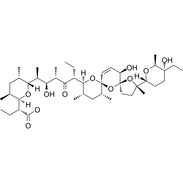
Narasin is a cationic ionophore antibiotic and coccidiostat agent. Narasin inhibits NF-κB signaling and induces tumor cells apoptosis. Narasin has antimicrobial, antiviral anticancer activity. Narasin inhibits tumor metastasis and growth of ERα‑positive breast cancer cells by inactivation of the TGF-β/SMAD3 and IL‑6/STAT3 signaling pathways.
Nur für Forschungszwecke. Wir verkaufen nicht an Patienten.
- Reinheit : 99.58%
- CAS. Nr.: 55134-13-9
- Formel: C43H72O11
- Molecular Weight:765.03
-
Speicherung:
-20°C, sealed storage, away from moisture and light
* In solvent : -80°C, 6 months; -20°C, 1 month (sealed storage, away from moisture and light)

To place orders, for customer services and technical support, please contact: MedChemExpress USA
Tel: 609-228-6898 E-mail: [email protected] [email protected]


-
 Biologische Aktivität
-
Chemical Information
-
Lösungsmittel & Löslichkeit
- Protokoll
- Reinheit & Dokumentation
- Verweise
-
 Help & FAQs


Publications Citing Use of MedChemExpress (MCE) Narasin
MoreAlle Parasite Isoform-spezifische Produkte anzeigen
MoreAlle Antibiotic Isoform-spezifische Produkte anzeigen
More

Biologische Aktivität
Beschreibung
IC50 & Target
|
Coccidia |
In Vitro
Narasin (1-25 ng/mL, 48 h) increases the percentage of phosphatidylserine exposing erythrocytes, cytosolic Ca2+ activity ([Ca2+]i) and the ceramide abundance at the erythrocyte surface[1].
Narasin (0-10 μM, 72 h) inhibits DENV2 infection with an IC50 value of 0.39 μM[2].
Narasin (0-10 μM, 72 h) significantly inhibits the cell viability of MCF-7, T47D and MDA-MB-231 cells, with respective IC50 values of 2.219, 3.562 and 11.76 µM[3].
Narasin (2.5-5 µM, 24 h) inhibits cell migration and invasion by suppressing EMT in ER+ BC cells[3].
MedChemExpress (MCE) has not independently confirmed the accuracy of these methods. They are for reference only.
-
Cell Line:MCF-7, T47D, MDA-MB-231
-
Concentration:0-10 μM
-
Incubation Time:72 h
-
Result:Significantly reduced the viability of MCF-7 and T47D cells, with IC50 values of 2.219 and 3.562 µM, respectively.
-
Cell Line:MCF-7 and T47D cells
-
Concentration:2.5-5 µM
-
Incubation Time:72 h
-
Result:Significantly inhibited the migration of MCF-7 and T47D cells.
In Vivo
MedChemExpress (MCE) has not independently confirmed the accuracy of these methods. They are for reference only.
-
Animal Model:Xenograft mice model with MCF-7 cells
-
Dosage:0.5 mg/kg or 1.5 mg/kg
-
Administration:i.p., every 2 days for 50 consecutive days
-
Result:Significantly decreased tumor volume and tumor weight in xenograft mice model with MCF-7 cells.


Chemical Information
-
CAS. Nr. 55134-13-9
-
Appearance Solid
-
Molecular Weight 765.03
-
Formel C43H72O11
-
Color White to off-white
-
SMILES
O[C@H]1[C@@]2(O[C@](C)([C@]3([H])O[C@H]([C@](CC)(O)CC3)C)CC2)O[C@@]4(O[C@]([C@@H](C)C[C@H]4C)([H])[C@@H](CC)C([C@@H](C)[C@@H](O)[C@@H]([C@]5([H])O[C@@]([C@@H](C)C[C@@H]5C)([H])[C@@H](CC)C(O)=O)C)=O)C=C1
-
Versand
Room temperature in continental US; may vary elsewhere.
-
Speicherung
-20°C, sealed storage, away from moisture and light
* In solvent : -80°C, 6 months; -20°C, 1 month (sealed storage, away from moisture and light)


Publications (2)
-
Journal Impact Factor
-
Most Recent
-
Cell Rep
Controlling mitochondrial membrane architecture via MIC60 determines viral replication to promote anti-viral immunity. [Abstract]2025 Jul 22;44(7):115922. PMID: 40628273 -
Talanta
Fluorescence-activated cell sorting-based efficient screening of monensin monoclonal antibodies and applications in lateral flow immunoassay. [Abstract]2025 Oct 1:293:128128. PMID: 40222095


Lösungsmittel & Löslichkeit
In Vitro:
DMSO : 12.5 mg/mL (16.34 mM; Need ultrasonic; Hygroscopic DMSO has a significant impact on the solubility of product, please use newly opened DMSO)
Please refer to the solubility information to select the appropriate solvent. Once prepared, please aliquot and store the solution to prevent product inactivation from repeated freeze-thaw cycles.
Storage method and period of stock solution: -80°C, 6 months; -20°C, 1 month (sealed storage, away from moisture and light). When stored at -80°C, please use it within 6 months. When stored at -20°C, please use it within 1 month.
Please refer to the solubility information to select the appropriate solvent. Once prepared, please aliquot and store the solution to prevent product inactivation from repeated freeze-thaw cycles.
Storage method and period of stock solution: -80°C, 6 months; -20°C, 1 month (sealed storage, away from moisture and light). When stored at -80°C, please use it within 6 months. When stored at -20°C, please use it within 1 month.
Konzentration (Stammlösung) × Volumen (Stammlösung) = Konzentration (Ziellösung) × Volumen (Ziellösung)
In Vivo:
Select the appropriate dissolution method based on your experimental animal and administration route.
- For the following dissolution methods, please ensure to first prepare a clear stock solution using an In Vitro approach and then sequentially add co-solvents:
- To ensure reliable experimental results, the clarified stock solution can be appropriately stored based on storage conditions. As for the working solution for In Vivo experiments, it is recommended to prepare freshly and use it on the same day.
- The percentages shown for the solvents indicate their volumetric ratio in the final prepared solution. If precipitation or phase separation occurs during preparation, heat and/or sonication can be used to aid dissolution.
Add each solvent one by one: 10% DMSO 40% PEG300 5% Tween-80 45% Saline
Solubility: ≥ 1.25 mg/mL (1.63 mM); Clear solution
This protocol yields a clear solution of ≥ 1.25 mg/mL (saturation unknown).
Taking 1 mL working solution as an example, add 100 μL DMSO stock solution (12.5 mg/mL) to 400 μL PEG300, and mix evenly; then add 50 μL Tween-80 and mix evenly; then add 450 μL Saline to adjust the volume to 1 mL.
Preparation of Saline: Dissolve 0.9 g sodium chloride in ddH₂O and dilute to 100 mL to obtain a clear Saline solution.
In Vivo Dissolution Calculator
Please enter the basic information of animal experiments:
-
-
-
-
Recommended: Prepare an additional quantity of animals to account for potential losses during experiments.
Please enter your animal formula composition:
-
%DMSO +
Recommended: Keep the proportion of DMSO in working solution below 2% if your animal is weak.
-
%+
-
+%Tween-80 + +
-
%Saline +
The co-solvents required include: DMSO, . All of co-solvents are available by MedChemExpress (MCE). , Tween 80. All of co-solvents are available by MedChemExpress (MCE).
Working solution concentration: 0.22 mg/mL
Method for preparing stock solution: mg drug dissolved in μL DMSO. Stock solution concentration: mg/mL. * In solvent : -80°C, 6 months; -20°C, 1 month (sealed storage, away from moisture and light)
1. Take μL DMSO stock solution;
2. Add μL .
μL , mix evenly;
3. Then add μL Tween 80, mix evenly;
4. Then add μL
Please ensure that the stock solution in the first step is dissolved to a clear state, and add co-solvents in sequence. You can use ultrasonic heating (ultrasonic cleaner, recommended frequency 20-40 kHz), vortexing, etc. to assist dissolution.


Reinheit & Dokumentation
-
Data Sheet (281 KB)
-
SDS (480 KB)
- English - EN (480 KB)
- Français - FR (480 KB)
- Deutsch - DE (480 KB)
- Norwegian - NO (480 KB)
- Español - ES (480 KB)
- Swedish - SV (480 KB)
- Italian - IT (480 KB)
- Korean - KR (480 KB)
- Portuguese - PT (480 KB)
-
Handling Instructions (2659 KB)
Verweise
[1]. Ghada Bouguerra, et al. Stimulation of Eryptosis by Narasin. Cell Physiol Biochem. 2015;37(5):1807-16. [Content Brief]
[2]. Low JS, et al. Narasin, a novel antiviral compound that blocks dengue virus protein expression. Antivir Ther. 2011;16(8):1203-18. [Content Brief]
[3]. Chen J, et al. Narasin inhibits tumor metastasis and growth of ERα‑positive breast cancer cells by inactivation of the TGF‑β/SMAD3 and IL‑6/STAT3 signaling pathways. Mol Med Rep. 2020 Dec;22(6):5113-5124. [Content Brief]


Complete Stock Solution Preparation Table
Please refer to the solubility information to select the appropriate solvent. Once prepared, please aliquot and store the solution to prevent product inactivation from repeated freeze-thaw cycles.
Storage method and period of stock solution: -80°C, 6 months; -20°C, 1 month (sealed storage, away from moisture and light). When stored at -80°C, please use it within 6 months. When stored at -20°C, please use it within 1 month.
| Optional Solvent | Concentration Solvent Mass | 1 mg | 5 mg | 10 mg | 25 mg |
|---|---|---|---|---|---|
| DMSO | 1 mM | 1.3071 mL | 6.5357 mL | 13.0714 mL | 32.6785 mL |
| 5 mM | 0.2614 mL | 1.3071 mL | 2.6143 mL | 6.5357 mL | |
| 10 mM | 0.1307 mL | 0.6536 mL | 1.3071 mL | 3.2678 mL | |
| 15 mM | 0.0871 mL | 0.4357 mL | 0.8714 mL | 2.1786 mL |


Narasin Related Classifications
HY-121410 Related Classifications
 Powered by Bioz