



Carboxymethyl chitosan

Based on 1 Customer Validation
Carboxymethyl chitosan is a derivative of chitosan. Carboxymethyl chitosan inhibits Apoptosis and ROS. Carboxymethyl chitosan increases the expression of Bcl-2 and reduces the expression of Bax, cytochrome c and caspase-3. Carboxymethyl chitosan inhibits the migration of various cells. Carboxymethyl chitosan exerts antitumor effects on Lewis tumors and hepatocarcinoma.
For research use only. We do not sell to patients.
- Assay : 90%
- CAS No.: 83512-85-0
-
Storage:
4°C, sealed storage, away from moisture
* In solvent : -80°C, 6 months; -20°C, 1 month (sealed storage, away from moisture)

To place orders, for customer services and technical support, please contact: MedChemExpress USA
Tel: 609-228-6898 E-mail: [email protected] [email protected]


-
 Biological Activity
-
Chemical Information
-
Solvent & Solubility
- Protocols
- Purity & Documentation
- References
-
 Help & FAQs


-
HY-B2144A
Chitosan (MW 150000) (≥85% deacetylated)Bacterial Inhibitor -
HY-135969
Glycol chitosanWater-Soluble Polymer -

HY-10182
LaduviglusibGSK-3 Inhibitor -
HY-B2144B
Chitosan (MW 30000) (≥85% deacetylated)Bacterial Inhibitor -

HY-B2144K
Chitosan (≥85% deacetylated, viscosity>90 mPa.s, MW 15000)Polysaccharid -
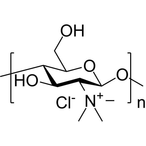
HY-148033
Trimethyl chitosanMultifunctional Polymer -
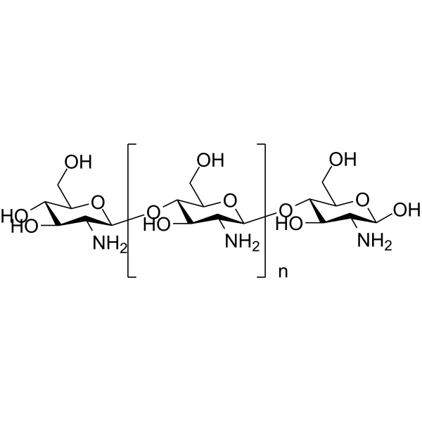
HY-B2144E
Chitosan (≥90% deacetylated, Low viscosity,<200mPa.s)Biochemical Assay Reagent -
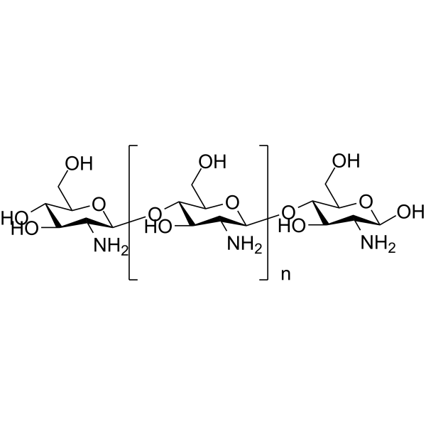
HY-B2144F
Chitosan (≥80% deacetylated, Medium viscosity,200-400mPa.s)Biochemical Assay Reagent -
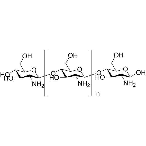
HY-B2144C
Chitosan (≥95% deacetylated,viscosity 100-200 mPa.s)Biochemical Assay Reagent -

HY-B2144J
Chitosan (≥90% deacetylated,viscosity 650-700 mPa.s)Biochemical Assay Reagent -

HY-B2144I
Chitosan (≥90% deacetylated,viscosity 10 mPa.s)Biochemical Assay Reagent -

HY-B2144N
Chitosan (≥98% deacetylated,mushroom)PEG Derivative
All Caspase Isoforms
More

Biological Activity
Description
|
Bax |
Bcl-2 |
Caspase-3 |
In Vitro
Carboxymethyl chitosan (100 μg/mL; 4 days) promotes the proliferation of normal skin fibroblasts but inhibits the proliferation of keloid fibroblasts[1].
Carboxymethyl chitosan hydrogels show enhanced cell migration compared with standard chitosan hydrogels[2].
Carboxymethylated chitosan (50-200 μg/mL; 8 h) increases the viability of Schwann cells exposed to H2O2 and decreases LDH release in a concentration-dependent manner[3].
Carboxymethyl chitosan (0.5-2.0 mg/mL; 24-48 h) significantly inhibits the migration of human umbilical vein endothelial cells (HUVECs)[4].
Carboxymethyl chitosan (50-200 μg/mL; 1 h) inhibits IL-1β-induced apoptosis of rabbit chondrocytes in a dose-dependent manner[5].
Carboxymethyl chitosan (4.0-5.0 mg/mL; 48 h) decreases viability of OS-RC-2, NCI-H1650, and HT-29 cells[6].
Carboxymethyl chitosan (1.0-5.0 mg/mL; 12-24 h) can significantly inhibit the two-dimensional migration of BEL-7402 cells in a dose-dependent manner[7].
MedChemExpress (MCE) has not independently confirmed the accuracy of these methods. They are for reference only. Further protocols information, click here.
-
Cell Line:Schwann cells
-
Concentration:5, 100, 200 μg/mL
-
Incubation Time:8 h
-
Result:Increased the expression of the anti-apoptotic protein Bcl-2 and decreased the expression of the pro-apoptotic proteins Bax, cytochrome c, and caspase-3.
Reduced cell apoptosis rates by 45.9%, 23.2%, and 9.3% at concentrations of 5 μg/mL, 100 μg/mL, and 200 μg/mL, respectively.
In Vivo
Carboxymethyl chitosan (20-120 mg/kg; i.v.; every other day; for seven times) can significantly inhibit the growth of Lewis tumor tissue, increase the spleen index of tumor-bearing mice, and make the liver and lung cells arrange more orderly, indicating its effectiveness in repressing Lewis tumor growth and metastasis[6].
Carboxymethyl chitosan (1350 mg/kg; i.p.) has no significant effects on the coagulation, anticoagulation, fibrinolysis, or hemorheology parameters of rats[8].
MedChemExpress (MCE) has not independently confirmed the accuracy of these methods. They are for reference only.
-
Animal Model:Kunming mice (female, 18-22 g, 6-8 weeks old); Mouse hepatocarcinoma H22-bearing tumor model[4]
-
Dosage:75 mg/kg, 150 mg/kg, 300 mg/kg (dissolved in normal saline)
-
Administration:Intraperitoneal injection, administered on days 1, 3, 5, 7, 9, 11, 13
-
Result:Inhibited the growth of H22 tumor tissue.
Showed inhibition rates of 32.63%, 51.43% and 29.89% at doses of 75 mg/kg, 150 mg/kg and 300 mg/kg, respectively.
Decreased CD34, VEGF and increased TIMP-1.
Increased the thymus index and spleen index of the mice, and enhanced serum IFN-γ and TNF-α levels.


Chemical Information
-
CAS No. 83512-85-0
-
Appearance Solid
-
Color White to light yellow
-
SMILES
[Carboxymethyl chitosan]
-
Shipping
Room temperature in continental US; may vary elsewhere.
-
Storage
4°C, sealed storage, away from moisture
* In solvent : -80°C, 6 months; -20°C, 1 month (sealed storage, away from moisture)


Solvent & Solubility
In Vitro:
H2O : 10 mg/mL (Need ultrasonic)

Protocols
-
RNA extraction experimental
By lysing cells, releasing RNA, and removing impurities such as proteins and DNA, high-purity RNA products are finally obtained. The commonly used traditional method is the guanidine isothiocyanate/phenol/chloroform method (Trizol), which is suitable for a variety of animal materials including animal tissues, microorganisms, cultured cells, etc., and most plant materials.
-
Apoptosis
Apoptosis, also called programmed cell death, is generally characterized by distinct morphological characteristics.
-
TUNEL staining for apoptotic DNA fragmentation
TUNEL staining detects DNA strand breaks by using terminal deoxynucleotidyl transferase to add labeled nucleotides to exposed 3′-OH DNA termini, generating either microscopic staining in fixed cells or tissue sections, or fluorescence/cytometric signal in cell suspensions. TUNEL positivity reflects DNA fragmentation but should not be interpreted alone as definitive apoptosis, because TUNEL can also label necrotic, autolytic, mechanically damaged, or DNA-repair-associated DNA breaks.
-
Annexin V plus membrane-impermeant dye apoptosis staining
Annexin V-based apoptosis assays rely on the detection of phosphatidylserine (PS) externalization from the inner leaflet of the plasma membrane to the outer leaflet, an early biochemical hallmark of apoptosis. Fluorescently labeled Annexin V binds PS in a calcium-dependent manner, enabling identification of early apoptotic cells by flow cytometry or fluorescence microscopy. When combined with a membrane-impermeant DNA-binding dye (e. g. , propidium iodide), this approach allows discrimination between viable (Annexin V−/dye−), early apoptotic (Annexin V+/dye−), and late apoptotic or necrotic (Annexin V+/dye+) cell populations by assessing membrane integrity and PS exposure.
-
ROS/oxidative-stress fluorescent staining
ROS/oxidative-stress fluorescent staining uses cell-permeant fluorogenic probes that become fluorescent after oxidation inside cells or tissues; commonly used examples include DCFH-DA/DCFDA for broad cellular oxidant detection, DHE for superoxide-related signal detection, MitoSOX for mitochondrial superoxide-related signal detection, and CellROX probes for oxidative-stress-associated fluorescence readouts. The assay detects probe oxidation rather than a single ROS species unless the probe and analysis method have been chemically validated for that species. DCFH-DA enters cells, is deacetylated by intracellular esterases to DCFH, and produces fluorescent DCF after oxidation, so the readout is used as an operational measure of total cellular oxidative stress rather than a species-specific ROS measurement. DHE and MitoSOX can report superoxide-related oxidation, but red fluorescence alone can include non-specific ethidium-like oxidation products; HPLC or optimized spectral approaches are
-
Cell migration
Cell migration is a method that plays an important role in wound healing, cell differentiation, embryonic development, etc.
-
Apoptosis Solutions
Apoptosis is a regulated, generally non-lytic cell-death pathway that removes unwanted, damaged, infected, or abnormal cells through coordinated morphological changes, caspase activation, DNA fragmentation, and membrane remodeling. The intrinsic apoptosis pathway is controlled mainly by mitochondrial outer membrane permeabilization, BCL-2 family proteins, cytochrome c release, apoptosome formation, caspase-9 activation, and downstream executioner caspase-3/7 activation. The extrinsic apoptosis pathway is initiated by death receptors such as Fas, TNFR, and TRAIL receptors, which recruit adaptor proteins and activate caspase-8 before engaging executioner caspases or mitochondrial amplification through BID cleavage. Apoptosis is linked to many phenotypes, including cancer cell killing, tissue homeostasis, immune regulation, neurodegeneration, infection response, and treatment-induced cytotoxicity; unresolved questions include how apoptosis interacts with necroptosis, pyroptosis, ferroptos


Purity & Documentation
-
Data Sheet (273 KB)
-
SDS (394 KB)
- English - EN (394 KB)
- Français - FR (394 KB)
- Deutsch - DE (394 KB)
- Norwegian - NO (394 KB)
- Español - ES (394 KB)
- Swedish - SV (394 KB)
- Italian - IT (394 KB)
- Korean - KR (394 KB)
- Portuguese - PT (394 KB)
-
Handling Instructions (2659 KB)
References
[1]. Chen XG, et al. The effect of carboxymethyl-chitosan on proliferation and collagen secretion of normal and keloid skin fibroblasts. Biomaterials. 2002 Dec;23(23):4609-14. [Content Brief]
[2]. Kruczkowska W, et al. Medical Applications and Cellular Mechanisms of Action of Carboxymethyl Chitosan Hydrogels. Molecules. 2024 Sep 13;29(18):4360. [Content Brief]
[3]. He B, et al. Carboxymethylated chitosan protects Schwann cells against hydrogen peroxide-induced apoptosis by inhibiting oxidative stress and mitochondria dependent pathway. Eur J Pharmacol. 2018 Apr 15;825:48-56. [Content Brief]
[4]. Jiang Z, et al. Carboxymethyl chitosan represses tumor angiogenesis in vitro and in vivo. Carbohydr Polym. 2015 Sep 20;129:1-8. [Content Brief]
[5]. Chen Q, et al. Carboxymethyl-chitosan protects rabbit chondrocytes from interleukin-1beta-induced apoptosis. Eur J Pharmacol. 2006 Jul 10;541(1-2):1-8. [Content Brief]
[6]. Jiang Z, et al. Evaluation on biological compatibility of carboxymethyl chitosan as biomaterials for antitumor drug delivery. J Biomater Appl. 2017 Feb;31(7):985-994. [Content Brief]
[7]. Jiang Z, et al. Preparation and anti-tumor metastasis of carboxymethyl chitosan. Carbohydr Polym. 2015 Jul 10;125:53-60. [Content Brief]
[8]. Yang Z, et al. Acute toxicity of high dosage carboxymethyl chitosan and its effect on the blood parameters in rats. J Mater Sci Mater Med. 2012 Feb;23(2):457-62. [Content Brief]

Calculators
Concentration (start) × Volume (start) = Concentration (final) × Volume (final)


Carboxymethyl chitosan Related Classifications
HY-W250111 Related Classifications
 Powered by Bioz