



CMIT/MIT (2.0-2.5% in water)

Based on 1 Customer Validation
CMIT/MIT (2.0-2.5% in water) (CMI/MI (2.0-2.5% in water)) is a mixture of CMIT and MIT, and is also a preservative and skin sensitizer. CMIT/MIT (2.0-2.5% in water) increases the levels of phosphorylated ERK1/2, p38, JNK1/2, p53, p21 and Bax, decreases the level of Bcl-2, and activates the Nrf-2/HO-1 signaling pathway. CMIT/MIT (2.0-2.5% in water) increases LDH release and lipid peroxidation levels, impairs antioxidant defense capacity, promotes pro-inflammatory cytokine release, induces apoptosis, triggers cell cycle arrest, exhibits neurotoxicity in neuroblastoma cells, and causes dysregulation of Th2/Th17 immune responses. CMIT/MIT (2.0-2.5% in water) can be used in studies related to allergy, atopic dermatitis and lung injury.
For research use only. We do not sell to patients.
- CAS No.: 55965-84-9
- Formula: C8H9ClN2O2S2
- Molecular Weight:264.75
-
Storage:
4°C, stored under nitrogen
* In solvent : -80°C, 6 months; -20°C, 1 month (stored under nitrogen)


To place orders, for customer services and technical support, please contact: MedChemExpress USA
Tel: 609-228-6898 E-mail: [email protected] [email protected]


-
 Biological Activity
-
Chemical Information
- Protocols
- Purity & Documentation
- References
-
 Help & FAQs


-

HY-W017982
CMIT/MIT (14.5% in water)Biochemical Assay Reagents -
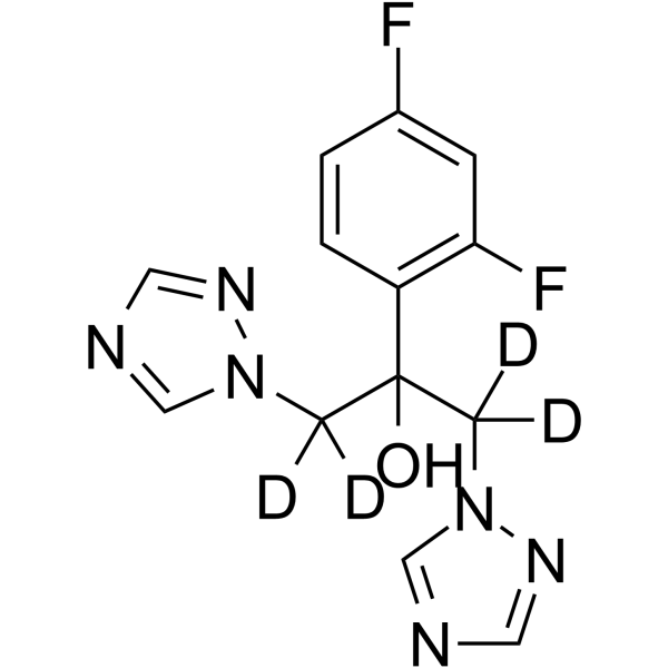
HY-B0101S
Fluconazole-d4Stable Isotope -
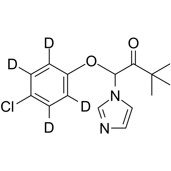
HY-B1151S
Climbazole-d4Stable Isotope -
HY-117517
NG-012NGF Potentiator -

HY-121003
Aspyrone -
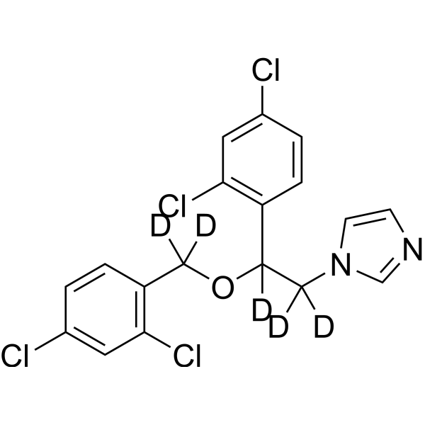
HY-B0454S
Miconazole-d5Stable Isotope -
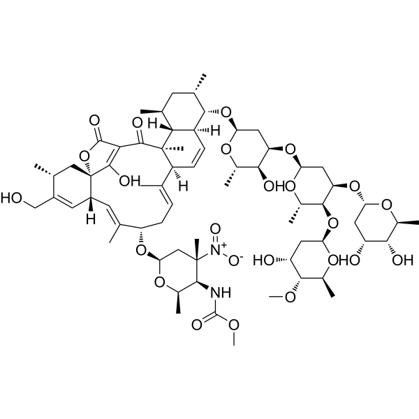
HY-125049
KijanimicinAntibiotic -
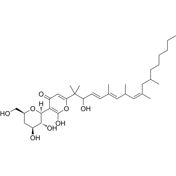
HY-N10273
DeoxyfusapyroneAntifungal Agent -

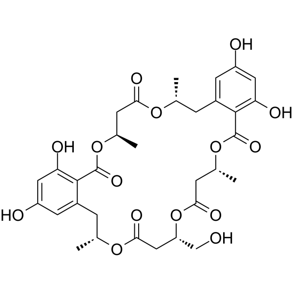
HY-125322
Reveromycin CPolyketide -
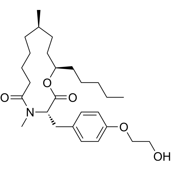
HY-123474
PF-1163BAntifungal Antibiotic -

HY-W793613
DrimendiolAntibiofilm Agent


Biological Activity
Description
|
ERK1 |
ERK2 |
JNK1 |
JNK2 |
Bax |
Bcl-2 |
IL-4 |
IL-17A |
IL-6 |
IL-1β |
In Vitro
CMIT/MIT (2.0-2.5% in water) can increase the levels of phosphorylated ERK1/2, p38, JNK1/2, p53, p21 and Bax, decrease the level of Bcl-2, and activate the Nrf-2/HO-1 signaling pathway[1].
CMIT/MIT (2.0-2.5% in water) increases the mutation frequency of mouse lymphoma cells by up to 10-fold[3].
CMIT/MIT (2.0-2.5% in water) does not induce chromosomal aberrations in Chinese hamster lung fibroblasts, although it causes cytotoxicity at concentrations ranging from 0.015 to 0.12 a.i. μg/mL[3].
CMIT/MIT (2.0-2.5% in water) exhibits high skin permeability in rat skin[3].
MedChemExpress (MCE) has not independently confirmed the accuracy of these methods. They are for reference only. Further protocols information, click here.
In Vivo
CMIT/MIT (1.5-15 a.i. mg/kg bw/day; oral administration; daily dosing from gestational day 5 to 15) (2.0-2.5% in water) causes dose-dependent maternal death in pregnant rats, but shows no teratogenicity or reproductive toxicity[3].
CMIT/MIT (1.5-13.3 a.i. mg/kg bw/day; administered orally; daily dosing on gestational days 6-18) (2.0-2.5% in water) causes severe dose-dependent maternal mortality and fetal loss in pregnant rabbits[3].
CMIT/MIT (2.0-2.5% in water) administered orally to dogs at doses ≥3.5 a.i. mg/kg bw/day over a two-week period triggers mild systemic adverse responses such as lowered food intake as well as changes in hematological and serum biochemical indicators[3].
CMIT/MIT (2.0-2.5% in water) causes dose-dependent gastric irritation in rats at doses of 100 and 300 a.i. ppm, but shows no carcinogenicity or systemic toxicity following 2 years of oral administration[3].
CMIT/MIT (2.0-2.5% in water) is a potent skin sensitizer in mice, with an EC3 range of 0.75-2.1 a.i. μg/cm2 in the local lymph node assay; dermal administration at a dose of 400 a.i. ppm for 30 months causes skin hyperplasia and inflammation in mice but no carcinogenicity; dermal administration at a dose of ≥ 0.56 a.i. mg/kg bw/day for 3 weeks induces moderate skin irritation in rabbits without associated systemic toxicity[3].
MedChemExpress (MCE) has not independently confirmed the accuracy of these methods. They are for reference only.
-
Animal Model:BALB/c (female, 5 weeks old, 16-20 g)[2]
-
Dosage:0.1875 mg/kg/day
-
Administration:epicutaneous; 5 days/week; 3 weeks
-
Result:Induced AD-like skin lesions with significantly increased transepidermal water loss (TEWL), total serum IgE levels, mast cell counts, and mast cell degranulation.
Significantly elevated skin mRNA expression of Th2-related cytokines/chemokines: thymic stromal lymphopoietin (TSLP), IL-6, IL-13, and CCL17.
Increased CD4+IL-4+ cell population in skin-draining lymph nodes.
-
Animal Model:BALB/c (female, 5 weeks old, 16-20 g, induced via 3 intraperitoneal sensitizations with ovalbumin (OVA) plus aluminum hydroxide at 1-week intervals, followed by 2 epicutaneous OVA sensitizations at 1-week intervals)[2]
-
Dosage:0.1875 mg/kg/day
-
Administration:epicutaneous; 5 days/week; 3 weeks
-
Result:Aggravated AD-like skin lesions with significantly higher TEWL, clinical scores, inflammatory cell infiltration, mast cell counts, and total serum IgE levels compared to mice treated with OVA alone.
Significantly increased skin mRNA expression of IL-4 and IL-17A compared to mice treated with OVA alone.
Increased CD4+IL-4+ and CD4+IL-17A+ cell populations in skin-draining lymph nodes compared to mice treated with OVA alone.


Chemical Information
-
CAS No. 55965-84-9
-
Appearance Liquid (Density: 1.19 g/cm3)
-
Molecular Weight 264.75
-
Formula C8H9ClN2O2S2
-
SMILES
O=C1C=CSN1C.O=C2C=C(SN2C)Cl
-
Synonyms
CMI/MI (2.0-2.5% in water)
-
Shipping
Room temperature in continental US; may vary elsewhere.
-
Storage
4°C, stored under nitrogen
* In solvent : -80°C, 6 months; -20°C, 1 month (stored under nitrogen)

Protocols
-
Neurotoxicity Study
This protocol assesses in vitro neurotoxicity by combining neuronal viability, mitochondrial/metabolic activity, neurite outgrowth, and optional neuronal network function readouts. Calcein-AM or resazurin/PrestoBlue readouts estimate viable or metabolically active cells; βIII-tubulin immunofluorescence detects neuronal morphology and neurite networks; TMRE detects mitochondrial membrane potential; and MEA recordings detect functional changes in neuronal network activity.
-
Annexin V plus membrane-impermeant dye apoptosis staining
Annexin V-based apoptosis assays rely on the detection of phosphatidylserine (PS) externalization from the inner leaflet of the plasma membrane to the outer leaflet, an early biochemical hallmark of apoptosis. Fluorescently labeled Annexin V binds PS in a calcium-dependent manner, enabling identification of early apoptotic cells by flow cytometry or fluorescence microscopy. When combined with a membrane-impermeant DNA-binding dye (e. g. , propidium iodide), this approach allows discrimination between viable (Annexin V−/dye−), early apoptotic (Annexin V+/dye−), and late apoptotic or necrotic (Annexin V+/dye+) cell populations by assessing membrane integrity and PS exposure.
-
Naïve CD4+ T-cell subset differentiation/polarization
Naïve CD4+ T-cell subset differentiation/polarization is an in vitro assay in which purified naïve CD4+ T cells are activated through TCR and CD28 costimulation and cultured with defined cytokines and neutralizing antibodies to generate Th0, Th1, Th2, Th17, or induced Treg-like populations. Differentiation is detected by subset-associated cytokines and transcription factors: IFN-γ/T-bet for Th1, IL-4/GATA3 for Th2, IL-17A/RORγt for Th17, and Foxp3 for induced Treg cells. The assay readout is usually generated by intracellular cytokine staining after restimulation, transcription-factor staining by flow cytometry, ELISA of secreted cytokines, or gene-expression analysis. The result reflects cytokine-directed lineage commitment or polarization rather than antigen-specific immune protection by itself.
-
BrdU Incorporation Assay
Bromodeoxyuridine (BrdU) incorporation assay is based on the principle that BrdU, a thymidine analog, is incorporated into newly synthesized DNA during the S phase of the cell cycle, thereby serving as a marker of DNA replication and cellular proliferation. Incorporated BrdU can be detected using anti-BrdU antibodies following DNA denaturation, enabling visualization or quantification of proliferating cells through immunochemical detection methods such as immunofluorescence or immunohistochemistry.
-
Protocol for Cell Cycle
Cell-cycle analysis by flow cytometry measures DNA content in single cells to estimate the fraction of cells in G0/G1, S, and G2/M phases. Propidium iodide intercalates into DNA, and after RNA removal with RNase, fluorescence intensity reflects cellular DNA content: 2N cells are assigned to G0/G1, cells between 2N and 4N to S phase, and 4N cells to G2/M. DNA-content analysis alone cannot reliably separate G0 from G1 or G2 from M. Ki-67 can distinguish quiescent G0 cells from cycling cells, EdU or BrdU incorporation marks active DNA synthesis in S phase, and phospho-histone H3 staining identifies mitotic cells within the 4N population.
-
Research Protocol for Inflammation-related Diseases
The NLRP3 inflammasome is a cytosolic innate immune signaling platform that integrates priming signals and danger-signal activation to promote caspase-1 activation, maturation of IL-1β and IL-18, and gasdermin D-mediated pyroptotic cell death. The core experimental logic is to determine whether inflammatory disease phenotypes are driven by increased NLRP3 expression, ASC-containing inflammasome assembly, caspase-1 cleavage, GSDMD cleavage, and extracellular release of IL-1β/IL-18 rather than by nonspecific cell injury alone. The pathway is strongly linked to inflammation-related disease phenotypes because monosodium urate crystals activate NALP3/NLRP3 inflammasome signaling in gout-like crystal inflammation, cholesterol crystals activate NLRP3 inflammasomes in atherogenesis models, and DSS-induced intestinal inflammation has been reported to involve NLRP3 inflammasome activity. However, experimental colitis studies also show context-dependent protective effects of NLRP3 inflammasome co
-
Apoptosis
Apoptosis, also called programmed cell death, is generally characterized by distinct morphological characteristics.
-
TUNEL staining for apoptotic DNA fragmentation
TUNEL staining detects DNA strand breaks by using terminal deoxynucleotidyl transferase to add labeled nucleotides to exposed 3′-OH DNA termini, generating either microscopic staining in fixed cells or tissue sections, or fluorescence/cytometric signal in cell suspensions. TUNEL positivity reflects DNA fragmentation but should not be interpreted alone as definitive apoptosis, because TUNEL can also label necrotic, autolytic, mechanically damaged, or DNA-repair-associated DNA breaks.
-
Flow cytometric DNA-content cell-cycle staining
Flow cytometric DNA-content cell-cycle staining measures the fluorescence intensity of DNA-bound fluorochromes in single cells or nuclei to estimate DNA content distributions, allowing assignment of populations to G0/G1, S, and G2/M phases by DNA histogram deconvolution. Propidium iodide (PI) intercalates into DNA, and PI fluorescence is proportional to cellular DNA content when staining is performed under conditions that make DNA accessible and minimize non-DNA signal. Cells with G2/M DNA content are expected to show approximately twice the fluorescence intensity of G0/G1 cells, while S-phase cells occupy intermediate fluorescence values. PI-based DNA-content analysis can also detect cells with fractional DNA content, often reported as sub-G1, when DNA fragmentation and extraction during staining reduce retained DNA signal in apoptotic cells. DAPI is an alternative DNA fluorochrome for univariate DNA-content analysis, while bivariate approaches combining DNA content with proliferation
-
TPA/Croton Oil Ear Edema and Dermatitis
The TPA (12-O-tetradecanoylphorbol-13-acetate) and croton oil-induced mouse ear edema model is a well-established acute cutaneous inflammation system used to evaluate topical anti-inflammatory activity by measuring edema formation, neutrophil infiltration, vascular permeability, and cytokine-mediated skin responses in vivo. The inflammatory response is triggered by topical application of phorbol esters (TPA) or croton oil constituents, leading to rapid activation of protein kinase C signaling, leukocyte recruitment, and increased vascular permeability, which can be quantified by ear thickness, weight, dye extravasation, and biochemical markers such as myeloperoxidase (MPO) activity and pro-inflammatory mediators in ear tissue homogenates. This model is widely used for screening anti-inflammatory agents, where reductions in edema and inflammatory biomarkers reflect suppression of acute dermal inflammation and immune cell infiltration. Histological evaluation typically confirms epidermal
-
Apoptosis Solutions
Apoptosis is a regulated, generally non-lytic cell-death pathway that removes unwanted, damaged, infected, or abnormal cells through coordinated morphological changes, caspase activation, DNA fragmentation, and membrane remodeling. The intrinsic apoptosis pathway is controlled mainly by mitochondrial outer membrane permeabilization, BCL-2 family proteins, cytochrome c release, apoptosome formation, caspase-9 activation, and downstream executioner caspase-3/7 activation. The extrinsic apoptosis pathway is initiated by death receptors such as Fas, TNFR, and TRAIL receptors, which recruit adaptor proteins and activate caspase-8 before engaging executioner caspases or mitochondrial amplification through BID cleavage. Apoptosis is linked to many phenotypes, including cancer cell killing, tissue homeostasis, immune regulation, neurodegeneration, infection response, and treatment-induced cytotoxicity; unresolved questions include how apoptosis interacts with necroptosis, pyroptosis, ferroptos
-
Ferroptosis Solutions
Ferroptosis is an iron-dependent, non-apoptotic form of regulated cell death characterized by lethal lipid peroxidation and sensitivity to suppression by iron chelators or lipophilic radical-trapping antioxidants. The core pathway links cystine uptake through system Xc−, glutathione availability, GPX4-dependent detoxification of phospholipid hydroperoxides, iron-dependent oxidative reactions, and polyunsaturated-phospholipid metabolism into a cell-death program that is biochemically and morphologically distinct from apoptosis, necrosis, and autophagy. The ferroptosis pathway is experimentally linked to phenotype through chemical and genetic perturbation. Erastin induces ferroptosis by inhibiting cystine uptake through system Xc− and weakening antioxidant defenses, while GPX4 inhibition or depletion causes lipid peroxide accumulation and ferroptotic cancer-cell death. ACSL4 and oxidizable arachidonoyl- or adrenoyl-containing phosphatidylethanolamines shape ferroptosis sensitivity by con
-
Inhalation Toxicity Study
Inhalation toxicity studies expose rodents to a controlled aerosol, vapor, gas, or smoke atmosphere and assess respiratory and systemic toxicity using exposure-atmosphere characterization, clinical observations, body and organ weights, bronchoalveolar lavage fluid, histopathology, blood chemistry, hematology, and, when included, molecular endpoints such as transcriptomics, proteomics, lipidomics, or tissue burden analysis. The primary biological readouts are airway irritation, pulmonary inflammation, cytotoxicity, altered surfactant or lipid homeostasis, impaired particle clearance, and tissue remodeling, reflected by BALF cell differentials, BALF protein, LDH, phosphatase activities, cytokines, lung weight, microscopic respiratory-tract lesions, and retained lung burden.


Purity & Documentation
-
Data Sheet (277 KB)
-
SDS (252 KB)
- Français - FR (252 KB)
- Deutsch - DE (252 KB)
- Norwegian - NO (252 KB)
- Español - ES (252 KB)
- Swedish - SV (252 KB)
- Italian - IT (252 KB)
- Korean - KR (252 KB)
- Portuguese - PT (252 KB)
-
Handling Instructions (2659 KB)
References
[1]. Molinari F, et al. Biocide mixture (CMIT/MIT) induces neurotoxicity through the upregulation of the MAPKs signaling pathways. Journal of neurophysiology. 2025 Jul 01;134(1):183-192. [Content Brief]
[2]. Go HN, et al. Effects of chloromethylisothiazolinone/methylisothiazolinone (CMIT/MIT) on Th2/Th17-related immune modulation in an atopic dermatitis mouse model. Scientific reports. 2020 Mar 05;10(1):4099. [Content Brief]

Calculators
Concentration (start) × Volume (start) = Concentration (final) × Volume (final)


CMIT/MIT (2.0-2.5% in water) Related Classifications
HY-W017982A Related Classifications
 Powered by Bioz
Keywords
- CMIT/MIT (2.0-2.5% in water)
- 55965-84-9
- CMI/MI (2.0-2.5% in water)
- ERK
- JNK
- Bcl-2 Family
- Keap1-Nrf2
- Apoptosis
- Interleukin Related
- Salmonella typhimurium strain TA100
- atopic dermatitis
- mouse lymphoma cells
- humidifier disinfectant-associated lung injury
- Th2/Th17 immune responses
- Chinese hamster lung fibroblasts
- BALB/c mice
- allergic contact dermatitis
- neuroblastoma cells
- primary rat hepatocytes
- Inhibitor
- inhibitor
- inhibit