



GEM144

Based on 1 Customer Validation
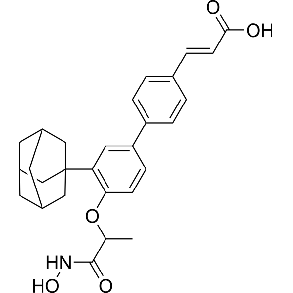
GEM144 is a potent and orally active DNA polymerase α (POLA1) and HDAC 11 dual inhibitor. GEM144 induces acetylation of p53, activation of p21, G1/S cell cycle arrest, and apoptosis. GEM144 has significant antitumor activity in human orthotopic malignant pleural mesothelioma xenografts.
For research use only. We do not sell to patients.
- CAS No.: 2487526-28-1
- Formula: C28H31NO5
- Molecular Weight:461.55
-
Storage:Powder -20°C, 3 years ; In solvent -80°C, 6 months , -20°C, 1 month

To place orders, for customer services and technical support, please contact: MedChemExpress USA
Tel: 609-228-6898 E-mail: [email protected] [email protected]


-
 Biological Activity
-
Chemical Information
-
Solvent & Solubility
- Protocols
- Purity & Documentation
- References
-
 Help & FAQs


-
HY-15654
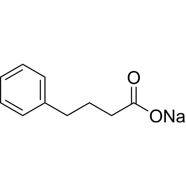
Sodium 4-phenylbutyrateHDAC/ERS Inhibitor -
HY-143412
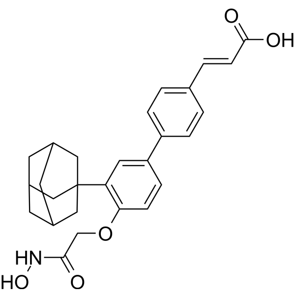
MIR002POLA1/HDAC 11 Inhibitor -

HY-15654R
Sodium 4-phenylbutyrate (Standard)HDAC/ERS Inhibitor -
HY-15433A
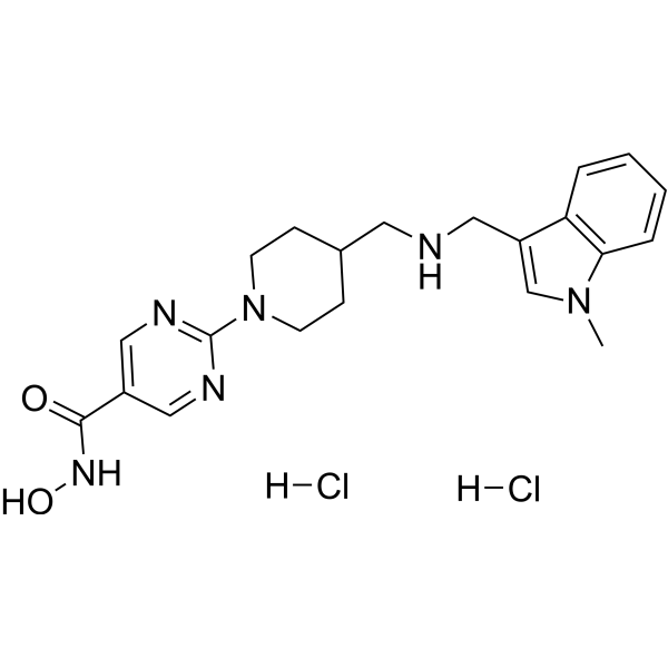
Quisinostat dihydrochloridePan-HDAC Inhibitor -
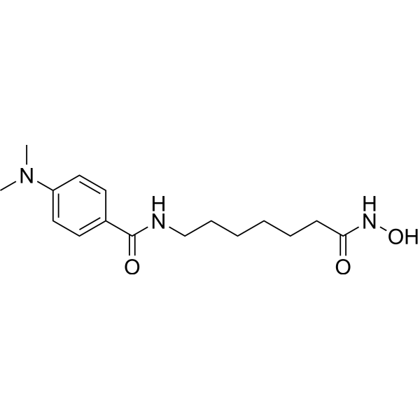
HY-13506
M344HDAC Inhibitor -
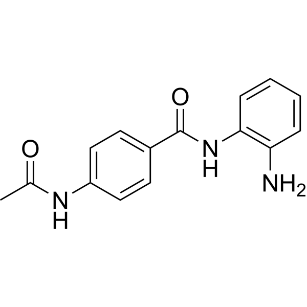
HY-50934
TacedinalineHDAC Inhibitor -
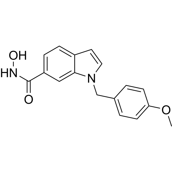
HY-15224
PCI-34051Apoptosis Inducer -
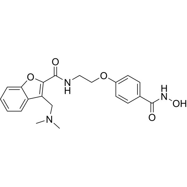
HY-10990
AbexinostatHDAC/MBLAC2 Inhibitor -
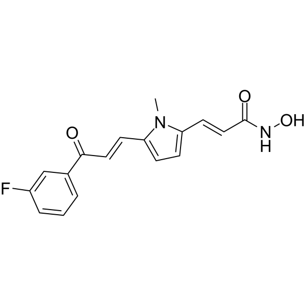
HY-16914
MC1568HDAC Inhibitor -
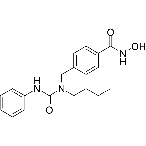
HY-16699
Nexturastat AHDAC6 Inhibitor -
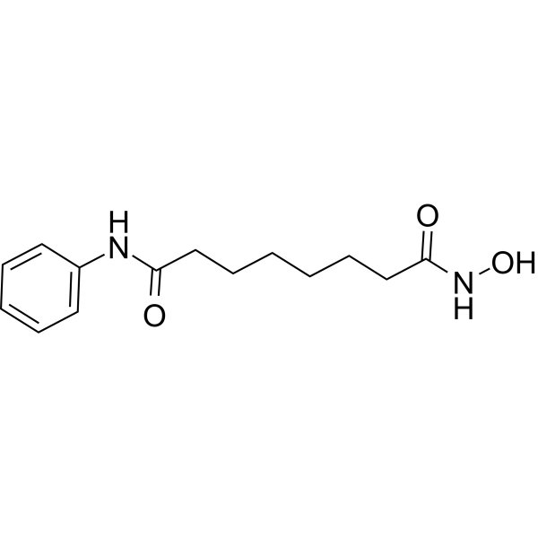
HY-10221
VorinostatHDAC Inhibitor -
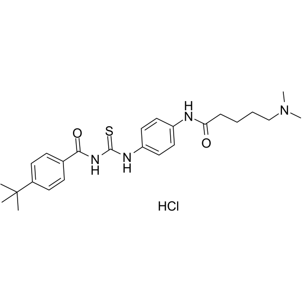
HY-15510B
Tenovin-6 Hydrochloridep53 Activator
All DNA/RNA Synthesis Isoforms
More

Biological Activity
Description
IC50 & Target
|
DNA Polymerase |
HDAC11 8.23 μM (IC50) |
In Vitro
GEM144 (0-10 μM; 24 hours) exhibits antiproliferative activity in tested cancer cell lines with IC50s of 0.26 ~ 2.2 μM[1].
GEM144 (0.1 - 0.4 μM; 24 hours) strongly increases levels of H2AX phosphorylation on Ser 39 (γH2AX), and strongly upregulates p21 expression in a dose-dependent manner[1].
GEM144 (0.26 μM in NCI-H4609, 0.95 μM in A2780 and 1.4 μM in MM473; 72 hours) induced G1/S cell cycle arrest[1].
MedChemExpress (MCE) has not independently confirmed the accuracy of these methods. They are for reference only.
-
Cell Line:NCI-H460, H460-R9A, A2780 and A2780-DX[1]
-
Concentration:0-10 μM
-
Incubation Time:24 hours (further 48 h growth in drug-free medium)
-
Result:Exhibited antiproliferative activity in tested cancer cell lines with IC50s of 0.26 ~ 2.2 μM.
-
Cell Line:NCI-H460[1]
-
Concentration:0.1 μM, 0.25 μM and 0.4 μM
-
Incubation Time:24 hours
-
Result:Strongly increased levels of H2AX phosphorylation on Ser 39 (γH2AX), and strongly upregulated p21 expression in a dose-dependent manner.
-
Cell Line:NCI-H4609, A2780 and MM473[1]
-
Concentration:0.26 μM in NCI-H4609, 0.95 μM in A2780 and 1.4 μM in MM473
-
Incubation Time:72 hours
-
Result:Induced G1/S cell cycle arrest.
In Vivo
MedChemExpress (MCE) has not independently confirmed the accuracy of these methods. They are for reference only.
-
Animal Model:Female CD-1 nude mice (injected with MM473 and MM487)[1]
-
Dosage:50 mg/kg
-
Administration:PO, bid, 5 days a week, for 3-4 weeks
-
Result:Significantly reduced tumor burden of MM487 with TGI of 72%.


Chemical Information
-
CAS No. 2487526-28-1
-
Appearance Solid
-
Molecular Weight 461.55
-
Formula C28H31NO5
-
Color White to off-white
-
SMILES
ONC(C(C)OC1=C(C=C(C2=CC=C(C=C2)/C=C/C(O)=O)C=C1)C34CC5CC(CC(C5)C4)C3)=O
-
Shipping
Room temperature in continental US; may vary elsewhere.
-
Storage
Powder -20°C 3 years In solvent -80°C 6 months -20°C 1 month


Solvent & Solubility
In Vitro:
DMSO : 200 mg/mL (433.32 mM; Need ultrasonic; Hygroscopic DMSO has a significant impact on the solubility of product, please use newly opened DMSO)
Please refer to the solubility information to select the appropriate solvent. Once prepared, please aliquot and store the solution to prevent product inactivation from repeated freeze-thaw cycles.
Storage method and period of stock solution: -80°C, 6 months; -20°C, 1 month. When stored at -80°C, please use it within 6 months. When stored at -20°C, please use it within 1 month.
Please refer to the solubility information to select the appropriate solvent. Once prepared, please aliquot and store the solution to prevent product inactivation from repeated freeze-thaw cycles.
Storage method and period of stock solution: -80°C, 6 months; -20°C, 1 month. When stored at -80°C, please use it within 6 months. When stored at -20°C, please use it within 1 month.
Concentration (start) × Volume (start) = Concentration (final) × Volume (final)
In Vivo:
Select the appropriate dissolution method based on your experimental animal and administration route.
- For the following dissolution methods, please ensure to first prepare a clear stock solution using an In Vitro approach and then sequentially add co-solvents:
- To ensure reliable experimental results, the clarified stock solution can be appropriately stored based on storage conditions. As for the working solution for In Vivo experiments, it is recommended to prepare freshly and use it on the same day.
- The percentages shown for the solvents indicate their volumetric ratio in the final prepared solution. If precipitation or phase separation occurs during preparation, heat and/or sonication can be used to aid dissolution.
In Vivo Dissolution Calculator
Please enter the basic information of animal experiments:
-
-
-
-
Recommended: Prepare an additional quantity of animals to account for potential losses during experiments.
Please enter your animal formula composition:
-
%DMSO +
Recommended: Keep the proportion of DMSO in working solution below 2% if your animal is weak.
-
%+
-
+%Tween-80 + +
-
%Saline +
The co-solvents required include: DMSO, . All of co-solvents are available by MedChemExpress (MCE). , Tween 80. All of co-solvents are available by MedChemExpress (MCE).
Working solution concentration: 0.22 mg/mL
Method for preparing stock solution: mg drug dissolved in μL DMSO. Stock solution concentration: mg/mL.
1. Take μL DMSO stock solution;
2. Add μL .
μL , mix evenly;
3. Then add μL Tween 80, mix evenly;
4. Then add μL
Please ensure that the stock solution in the first step is dissolved to a clear state, and add co-solvents in sequence. You can use ultrasonic heating (ultrasonic cleaner, recommended frequency 20-40 kHz), vortexing, etc. to assist dissolution.


Purity & Documentation
-
Data Sheet (277 KB)
-
SDS (252 KB)
- Français - FR (252 KB)
- Deutsch - DE (252 KB)
- Norwegian - NO (252 KB)
- Español - ES (252 KB)
- Swedish - SV (252 KB)
- Italian - IT (252 KB)
- Korean - KR (252 KB)
- Portuguese - PT (252 KB)
-
Handling Instructions (2659 KB)
References


Complete Stock Solution Preparation Table
Please refer to the solubility information to select the appropriate solvent. Once prepared, please aliquot and store the solution to prevent product inactivation from repeated freeze-thaw cycles.
Storage method and period of stock solution: -80°C, 6 months; -20°C, 1 month. When stored at -80°C, please use it within 6 months. When stored at -20°C, please use it within 1 month.
| Optional Solvent | Concentration Solvent Mass | 1 mg | 5 mg | 10 mg | 25 mg |
|---|---|---|---|---|---|
| DMSO | 1 mM | 2.1666 mL | 10.8331 mL | 21.6661 mL | 54.1653 mL |
| 5 mM | 0.4333 mL | 2.1666 mL | 4.3332 mL | 10.8331 mL | |
| 10 mM | 0.2167 mL | 1.0833 mL | 2.1666 mL | 5.4165 mL | |
| 15 mM | 0.1444 mL | 0.7222 mL | 1.4444 mL | 3.6110 mL | |
| 20 mM | 0.1083 mL | 0.5417 mL | 1.0833 mL | 2.7083 mL | |
| 25 mM | 0.0867 mL | 0.4333 mL | 0.8666 mL | 2.1666 mL | |
| 30 mM | 0.0722 mL | 0.3611 mL | 0.7222 mL | 1.8055 mL | |
| 40 mM | 0.0542 mL | 0.2708 mL | 0.5417 mL | 1.3541 mL | |
| 50 mM | 0.0433 mL | 0.2167 mL | 0.4333 mL | 1.0833 mL | |
| 60 mM | 0.0361 mL | 0.1806 mL | 0.3611 mL | 0.9028 mL | |
| 80 mM | 0.0271 mL | 0.1354 mL | 0.2708 mL | 0.6771 mL | |
| 100 mM | 0.0217 mL | 0.1083 mL | 0.2167 mL | 0.5417 mL |


GEM144 Related Classifications
HY-143411 Related Classifications
 Powered by Bioz