



Erenapurstat
Based on 4 publication(s) in Google Scholar
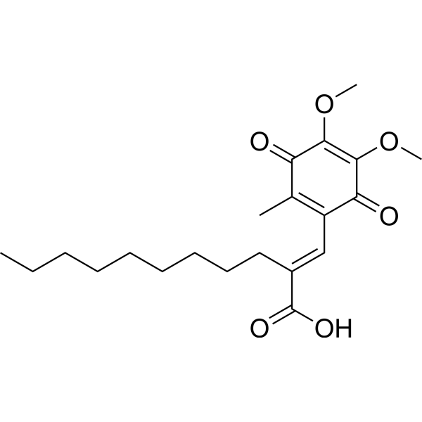
Erenapursta (E3330) is a direct, orally active and selective inhibitor of Ape-1 (apurinic/apyrimidinic endonuclease 1)/Ref-1 (redox factor-1) redox. Erenapursta is able to impair tumor growth and blocks the activity of NF-κB, AP-1, and HIF-1α in pancreatic cancer. Erenapursta shows anticancer activities.
For research use only. We do not sell to patients.
- Purity : 99.67%
- CAS No.: 136164-66-4
- Formula: C21H30O6
- Molecular Weight:378.46
-
Storage:Powder -20°C, 3 years , 4°C, 2 years ; In solvent -80°C, 2 years , -20°C, 1 year

To place orders, for customer services and technical support, please contact: MedChemExpress USA
Tel: 609-228-6898 E-mail: [email protected] [email protected]


-
 Biological Activity
-
Chemical Information
-
Solvent & Solubility
- Protocols
- Purity & Documentation
- References
-
 Help & FAQs


-
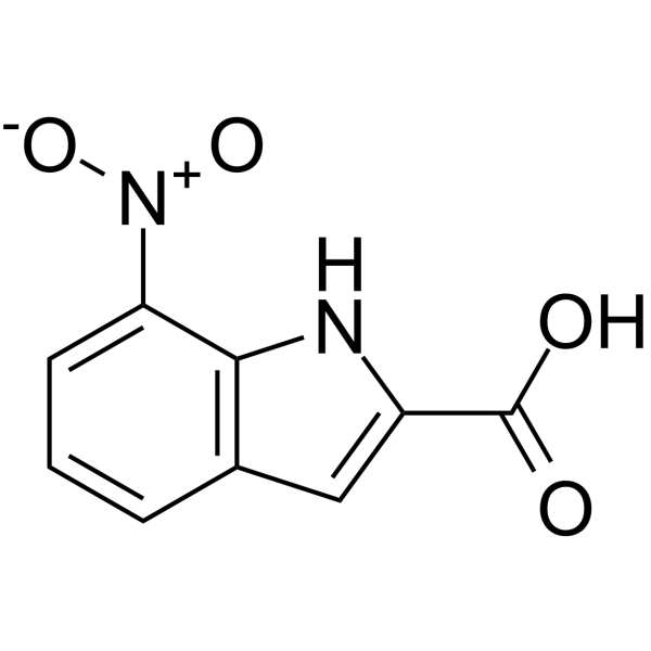
HY-W014622
CRT0044876APE1 Inhibitor -
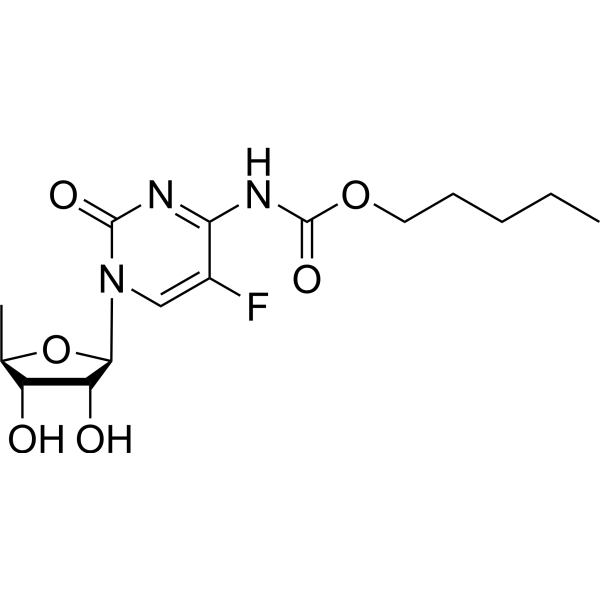
HY-B0016
CapecitabineDNA/RNA Synthesis Inhibitor -
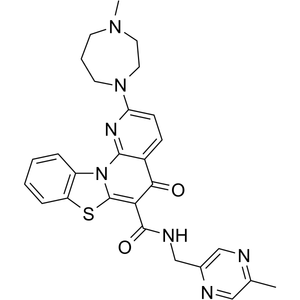
HY-13323
CX-5461RNA Polymerase I Inhibitor -
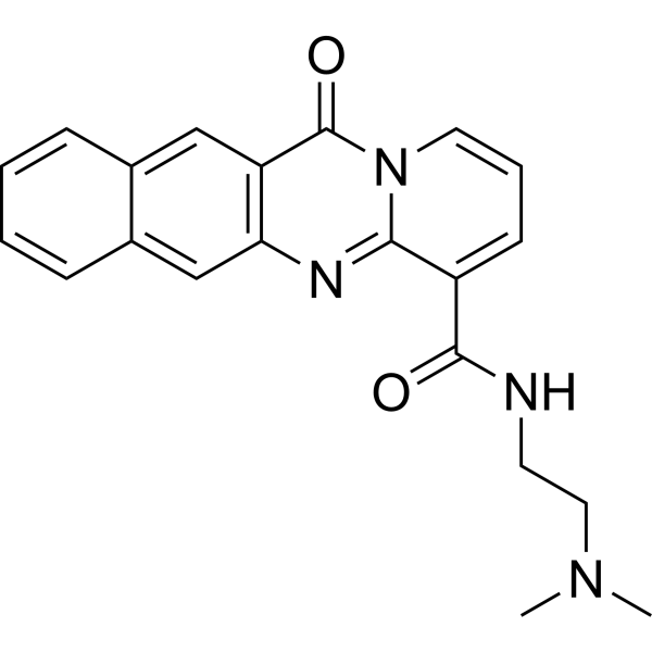
HY-12484
BMH-21DNA Intercalator/Pol I Elongation Inhibitor -

HY-100579
Ferrostatin-1Ferroptosis Inhibitor -
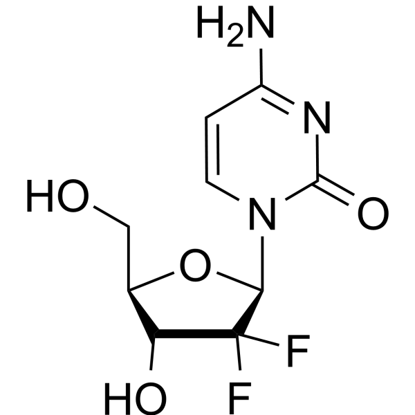
HY-17026
GemcitabineDNA Synthesis Inhibitor -
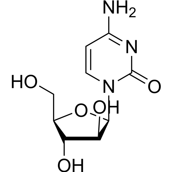
HY-13605
CytarabineDNA Synthesis Inhibitor -

HY-10219
RapamycinmTOR Inhibitor -
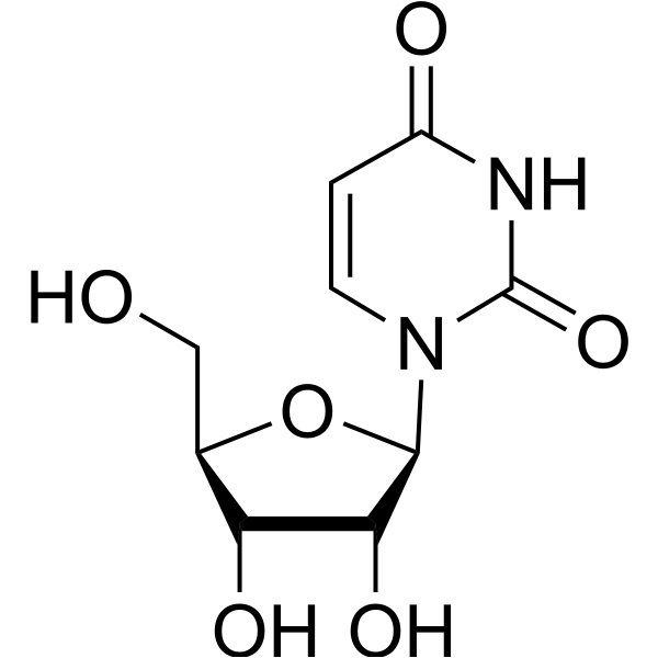
HY-B1449
UridineNucleoside -
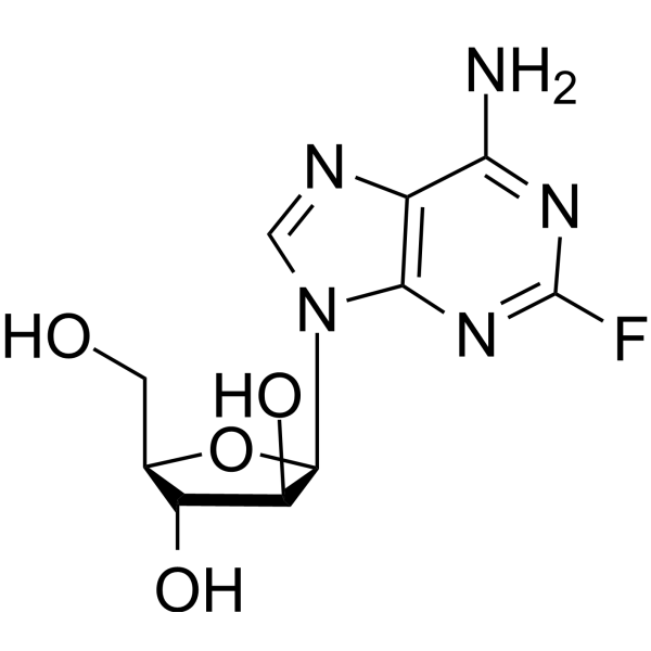
HY-B0069
FludarabineDNA Synthesis Inhibitor -
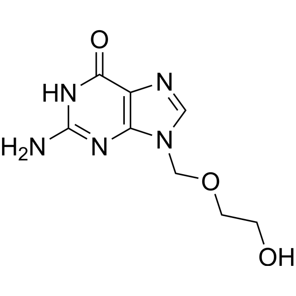
HY-17422
AcyclovirHSV Inhibitor -
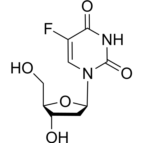
HY-B0097
FloxuridineOncology Antimetabolite
Publications Citing Use of MedChemExpress (MCE) Erenapurstat
More Customer Validation & Images

-
Histological Imaging/Staining

-
Cell Imaging/Staining

-
WB
-
Flow Cytometry
-
Cell Proliferation/Viability Assay
All DNA/RNA Synthesis Isoforms
MoreAll AP-1 Isoforms
MoreAll VEGFR Isoforms
More

Biological Activity
Description
IC50 & Target
Ape-1, Ref-1[1]
In Vitro
Erenapursta (E3330) (0-50 μM, 48 h) inhibits the growth of HUVECs, PCECs and EPCs[1].
Erenapursta (0-5 μM) reduces secreted and intracellular VEGF (vascular endothelial growth factor) expression by pancreatic cancer cells, while concomitantly downregulating the cognate receptor Flk-1/KDR on PCECs[1].
Erenapursta (0-1 μM) inhibits the differentiation of bone marrow mesenchymal stem cells (BM-MSCs) into CD31+ cells of endothelial lineage[1].
Erenapursta (0-50 μM, 72 h) decreases cell viability in H1975 cells about 45% at 50 μM[2].
Erenapursta (0-30 μM) inhibits the growth and migration of pancreatic cancer cells[3].
Erenapursta (0-30 μM) significantly enhances intracellular ROS level and inhibits CD44 expression in PANC1 cells[3].
MedChemExpress (MCE) has not independently confirmed the accuracy of these methods. They are for reference only. Further protocols information, click here.
-
Cell Line:Human umbilical vein endothelial cells (HUVECs), murine pancreatic cancer associated endothelial cells (mPCECs), human endothelial progenitor cells (hEPCs)
-
Concentration:1, 5, and 10 μM (HUVECs); 1. 5, 10. 20. 30, 40, and 50 μM (mPCECs); 1, 5, 10, 15, 20, 25, and 30 μM (hEPCs)
-
Incubation Time:48 h
-
Result:Inhibited the growth of HUVECs, PCECs and EPCs.
-
Cell Line:mPCECs
-
Concentration:1, 5, and 10 μM
-
Incubation Time:48 h
-
Result:Inhibited the growth of HUVECs, PCECs and EPCs.
-
Cell Line:H1975 cells
-
Concentration:0, 5, 10, 20, 30, 40, and 50 μM
-
Incubation Time:72 h
-
Result:Showed decreased cell viability in about 45% at 50 µM.
In Vivo
Erenapursta (0-100 mg/kg, Orally, once) attenuates the liver injury when given at 1 h, 6 h or 12 h after galactosamine challenge[5].
MedChemExpress (MCE) has not independently confirmed the accuracy of these methods. They are for reference only.
-
Animal Model:Sprague-Dawley rats (adult male, 150-175 g)[4]
-
Dosage:25 mg/kg
-
Administration:Orally, 5 daily, five days each week for three weeks
-
Result:Attenuated the cisplatin-induced decrease in capsaicin-induced vasodilatation in the rat hindpaw.
-
Animal Model:Male Fischer (F344/DuCrj) rats (160-190 g)[5]
-
Dosage:0, 10, 30, and 100 mg/kg
-
Administration:Orally, 1 h, 6 h or 12 h after galactosamine challenge
-
Result:Attenuated the liver injury when given at 1 h, 6 h or 12 h after galactosamine challenge.
Clinical Trial
| NCT Number | Sponsor | Condition | Start Date |
Phase

|
|---|---|---|---|---|
| NCT01329991 | Plexxikon| | 2011-05 | PHASE1 |


Chemical Information
-
CAS No. 136164-66-4
-
Appearance Solid
-
Molecular Weight 378.46
-
Formula C21H30O6
-
Color Yellow to orange
-
SMILES
CCCCCCCCC/C(C(O)=O)=C\C1=C(C)C(C(OC)=C(OC)C1=O)=O
-
Synonyms
E3330; APX-3330
-
Shipping
Room temperature in continental US; may vary elsewhere.
-
Storage
Powder -20°C 3 years 4°C 2 years In solvent -80°C 2 years -20°C 1 year


Publications (4)
-
Journal Impact Factor
-
Most Recent
-
Cell Death Dis
Cephalomannine inhibits hypoxia-induced cellular function via the suppression of APEX1/HIF-1α interaction in lung cancer. [Abstract]2021 May 14;12(5):490. PMID: 33990544
Erenapurstat purchased from MedChemExpress. Usage Cited in: Cell Death Dis. 2021 May 14;12(5):490. [Abstract]
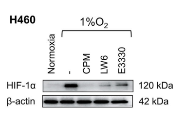
H460 cells were treated with CPM, 10 μM APEX1 inhibitor Erenapurstat (E3330), and 5 μM LW6 in normoxia and hypoxia. Protein expression of HIF-1α was assessed by western blotting.
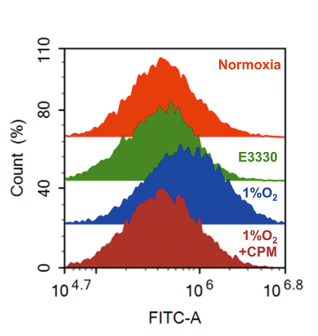
Erenapurstat purchased from MedChemExpress. Usage Cited in: Cell Death Dis. 2021 May 14;12(5):490. [Abstract]
Intracellular pH determination of H460 cell line treated with APEX1 inhibitor Erenapurstat (E3330) under normoxia and hypoxia using flow cytometry.
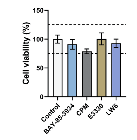
Erenapurstat purchased from MedChemExpress. Usage Cited in: Cell Death Dis. 2021 May 14;12(5):490. [Abstract]
HUVEC cell viability assay by treating with 5 μM BAY-85-3934, 25 nM CPM, 10 μM Erenapurstat (E3330), and 5 μM LW6.
-
J Allergy Clin Immunol
Role of APE1 redox function in chronic rhinosinusitis pathogenesis: Implications for targeted therapy. [Abstract]2025 Jul 17:S0091-6749(25)00775-4. PMID: 40683569
Erenapurstat purchased from MedChemExpress. Usage Cited in: J Allergy Clin Immunol. 2025 Jul 17:S0091-6749(25)00775-4. [Abstract]
Erenapurstat (E3330: 10 mg/kg). Representative HE staining images and quantitative analysis of eosinophils in nasal tissue. Each dot represents the sum of 10 randomly selected high-power fields for each sample. Black arrows indicate eosinophils.
Erenapurstat purchased from MedChemExpress. Usage Cited in: J Allergy Clin Immunol. 2025 Jul 17:S0091-6749(25)00775-4. [Abstract]
Erenapurstat (E3330). Representative images of ROS staining with dichlorodihydrofluorescein diacetate in HNEC cells.
-
Brain Behav Immun
Schwann cell-derived CXCL2 contributes to cancer pain by modulating macrophage infiltration in a mouse breast cancer model. [Abstract]2023 Mar:109:308-320. PMID: 36754246 -
Int Immunopharmacol
Inhibitors of APE1 redox and ATM synergistically sensitize osteosarcoma cells to ionizing radiation by inducing ferroptosis. [Abstract]2024 Sep 30:139:112672. PMID: 39032469


Solvent & Solubility
In Vitro:
DMSO : 120 mg/mL (317.07 mM; Need ultrasonic; Hygroscopic DMSO has a significant impact on the solubility of product, please use newly opened DMSO)
Please refer to the solubility information to select the appropriate solvent. Once prepared, please aliquot and store the solution to prevent product inactivation from repeated freeze-thaw cycles.
Storage method and period of stock solution: -80°C, 2 years; -20°C, 1 year. When stored at -80°C, please use it within 2 years. When stored at -20°C, please use it within 1 year.
Please refer to the solubility information to select the appropriate solvent. Once prepared, please aliquot and store the solution to prevent product inactivation from repeated freeze-thaw cycles.
Storage method and period of stock solution: -80°C, 2 years; -20°C, 1 year. When stored at -80°C, please use it within 2 years. When stored at -20°C, please use it within 1 year.
Concentration (start) × Volume (start) = Concentration (final) × Volume (final)
In Vivo:
Select the appropriate dissolution method based on your experimental animal and administration route.
- For the following dissolution methods, please ensure to first prepare a clear stock solution using an In Vitro approach and then sequentially add co-solvents:
- To ensure reliable experimental results, the clarified stock solution can be appropriately stored based on storage conditions. As for the working solution for In Vivo experiments, it is recommended to prepare freshly and use it on the same day.
- The percentages shown for the solvents indicate their volumetric ratio in the final prepared solution. If precipitation or phase separation occurs during preparation, heat and/or sonication can be used to aid dissolution.
Add each solvent one by one: 10% DMSO 40% PEG300 5% Tween-80 45% Saline
Solubility: ≥ 2.08 mg/mL (5.50 mM); Clear solution
This protocol yields a clear solution of ≥ 2.08 mg/mL (saturation unknown).
Taking 1 mL working solution as an example, add 100 μL DMSO stock solution (20.8 mg/mL) to 400 μL PEG300, and mix evenly; then add 50 μL Tween-80 and mix evenly; then add 450 μL Saline to adjust the volume to 1 mL.
Preparation of Saline: Dissolve 0.9 g sodium chloride in ddH₂O and dilute to 100 mL to obtain a clear Saline solution.
Add each solvent one by one: 10% DMSO 90% (20% SBE-β-CD in Saline)
Solubility: ≥ 2.08 mg/mL (5.50 mM); Clear solution
This protocol yields a clear solution of ≥ 2.08 mg/mL (saturation unknown).
Taking 1 mL working solution as an example, add 100 μL DMSO stock solution (20.8 mg/mL) to 900 μL 20% SBE-β-CD in Saline, and mix evenly.
Preparation of 20% SBE-β-CD in Saline (4°C, storage for one week): 2 g SBE-β-CD powder is dissolved in 10 mL Saline, completely dissolve until clear.
In Vivo Dissolution Calculator
Please enter the basic information of animal experiments:
-
-
-
-
Recommended: Prepare an additional quantity of animals to account for potential losses during experiments.
Please enter your animal formula composition:
-
%DMSO +
Recommended: Keep the proportion of DMSO in working solution below 2% if your animal is weak.
-
%+
-
+%Tween-80 + +
-
%Saline +
The co-solvents required include: DMSO, . All of co-solvents are available by MedChemExpress (MCE). , Tween 80. All of co-solvents are available by MedChemExpress (MCE).
Working solution concentration: 0.22 mg/mL
Method for preparing stock solution: mg drug dissolved in μL DMSO. Stock solution concentration: mg/mL.
1. Take μL DMSO stock solution;
2. Add μL .
μL , mix evenly;
3. Then add μL Tween 80, mix evenly;
4. Then add μL
Please ensure that the stock solution in the first step is dissolved to a clear state, and add co-solvents in sequence. You can use ultrasonic heating (ultrasonic cleaner, recommended frequency 20-40 kHz), vortexing, etc. to assist dissolution.

Protocols
-
ROS/oxidative-stress fluorescent staining
ROS/oxidative-stress fluorescent staining uses cell-permeant fluorogenic probes that become fluorescent after oxidation inside cells or tissues; commonly used examples include DCFH-DA/DCFDA for broad cellular oxidant detection, DHE for superoxide-related signal detection, MitoSOX for mitochondrial superoxide-related signal detection, and CellROX probes for oxidative-stress-associated fluorescence readouts. The assay detects probe oxidation rather than a single ROS species unless the probe and analysis method have been chemically validated for that species. DCFH-DA enters cells, is deacetylated by intracellular esterases to DCFH, and produces fluorescent DCF after oxidation, so the readout is used as an operational measure of total cellular oxidative stress rather than a species-specific ROS measurement. DHE and MitoSOX can report superoxide-related oxidation, but red fluorescence alone can include non-specific ethidium-like oxidation products; HPLC or optimized spectral approaches are
-
Patient-Derived Xenograft (PDX)
Patient-derived xenograft (PDX) models are generated by engrafting primary human tumor tissue directly into immunodeficient mice, allowing in vivo propagation of patient tumor biology without initial in vitro adaptation. These models are used to preserve key histopathological and molecular characteristics of the original tumor and enable assessment of tumor growth dynamics and therapeutic response in a living organism. The biological readout is tumor engraftment and subsequent growth in the murine host, which reflects the ability of human tumor cells to survive, vascularize, and expand in an immunocompromised microenvironment.
-
Patient-Derived Orthotopic Xenograft (PDOX)
Patient-derived orthotopic xenograft (PDOX) modeling implants fresh patient tumor tissue or patient-derived tumor cells into the anatomically corresponding organ or tissue site of immunodeficient mice, usually by surgical orthotopic implantation, to preserve patient tumor histology, local microenvironmental context, invasion, metastatic behavior, and treatment-response features better than subcutaneous implantation. PDOX readouts include tumor engraftment, orthotopic tumor growth, local invasion, metastasis, recurrence after resection, histologic similarity to the donor tumor, biomarker retention, molecular concordance, survival, and response or resistance to therapy. PDOX models are used for preclinical drug testing and individualized therapy evaluation, but engraftment success varies by tumor type and specimen quality.
-
Research Protocol for Inflammation-related Diseases
The NLRP3 inflammasome is a cytosolic innate immune signaling platform that integrates priming signals and danger-signal activation to promote caspase-1 activation, maturation of IL-1β and IL-18, and gasdermin D-mediated pyroptotic cell death. The core experimental logic is to determine whether inflammatory disease phenotypes are driven by increased NLRP3 expression, ASC-containing inflammasome assembly, caspase-1 cleavage, GSDMD cleavage, and extracellular release of IL-1β/IL-18 rather than by nonspecific cell injury alone. The pathway is strongly linked to inflammation-related disease phenotypes because monosodium urate crystals activate NALP3/NLRP3 inflammasome signaling in gout-like crystal inflammation, cholesterol crystals activate NLRP3 inflammasomes in atherogenesis models, and DSS-induced intestinal inflammation has been reported to involve NLRP3 inflammasome activity. However, experimental colitis studies also show context-dependent protective effects of NLRP3 inflammasome co
-
Subcutaneous Cell-Line-Derived Xenograft
Subcutaneous cell-line-derived xenograft (CDX) models are established by implanting cultured human cancer cell lines into immunodeficient mice, where the injected cells form localized tumors that can be monitored in vivo as a measure of tumorigenic potential, growth kinetics, and treatment response. These models are widely used in oncology research because they allow reproducible tumor formation and enable comparative assessment of tumor growth between different cell lines or genetic manipulations in a controlled in vivo microenvironment. Subcutaneous implantation of cancer cells in immunodeficient mice is a standard approach for evaluating tumor growth behavior and therapeutic response across multiple cancer types, including prostate, esophageal, pancreatic, and colon cancer models.
-
Orthotopic Cell-Line Xenograft
Orthotopic cell-line xenograft models involve implantation of human cancer cell lines into the anatomically corresponding organ of immunodeficient mice to reproduce tumor growth within a native microenvironment, enabling more clinically relevant tumor behavior compared with subcutaneous models. These models are widely used because orthotopic placement better recapitulates tumor progression, including invasion and metastatic spread, which are often underrepresented in heterotopic implantation systems. Compared with conventional xenografts, orthotopic implantation is described as more technically complex but provides improved simulation of tumor-microenvironment interactions and metastatic behavior, making it particularly valuable for translational oncology research. Surgical orthotopic implantation approaches have been emphasized as enabling faithful reproduction of clinical cancer features, including metastasis and disease progression patterns that align with the tumor’s organ of origi


Purity & Documentation
-
Data Sheet (280 KB)
-
SDS (536 KB)
- English - EN (536 KB)
- Français - FR (536 KB)
- Deutsch - DE (536 KB)
- Norwegian - NO (536 KB)
- Español - ES (536 KB)
- Swedish - SV (536 KB)
- Italian - IT (536 KB)
- Korean - KR (536 KB)
- Portuguese - PT (536 KB)
-
Handling Instructions (2659 KB)
References
[1]. Zou GM, et al. The Ape-1/Ref-1 redox antagonist E3330 inhibits the growth of tumor endothelium and endothelial progenitor cells: therapeutic implications in tumor angiogenesis. J Cell Physiol. 2009 Apr;219(1):209-18. [Content Brief]
[2]. Manguinhas R, et al. Impact of the APE1 Redox Function Inhibitor E3330 in Non-small Cell Lung Cancer Cells Exposed to Cisplatin: Increased Cytotoxicity and Impairment of Cell Migration and Invasion. Antioxidants (Basel). 2020 Jun 24;9(6):550. [Content Brief]
[3]. Zou GM, et al. Small-molecule inhibitor of the AP endonuclease 1/REF-1 E3330 inhibits pancreatic cancer cell growth and migration. Mol Cancer Ther. 2008 Jul;7(7):2012-21. [Content Brief]
[4]. Kelley MR, et al. Role of the DNA base excision repair protein, APE1 in cisplatin, oxaliplatin, or carboplatin induced sensory neuropathy. PLoS One. 2014 Sep 4;9(9):e106485. [Content Brief]
[5]. Nagakawa J, et al. Protective effect of E3330, a novel quinone derivative, in galactosamine-induced hepatitis in rats. J Pharmacol Exp Ther. 1993 Jan;264(1):496-500. [Content Brief]


Complete Stock Solution Preparation Table
Please refer to the solubility information to select the appropriate solvent. Once prepared, please aliquot and store the solution to prevent product inactivation from repeated freeze-thaw cycles.
Storage method and period of stock solution: -80°C, 2 years; -20°C, 1 year. When stored at -80°C, please use it within 2 years. When stored at -20°C, please use it within 1 year.
| Optional Solvent | Concentration Solvent Mass | 1 mg | 5 mg | 10 mg | 25 mg |
|---|---|---|---|---|---|
| DMSO | 1 mM | 2.6423 mL | 13.2114 mL | 26.4229 mL | 66.0572 mL |
| 5 mM | 0.5285 mL | 2.6423 mL | 5.2846 mL | 13.2114 mL | |
| 10 mM | 0.2642 mL | 1.3211 mL | 2.6423 mL | 6.6057 mL | |
| 15 mM | 0.1762 mL | 0.8808 mL | 1.7615 mL | 4.4038 mL | |
| 20 mM | 0.1321 mL | 0.6606 mL | 1.3211 mL | 3.3029 mL | |
| 25 mM | 0.1057 mL | 0.5285 mL | 1.0569 mL | 2.6423 mL | |
| 30 mM | 0.0881 mL | 0.4404 mL | 0.8808 mL | 2.2019 mL | |
| 40 mM | 0.0661 mL | 0.3303 mL | 0.6606 mL | 1.6514 mL | |
| 50 mM | 0.0528 mL | 0.2642 mL | 0.5285 mL | 1.3211 mL | |
| 60 mM | 0.0440 mL | 0.2202 mL | 0.4404 mL | 1.1010 mL | |
| 80 mM | 0.0330 mL | 0.1651 mL | 0.3303 mL | 0.8257 mL | |
| 100 mM | 0.0264 mL | 0.1321 mL | 0.2642 mL | 0.6606 mL |


Erenapurstat Related Classifications
HY-19357 Related Classifications
 Powered by Bioz
Keywords
- Erenapurstat
- 136164-66-4
- E3330
- APX-3330
- E 3330
- E-3330
- APX3330
- APX 3330
- APX-3330
- DNA/RNA Synthesis
- NF-κB
- AP-1
- HIF/HIF Prolyl-Hydroxylase
- VEGFR
- Reactive Oxygen Species (ROS)
- H1975
- PANC1
- HUVECs
- PCECs
- EPCs
- orally
- AP
- endonuclease
- 1
- APE1
- REF-1
- DNA-binding
- TNF-α
- IL-8
- NSCLC cells
- anticancer
- pancreatic cancer
- neuroprotective
- Inhibitor
- inhibitor
- inhibit