



Citicoline
Based on 5 publication(s) in Google Scholar
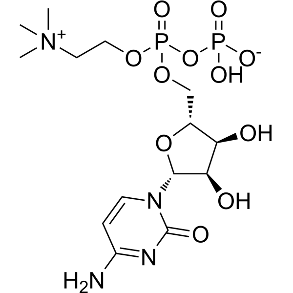
Citicoline is an endogenous intermediate in the synthesis of phosphatidylcholine which is a component of cell membranes. Citicoline inhibits reactive oxygen species (ROS) and apoptosis. Citicoline can be used for neurological disease and hearing loss study.
For research use only. We do not sell to patients.
- Purity : 99.57%
- CAS No.: 987-78-0
- Formula: C14H26N4O11P2
- Molecular Weight:488.32
-
Storage:Powder -20°C, 3 years , 4°C, 2 years
* The compound is unstable in solutions, freshly prepared is recommended.

To place orders, for customer services and technical support, please contact: MedChemExpress USA
Tel: 609-228-6898 E-mail: [email protected] [email protected]


-
 Biological Activity
-
Chemical Information
-
Solvent & Solubility
- Protocols
- Purity & Documentation
- References
-
 Help & FAQs


-
HY-W009162
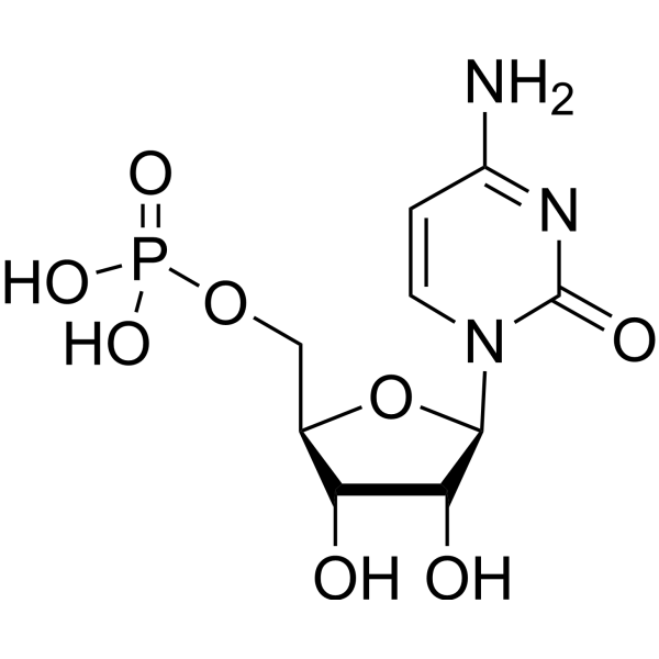
Cytidine 5'-monophosphateEndogenous Metabolite -
HY-B2235
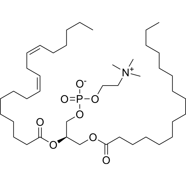
LecithinEndogenous Metabolite -
HY-W009216
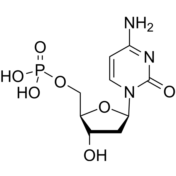
2'-Deoxycytidine-5'-monophosphate80,000-dalton adenovirus terminal protein Binder -
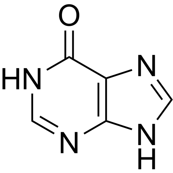
HY-N0091
HypoxanthineEndogenous Metabolite -
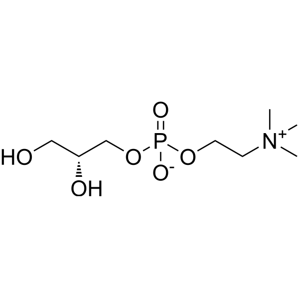
HY-17552
sn-Glycero-3-phosphocholineCholinesterase (ChE) Activator -
HY-K1006
Penicillin-Streptomycin (100×), Sterile -

HY-B0215
AcetylcysteineMucolytic Agent -
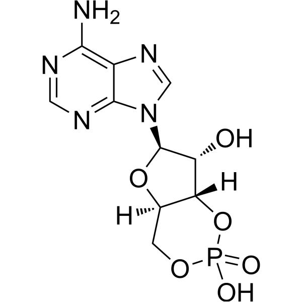
HY-B1511
Cyclic AMPSecond Messenger -

HY-10071
Y-27632ROCK Inhibitor -

HY-125854
Phosphatidylcholines, eggPhosphatidylcholine -

HY-113498
SphingomyelinEndogenous Metabolite -
HY-B0282
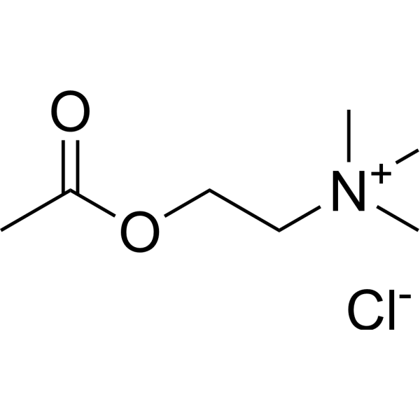
Acetylcholine chlorideCholinergic Agonist
Publications Citing Use of MedChemExpress (MCE) Citicoline
More Customer Validation & Images

-
Others
All Endogenous Metabolite Isoforms
MoreAll Caspase Isoforms
More

Biological Activity
Description
IC50 & Target
|
Microbial Metabolite |
Human Endogenous Metabolite |
Caspase-8 |
Caspase-9 |
Caspase 3 |
In Vitro
Citicoline (1-100 μM, 24 h or 24 h before glutamate treatment) increases rats retinal cells viability after glutamate-induced excitotoxicity, and has synergistic effects with Tramiprosate (Homotaurine) (HY-14602)[1].
Citicoline (100 μM, 24 h before glutamate treatment and 30 min before HG treatment) decreases proapoptotic effects associated with neuroretinal degeneration such as glutamate-induced excitotoxicity and high glucose (HG)-induced neurotoxicity, and has synergistic effects with Tramiprosate in rats retinal cells[1].
Citicoline (100 μM, 36-48 h) reduces apoptosis in mice cochlear hair cells (HCs) and HEI-OC-1 cells after Neomycin (HY-150520) exposure, decreases the mRNA level of pro-apoptotic factor and increases anti-apoptotic factor, suppresses proapoptotic protein expression[2][3].
Citicoline (100 μM, 36-48 h) attenuates oxidative stress in mice cochlear HCs and HEI-OC-1 cells after Neomycin injury[3].
MedChemExpress (MCE) has not independently confirmed the accuracy of these methods. They are for reference only.
-
Cell Line:Rat retinal cells
-
Concentration:1, 10, 100 μM
-
Incubation Time:24 h or 24 h before glutamate treatment (100 μM)
-
Result:Did not affect viability of primary retinal cultures.
Significantly increased cell viability after glutamate-induced excitotoxicity, and had synergistic effects with homotaurine
-
Cell Line:Rat retinal cells
-
Concentration:100 μM
-
Incubation Time:24 h before glutamate treatment and 30 min before HG treatment
-
Result:Reduced apoptosis induced by administration of glutamate and HG, and had synergistic effects with homotaurine.
-
Cell Line:HEI-OC-1, mice cochlear tissue
-
Concentration:10 μM
-
Incubation Time:36 or 48 h (pretreatment for 12 h and then treated with 10 mM neomycin together for 12 and 24 h in cochlear tissue and HEI-OC-1, respectively, then allowed to recover in culture medium for another 12
-
Result:Showed significantly lower numbers of cleaved caspase 3-positive cells and TUNEL-positive cells than the neomycin-only group.
Significantly reduced the percentage of apoptosis.
The protein levels of cleaved caspase 3 were decreased compared with the Neomycin-only group.
Decreased the pro-apoptotic factor Bax, Casp3, Casp8, and Casp9, and increased anti-apoptotic factor Bcl-2 by qPT-PCR analysis.
In Vivo
MedChemExpress (MCE) has not independently confirmed the accuracy of these methods. They are for reference only.
-
Animal Model:Male C57Bl/6 mice (23-27 g) (The solution of convulsant pentylenetetrazole (1%) was infused intravenously at a flow rate of 0.01 ml/sec.)[2]
-
Dosage:500, 1000 mg/kg
-
Administration:Intraperitoneal injection (i.p.); 1 h before test
-
Result:Increased the threshold of clonic seizures and tonic phase of seizures with lethal outcome in a dose of 500 mg/kg.
The anticonvulsant effect was most pronounced in a dose of 1000 mg/kg.


Chemical Information
-
CAS No. 987-78-0
-
Appearance Solid
-
Molecular Weight 488.32
-
Formula C14H26N4O11P2
-
Color White to off-white
-
SMILES
NC(C=CN1[C@H]2[C@H](O)[C@H](O)[C@@H](COP(OP(OCC[N+](C)(C)C)([O-])=O)(O)=O)O2)=NC1=O
-
Synonyms
Cytidine diphosphate-choline; CDP-Choline; Cytidine 5'-diphosphocholine
-
Structure Classification
-
Initial Source
-
Shipping
Room temperature in continental US; may vary elsewhere.
-
Storage
Powder -20°C 3 years 4°C 2 years * The compound is unstable in solutions, freshly prepared is recommended.


Publications (5)
-
Journal Impact Factor
-
Most Recent
-
Nat Neurosci
2023 Apr;26(4):542-554. PMID: 36941428
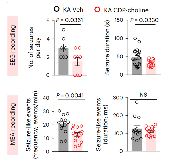
Citicoline purchased from MedChemExpress. Usage Cited in: Nat Neurosci. 2023 Apr;26(4):542-554. [Abstract]
In vivo EEG and ex vivo MEA recordings of KA epileptic mice treated with CDP-choline (Citicoline: 100 mg/kg) or vehicle control.
-
J Neuroinflammation
Microglial IRF7-induced lipophagy impairment aggravates lipid droplet overload and impedes neurological recovery after ischemic stroke. [Abstract]2026 Jun 17. PMID: 42310673 -
Int J Biol Macromol
2025 Mar:294:139536. PMID: 39765299 -
-


Solvent & Solubility
In Vitro:
H2O : 50 mg/mL (102.39 mM; Need ultrasonic)
DMSO : < 1 mg/mL (insoluble or slightly soluble)
Please refer to the solubility information to select the appropriate solvent. The compound is unstable in solutions, freshly prepared is recommended.
* Note: If you choose water as the stock solution, please dilute it to the working solution, then filter and sterilize it with a 0.22 μm filter before use.
Please refer to the solubility information to select the appropriate solvent. The compound is unstable in solutions, freshly prepared is recommended.
* Note: If you choose water as the stock solution, please dilute it to the working solution, then filter and sterilize it with a 0.22 μm filter before use.
Concentration (start) × Volume (start) = Concentration (final) × Volume (final)
In Vivo:
For the following dissolution methods, please prepare the working solution directly:
It is recommended to prepare fresh solutions and use them promptly within a short period of time.
The percentages shown for the solvents indicate their volumetric ratio in the final prepared solution. If precipitation or phase separation occurs during preparation, heat and/or sonication can be used to aid dissolution.
Add each solvent one by one: PBS
Solubility: 50 mg/mL (102.39 mM); Clear solution; Need ultrasonic


Purity & Documentation
-
Data Sheet (279 KB)
-
SDS (396 KB)
- English - EN (396 KB)
- Français - FR (396 KB)
- Deutsch - DE (396 KB)
- Norwegian - NO (396 KB)
- Español - ES (396 KB)
- Swedish - SV (396 KB)
- Italian - IT (396 KB)
- Korean - KR (396 KB)
- Portuguese - PT (396 KB)
-
Handling Instructions (2659 KB)
References
[1]. Davinelli S, et al. Cytoprotective Effects of Citicoline and Homotaurine against Glutamate and High Glucose Neurotoxicity in Primary Cultured Retinal Cells. Oxid Med Cell Longev. 2017;2017:2825703. [Content Brief]
[2]. Karpova MN, et al. Increase of the seizure threshold in C57BL/6 mice after citicoline administration. Bull Exp Biol Med. 2015 Jan;158(3):315-7. [Content Brief]
[3]. Zhong Z, et al. Citicoline Protects Auditory Hair Cells Against Neomycin-Induced Damage. Front Cell Dev Biol. 2020 Aug 31;8:712. [Content Brief]


Complete Stock Solution Preparation Table
Please refer to the solubility information to select the appropriate solvent. The compound is unstable in solutions, freshly prepared is recommended.
| Optional Solvent | Concentration Solvent Mass | 1 mg | 5 mg | 10 mg | 25 mg |
|---|---|---|---|---|---|
| H2O | 1 mM | 2.0478 mL | 10.2392 mL | 20.4784 mL | 51.1959 mL |
| 5 mM | 0.4096 mL | 2.0478 mL | 4.0957 mL | 10.2392 mL | |
| 10 mM | 0.2048 mL | 1.0239 mL | 2.0478 mL | 5.1196 mL | |
| 15 mM | 0.1365 mL | 0.6826 mL | 1.3652 mL | 3.4131 mL | |
| 20 mM | 0.1024 mL | 0.5120 mL | 1.0239 mL | 2.5598 mL | |
| 25 mM | 0.0819 mL | 0.4096 mL | 0.8191 mL | 2.0478 mL | |
| 30 mM | 0.0683 mL | 0.3413 mL | 0.6826 mL | 1.7065 mL | |
| 40 mM | 0.0512 mL | 0.2560 mL | 0.5120 mL | 1.2799 mL | |
| 50 mM | 0.0410 mL | 0.2048 mL | 0.4096 mL | 1.0239 mL | |
| 60 mM | 0.0341 mL | 0.1707 mL | 0.3413 mL | 0.8533 mL | |
| 80 mM | 0.0256 mL | 0.1280 mL | 0.2560 mL | 0.6399 mL | |
| 100 mM | 0.0205 mL | 0.1024 mL | 0.2048 mL | 0.5120 mL |
* Note: If you choose water as the stock solution, please dilute it to the working solution, then filter and sterilize it with a 0.22 μm filter before use.


Citicoline Related Classifications
HY-B0739 Related Classifications
 Powered by Bioz