



CMC2.24

Based on 1 Customer Validation
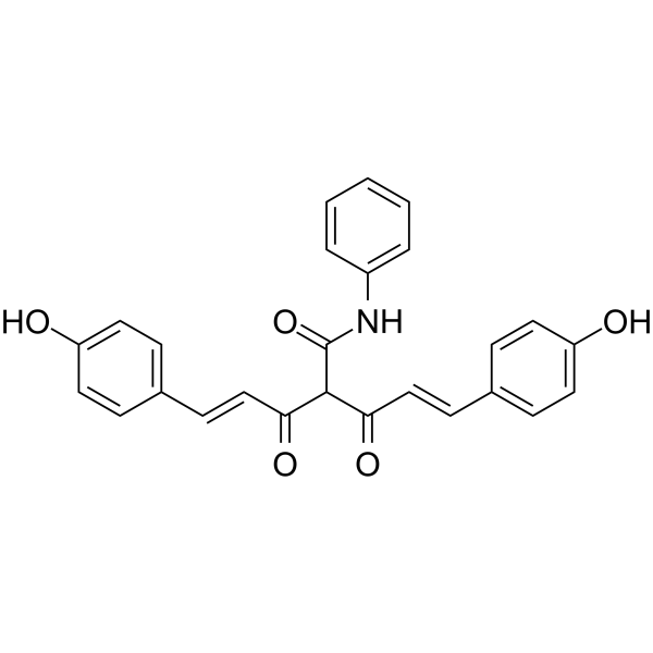
CMC2.24 (TRB-N0224), an orally active tricarbonylmethane agent, is effective against pancreatic tumor in mice by inhibiting Ras activation and its downstream effector ERK1/2 pathway. CMC2.24 is also a potent inhibitor of zinc-dependent MMPs with IC50s ranging from 2.0-69 μM. CMC2.24 alleviates osteoarthritis progression by restoring cartilage homeostasis and inhibiting chondrocyte apoptosis via the NF-κB/HIF-2α axis.
For research use only. We do not sell to patients.
- Purity : 97.11%
- CAS No.: 1255639-43-0
- Formula: C26H21NO5
- Molecular Weight:427.45
-
Storage:
-20°C, protect from light
* In solvent : -80°C, 6 months; -20°C, 1 month (protect from light)

To place orders, for customer services and technical support, please contact: MedChemExpress USA
Tel: 609-228-6898 E-mail: [email protected] [email protected]


-
 Biological Activity
-
Chemical Information
-
Solvent & Solubility
- Protocols
- Purity & Documentation
- References
-
 Help & FAQs


-
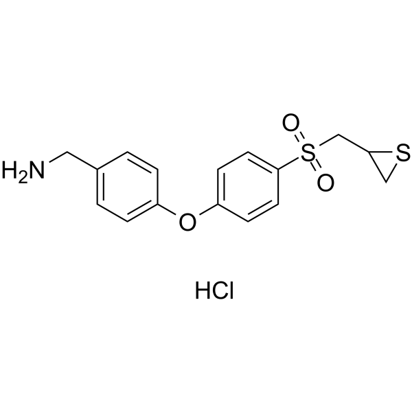
HY-124373
ND-336MMP Inhibitor -
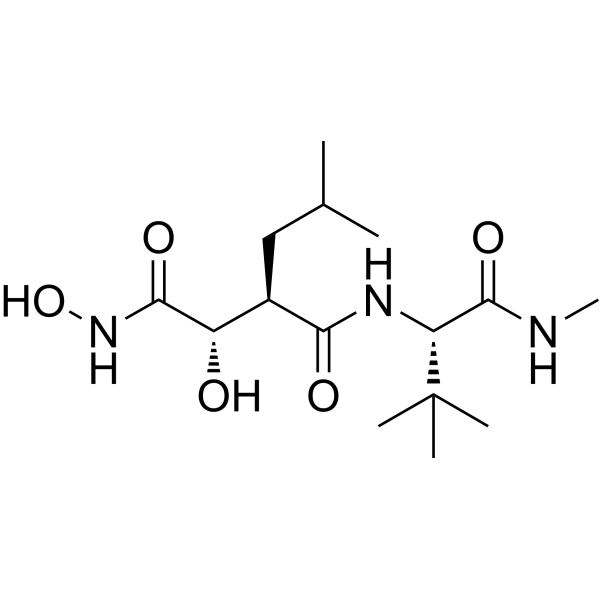
HY-12169
MarimastatMMP Inhibitor -
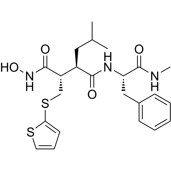
HY-13564
BatimastatMMP Inhibitor -
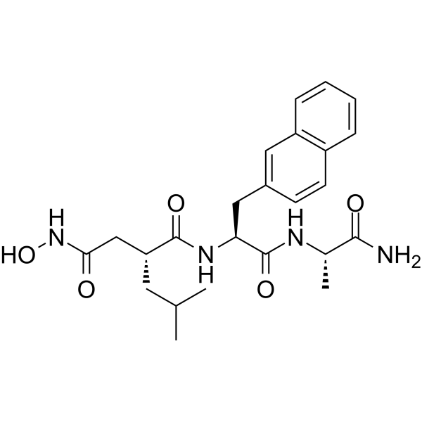
HY-118694
TAPI-0TACE/MMP Inhibitor -
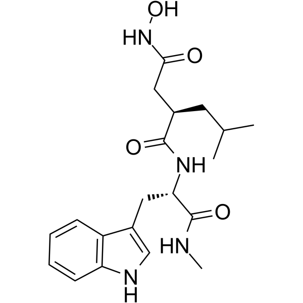
HY-15768
IlomastatMMP Inhibitor -
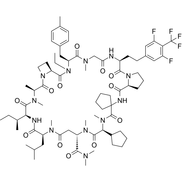
HY-156002
LUNA18KRAS Inhibitor, ERK Inhibitor, RAS Inhibitor -
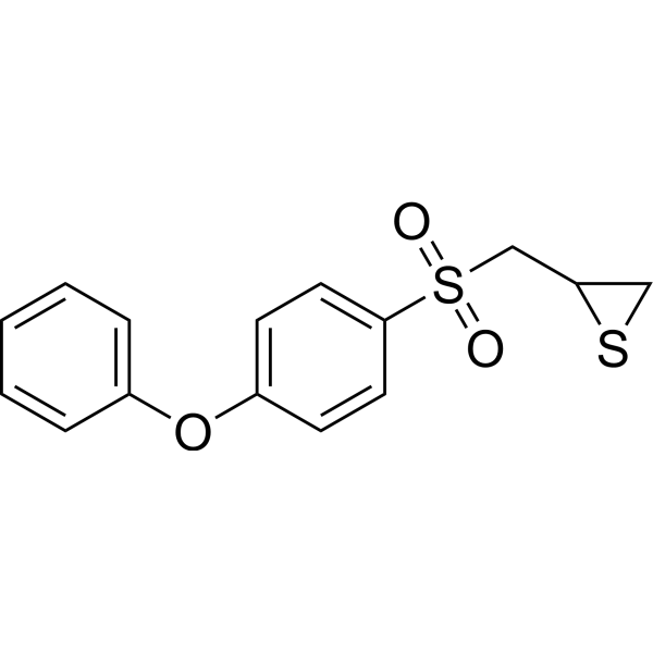
HY-12354
SB-3CTMMP2/9 Inhibitor -
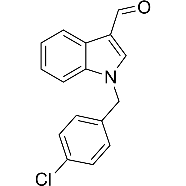
HY-16662
Oncrasin-1RNA Polymerase Inhibitor -
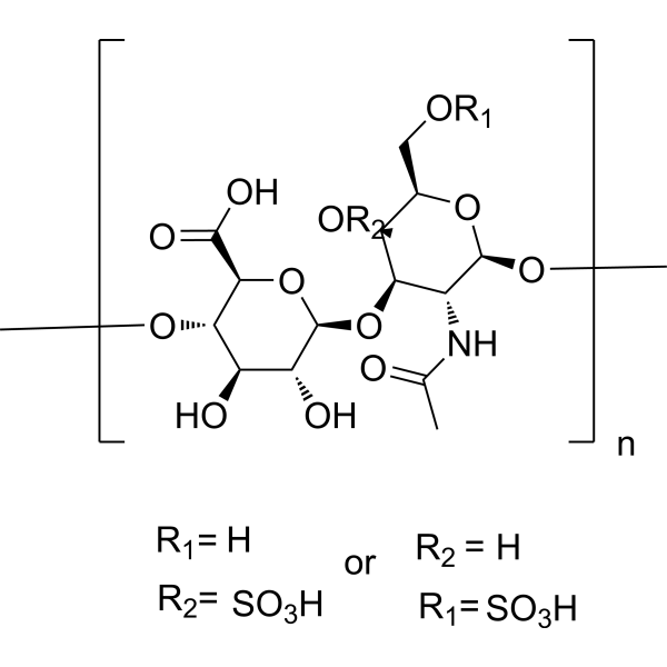
HY-B2162
Chondroitin sulfateGlycosaminoglycan -
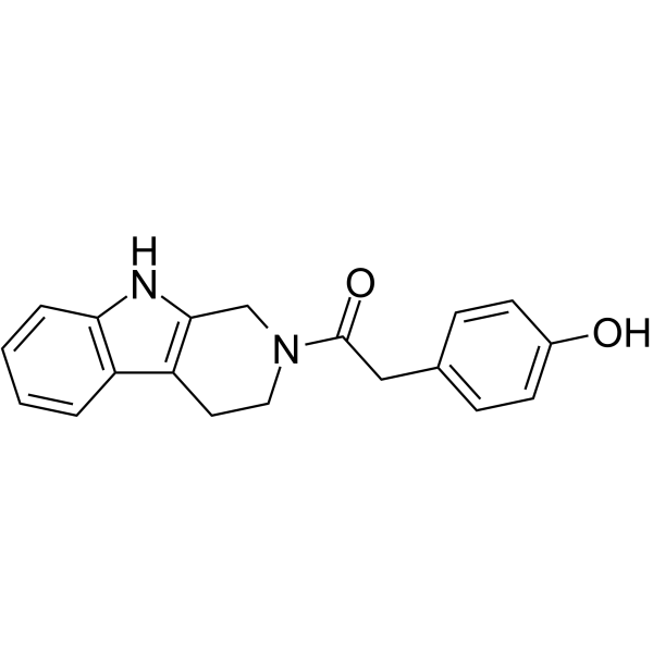
HY-120213
YH-306FAK Inhibitor -
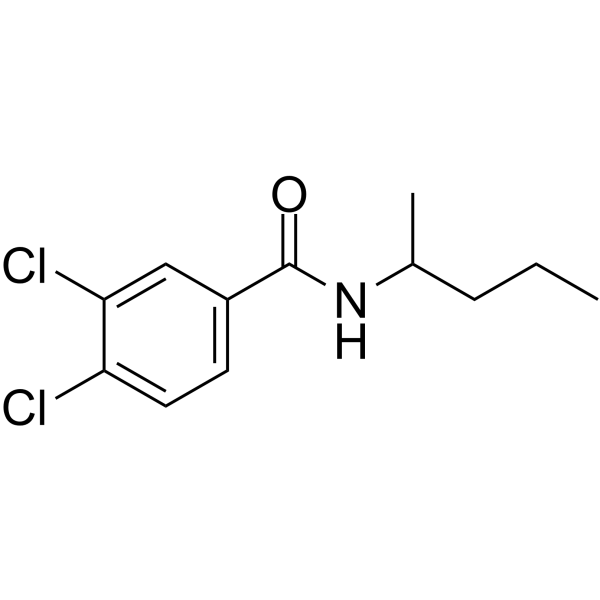
HY-15827
NSC 405020MMP14 Inhibitor -
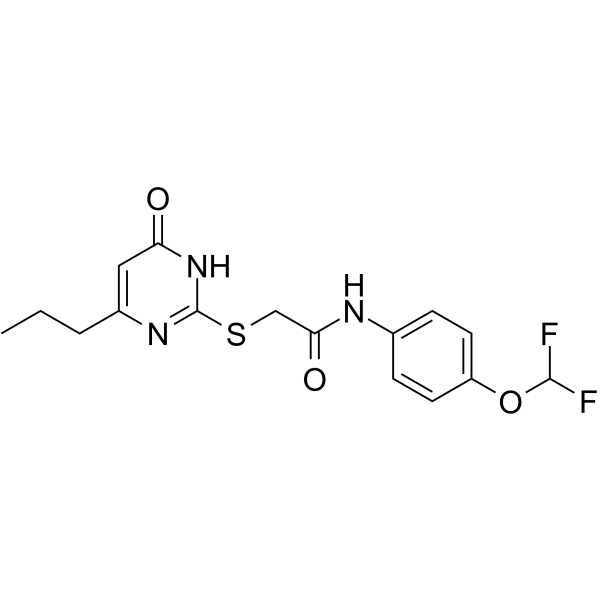
HY-135232
MMP-9-IN-1MMP-9 Inhibitor


Biological Activity
Description
|
RAS |
MMP-1 69.8 μM (IC50) |
MMP-2 4.8 μM (IC50) |
MMP-3 2.9 μM (IC50) |
MMP-7 5 μM (IC50) |
MMP-8 4.5 μM (IC50) |
MMP-9 8 μM (IC50) |
MMP-12 2 μM (IC50) |
MMP-13 2.7 μM (IC50) |
MMP-14 15.3 μM (IC50) |
In Vitro
CMC2.24 (0-60 μM; 24 hours) inhibits pancreatic cancer growth in vitro[1].
CMC2.24 reduces STAT3Ser727 phosphorylation levels, induces mitochondrial reactive oxygen species and mitochondrial cell death in pancreatic cancer cells. CMC2.24 induces mitochondrial reactive oxygen species and intrinsic apoptosis[1].
MedChemExpress (MCE) has not independently confirmed the accuracy of these methods. They are for reference only.
-
Cell Line:AsPC-1, Panc-1, MIA PaCa-2 and BxPC-3 PC cells
-
Concentration:0-60 μM
-
Incubation Time:24 hours
-
Result:Inhibits PC cell growth in a concentration-dependent manner.
In Vivo
CMC2.24 inhibits the growth of human PC through a strong cytokinetic effect. CMC2.24 inhibits ERK signaling pathway in PC cells and xenografts[1].
MedChemExpress (MCE) has not independently confirmed the accuracy of these methods. They are for reference only.
-
Animal Model:Female immune deficient BALB/c nude mice[1]
-
Dosage:50 mg/kg
-
Administration:P.o.; five times per week during 17 days
-
Result:Reduced the rate of growth over baseline by 66.9%.


Chemical Information
-
CAS No. 1255639-43-0
-
Appearance Solid
-
Molecular Weight 427.45
-
Formula C26H21NO5
-
Color Light yellow to orange
-
SMILES
O=C(NC1=CC=CC=C1)C(C(/C=C/C2=CC=C(O)C=C2)=O)C(/C=C/C3=CC=C(O)C=C3)=O
-
Synonyms
TRB-N0224
-
Shipping
Room temperature in continental US; may vary elsewhere.
-
Storage
-20°C, protect from light
* In solvent : -80°C, 6 months; -20°C, 1 month (protect from light)


Solvent & Solubility
In Vitro:
DMSO : < 1 mg/mL (insoluble or slightly soluble)


Purity & Documentation
-
Data Sheet (273 KB)
-
SDS (394 KB)
- English - EN (394 KB)
- Français - FR (394 KB)
- Deutsch - DE (394 KB)
- Norwegian - NO (394 KB)
- Español - ES (394 KB)
- Swedish - SV (394 KB)
- Italian - IT (394 KB)
- Korean - KR (394 KB)
- Portuguese - PT (394 KB)
-
Handling Instructions (2659 KB)
References
[1]. Mallangada NA, et al. A novel tricarbonylmethane agent (CMC2.24) reduces human pancreatic tumor growth in mice by targeting Ras. Mol Carcinog. 2018;57(9):1130-1143. [Content Brief]
[2]. Zhou Y, et al. Chemically modified curcumin (CMC2.24) alleviates osteoarthritis progression by restoring cartilage homeostasis and inhibiting chondrocyte apoptosis via the NF-κB/HIF-2α axis. J Mol Med (Berl). 2020;98(10):1479-1491. [Content Brief]
[3]. Zhang Y, et al. Design, synthesis and biological activity of new polyenolic inhibitors of matrix metalloproteinases: a focus on chemically-modified curcumins. Curr Med Chem. 2012;19(25):4348-4358. [Content Brief]

Calculators
Concentration (start) × Volume (start) = Concentration (final) × Volume (final)


CMC2.24 Related Classifications
HY-120793 Related Classifications
 Powered by Bioz