



Khasianine
Based on 3 publication(s) in Google Scholar
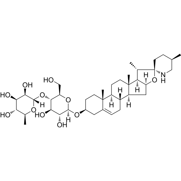
Khasianine is a steroidal glycoalkaloid. Khasianine alleviates psoriasis-like skin inflammation by inhibiting the TNF-α/NF-κB axis. Khasianine can downregulate the RhoA pathway and exhibits potential antimetastatic activity. Khasianine upregulates the expression of TNFR I and TNFR II without inducing apoptotic effects. Khasianine can be used in studies related to psoriasis, pancreatic ductal adenocarcinoma, and liver cancer.
For research use only. We do not sell to patients.
- Purity : 99.11%
- CAS No.: 32449-98-2
- Formula: C39H63NO11
- Molecular Weight:721.92
-
Storage:
4°C, protect from light
* In solvent : -80°C, 6 months; -20°C, 1 month (protect from light)

To place orders, for customer services and technical support, please contact: MedChemExpress USA
Tel: 609-228-6898 E-mail: [email protected] [email protected]


-
 Biological Activity
-
Chemical Information
-
Solvent & Solubility
- Protocols
- Purity & Documentation
- References
-
 Help & FAQs



Publications Citing Use of MedChemExpress (MCE) Khasianine
More Customer Validation & Images

-
IF

-
IHC

-
WB

-
RT-PCR

-
Cell Proliferation/Viability Assay


Biological Activity
Description
IC50 & Target
IC50: antitumor activity[1]
Cellular Effect
|
Cell Line

|
Type | Value | Description | References |
|---|---|---|---|---|
| PLC-PRF-5 | ED50 |
8.6 μg/mL
Compound: khasianine
|
Cytotoxicity against human PLC/PRF/5 cells by microassay
Cytotoxicity against human PLC/PRF/5 cells by microassay
|
[PMID: 2380724] |
In Vitro
Khasianine (3.2-50 nM; 24 h) dose-dependently inhibits TNF-α-induced phosphorylation of NF-κB p65 at the Ser536 site in HaCaT cells without altering the level of total NF-κB p65[1].
Khasianine (3.2-50 nM; 24 h) dose-dependently inhibits TNF-α-induced IL-17A and IL-33 protein expression in HaCaT cells[1].
Khasianine (50 nM) blocks TNF-α-induced nuclear translocation of NF-κB p65 in HaCaT cells[1].
Khasianine (50 nM) displaces NF-κB p65 from the promoter regions of IL-17A and IL-33, thereby inhibiting TNF-α-induced transactivation of these cytokines in HaCaT cells[1].
Khasianine (3.2-12.5 nM) inhibits TNF-α-induced IL-17A mRNA expression in HaCaT cells in a dose-dependent manner, and significantly reduces TNF-α-induced IL-33 mRNA expression in these cells at a concentration of 12.5 nM[1].
Khasianine (50-200 μM; 6 days) shows no obvious cytotoxicity against HaCaT cells at concentrations up to 200 μM during the 6-day incubation period[1].
Khasianine (30-60 μg/mL) does not induce apoptotic cell death (characterized by a sub-G1 peak) in human hepatocellular carcinoma Hep3B cells even at concentrations as high as 60 μg/mL[3].
Khasianine (30-100 μg/mL; 48 h) inhibits the proliferation of human Suit2-007 cells and rat ASML pancreatic cancer cells, with IC50 values of 50 μg/mL and 54 μg/mL, respectively[2].
Khasianine (up to 20 μg/mL; 16 h) exhibits extremely low cytotoxicity against human hepatocellular carcinoma Hep3B cells, with an IC50 greater than 20 μg/mL[3].
Khasianine (50 μg/mL; 48 h) regulates the mRNA expression of glucose-sensitive LSBP in human Suit2-007 pancreatic cancer cells, with the most significant downregulation observed in lactose-sensitive LSBP, and downregulates the Sirtuin, RhoGTPase and RhoA signaling pathways[2].
Khasianine (50 μg/mL; 48 h) regulates the protein expression of glucose-sensitive LSBP in human Suit2-007 pancreatic cancer cells, with the most significant down-regulation observed in lactose-sensitive LSBP[2].
MedChemExpress (MCE) has not independently confirmed the accuracy of these methods. They are for reference only. Further protocols information, click here.
-
Cell Line:HaCaT
-
Concentration:3.2, 12.5, 50 nM
-
Incubation Time:24 h
-
Result:Inhibited TNF-α-induced phosphorylation of NF-κB p65 at ser536 in a dose-dependent manner.
Had no effect on total NF-κB p65 expression.\nDownregulated TNF-α-induced IL-17A and IL-33 protein expression in a dose-dependent manner.
-
Cell Line:HaCaT
-
Concentration:50, 100, 200 μM
-
Incubation Time:up to 6 days
-
Result:Showed no obvious cytotoxicity in HaCaT cells treated at concentrations up to 200 μM over the 6-day incubation period.
Maintained cell viability comparable to DMSO-treated controls.
-
Cell Line:human pancreatic ductal adenocarcinoma (PDAC) Suit2-007 cells, rat PDAC ASML cells
-
Concentration:30, 40, 50, 60, 80, 100 μg/mL
-
Incubation Time:48 h
-
Result:Inhibited proliferation of Suit2-007 cells with an IC50 value of 50 μg/mL.
Inhibited proliferation of ASML cells with an IC50 value of 54 μg/mL.
-
Cell Line:human hepatoma Hep3B cells
-
Concentration:20 μg/mL
-
Incubation Time:30 min
-
Result:Up-regulated TNFR I gene expression to the highest extent among the tested alkaloids, resulting in a TNFR II/I expression ratio of 0.95 (compared to a control ratio of 1.37).
In Vivo
MedChemExpress (MCE) has not independently confirmed the accuracy of these methods. They are for reference only.
-
Animal Model:C57BL/6 (8 to 10 weeks of age; n=5 per treatment group; imiquimod-induced psoriasis-like skin inflammation)[1]
-
Dosage:0.1 μg/g, 1 μg/g, 5 μg/g, 25 μg/g; 5 μg/g (most effective)
-
Administration:topical; 3 times daily; 4 days
-
Result:Reduced clinical features of psoriasis-like inflammation, including scales, epidermal thickness, and immune cell infiltration.
Significantly reduced CD4+ T helper cells and macrophages in skin lesions.
Markedly decreased TNF-α-positive cells, total NF-κB p65-positive cells, phosphorylated NF-κB p65 (ser536)-positive cells, IL-17A-positive cells, and IL-33-positive cells in both epidermis and dermis.


Chemical Information
-
CAS No. 32449-98-2
-
Appearance Solid
-
Molecular Weight 721.92
-
Formula C39H63NO11
-
Color White to off-white
-
SMILES
C[C@@H]1[C@@]2(CC[C@@H](C)CN2)O[C@](C[C@@]3([H])[C@@](CC=C4[C@@]5(CC[C@H](O[C@]([C@@H]([C@@H](O)[C@@H]6O[C@@](O[C@@H](C)[C@H](O)[C@H]7O)([H])[C@@H]7O)O)([H])O[C@@H]6CO)C4)C)([H])[C@]5([H])CC8)([H])[C@@]1([H])[C@]38C
-
Structure Classification
-
Initial Source
-
Shipping
Room temperature in continental US; may vary elsewhere.
-
Storage
4°C, protect from light
* In solvent : -80°C, 6 months; -20°C, 1 month (protect from light)


Publications (3)
-
Journal Impact Factor
-
Most Recent
-
Phytomedicine
Total alkaloids of Fritillaria unibracteata var. wabuensis bulbus ameliorate chronic asthma via the TRPV1/Ca2+/NFAT pathway. [Abstract]2023 Sep:118:154946. PMID: 37421766 -
J Ethnopharmacol
Khasianine ameliorates psoriasis-like skin inflammation and represses TNF-α/NF-κB axis mediated transactivation of IL-17A and IL-33 in keratinocytes. [Abstract]2022 Jun 28:292:115124. PMID: 35183690
Khasianine purchased from MedChemExpress. Usage Cited in: J Ethnopharmacol. 2022 Jun 28:292:115124. [Abstract]
Khasianine (0.5 μg/g) reduces immune cells infiltration. Skin samples were immunofluorescence stained by CD45 for total leukocytes, CD4 for Th cells and F4/80 for macrophages.
Khasianine purchased from MedChemExpress. Usage Cited in: J Ethnopharmacol. 2022 Jun 28:292:115124. [Abstract]
Representative immunohistochemical staining of consecutive sections for TNF-α, NF-κB p65 and NF-κB p-p65 ser536 in normal skin and psoriatic skin lesions after 4 days of treatment with or without Khasianine (0.5 μg/g).
Khasianine purchased from MedChemExpress. Usage Cited in: J Ethnopharmacol. 2022 Jun 28:292:115124. [Abstract]
Khasianine (0, 3.2, 12.5, 50 nM) decreased phosphorylation level of NF-κB p65 at ser536 induced by TNF-α in a dose-dependent manner in HaCaT cells.
Khasianine purchased from MedChemExpress. Usage Cited in: J Ethnopharmacol. 2022 Jun 28:292:115124. [Abstract]
Khasianine (3.2, 12.5 nM) repressed TNF-α induced mRNA and protein expression of IL-17A and IL-33 in HaCaT cells in a dose-dependent manner.
Khasianine purchased from MedChemExpress. Usage Cited in: J Ethnopharmacol. 2022 Jun 28:292:115124. [Abstract]
Growth curves of HaCaT cells treated with 0.1% Dimethyl sulfoxide (DMSO) or different dosages of Khasianine (50, 100, 200 nM), respectively.
-
Molecules
Metabolite Profiling and Anti-Inflammatory Activities of Fritillaria cirrhosa D. Don Bulbs Derived from Tissue Culture. [Abstract]2025 Jan 31;30(3):623. PMID: 39942727


Solvent & Solubility
In Vitro:
DMSO : 50 mg/mL (69.26 mM; Need ultrasonic; Hygroscopic DMSO has a significant impact on the solubility of product, please use newly opened DMSO)
Please refer to the solubility information to select the appropriate solvent. Once prepared, please aliquot and store the solution to prevent product inactivation from repeated freeze-thaw cycles.
Storage method and period of stock solution: -80°C, 6 months; -20°C, 1 month (protect from light). When stored at -80°C, please use it within 6 months. When stored at -20°C, please use it within 1 month.
Please refer to the solubility information to select the appropriate solvent. Once prepared, please aliquot and store the solution to prevent product inactivation from repeated freeze-thaw cycles.
Storage method and period of stock solution: -80°C, 6 months; -20°C, 1 month (protect from light). When stored at -80°C, please use it within 6 months. When stored at -20°C, please use it within 1 month.
Concentration (start) × Volume (start) = Concentration (final) × Volume (final)
In Vivo:
Select the appropriate dissolution method based on your experimental animal and administration route.
- For the following dissolution methods, please ensure to first prepare a clear stock solution using an In Vitro approach and then sequentially add co-solvents:
- To ensure reliable experimental results, the clarified stock solution can be appropriately stored based on storage conditions. As for the working solution for In Vivo experiments, it is recommended to prepare freshly and use it on the same day.
- The percentages shown for the solvents indicate their volumetric ratio in the final prepared solution. If precipitation or phase separation occurs during preparation, heat and/or sonication can be used to aid dissolution.
Add each solvent one by one: 10% DMSO 40% PEG300 5% Tween-80 45% Saline
Solubility: ≥ 2.5 mg/mL (3.46 mM); Clear solution
This protocol yields a clear solution of ≥ 2.5 mg/mL (saturation unknown).
Taking 1 mL working solution as an example, add 100 μL DMSO stock solution (25.0 mg/mL) to 400 μL PEG300, and mix evenly; then add 50 μL Tween-80 and mix evenly; then add 450 μL Saline to adjust the volume to 1 mL.
Preparation of Saline: Dissolve 0.9 g sodium chloride in ddH₂O and dilute to 100 mL to obtain a clear Saline solution.
Add each solvent one by one: 10% DMSO 90% (20% SBE-β-CD in Saline)
Solubility: ≥ 2.5 mg/mL (3.46 mM); Clear solution
This protocol yields a clear solution of ≥ 2.5 mg/mL (saturation unknown).
Taking 1 mL working solution as an example, add 100 μL DMSO stock solution (25.0 mg/mL) to 900 μL 20% SBE-β-CD in Saline, and mix evenly.
Preparation of 20% SBE-β-CD in Saline (4°C, storage for one week): 2 g SBE-β-CD powder is dissolved in 10 mL Saline, completely dissolve until clear.
In Vivo Dissolution Calculator
Please enter the basic information of animal experiments:
-
-
-
-
Recommended: Prepare an additional quantity of animals to account for potential losses during experiments.
Please enter your animal formula composition:
-
%DMSO +
Recommended: Keep the proportion of DMSO in working solution below 2% if your animal is weak.
-
%+
-
+%Tween-80 + +
-
%Saline +
The co-solvents required include: DMSO, . All of co-solvents are available by MedChemExpress (MCE). , Tween 80. All of co-solvents are available by MedChemExpress (MCE).
Working solution concentration: 0.22 mg/mL
Method for preparing stock solution: mg drug dissolved in μL DMSO. Stock solution concentration: mg/mL. * In solvent : -80°C, 6 months; -20°C, 1 month (protect from light)
1. Take μL DMSO stock solution;
2. Add μL .
μL , mix evenly;
3. Then add μL Tween 80, mix evenly;
4. Then add μL
Please ensure that the stock solution in the first step is dissolved to a clear state, and add co-solvents in sequence. You can use ultrasonic heating (ultrasonic cleaner, recommended frequency 20-40 kHz), vortexing, etc. to assist dissolution.

Protocols
-
RNA extraction experimental
By lysing cells, releasing RNA, and removing impurities such as proteins and DNA, high-purity RNA products are finally obtained. The commonly used traditional method is the guanidine isothiocyanate/phenol/chloroform method (Trizol), which is suitable for a variety of animal materials including animal tissues, microorganisms, cultured cells, etc., and most plant materials.
-
Apoptosis
Apoptosis, also called programmed cell death, is generally characterized by distinct morphological characteristics.
-
Imiquimod-Induced Psoriasiform Dermatitis
Imiquimod (IMQ)-induced psoriasiform dermatitis is a widely used murine model in which topical application of IMQ, a Toll-like receptor 7 (TLR7) agonist, triggers innate immune activation in the skin and induces a psoriasis-like inflammatory cascade characterized by epidermal hyperplasia, immune cell infiltration, and cytokine production dominated by the IL-23/IL-17 axis. This inflammatory response is mediated through activation of dendritic cells and downstream induction of IL-23, IL-17A, IL-22, and related pro-inflammatory mediators, recapitulating key features of human plaque psoriasis and enabling mechanistic and therapeutic studies. The model is commonly induced using Aldara (5% IMQ cream) applied topically to murine skin, resulting in rapid onset of erythema, scaling, and thickening that can be quantified as disease severity indices and validated histologically.
-
TPA/Croton Oil Ear Edema and Dermatitis
The TPA (12-O-tetradecanoylphorbol-13-acetate) and croton oil-induced mouse ear edema model is a well-established acute cutaneous inflammation system used to evaluate topical anti-inflammatory activity by measuring edema formation, neutrophil infiltration, vascular permeability, and cytokine-mediated skin responses in vivo. The inflammatory response is triggered by topical application of phorbol esters (TPA) or croton oil constituents, leading to rapid activation of protein kinase C signaling, leukocyte recruitment, and increased vascular permeability, which can be quantified by ear thickness, weight, dye extravasation, and biochemical markers such as myeloperoxidase (MPO) activity and pro-inflammatory mediators in ear tissue homogenates. This model is widely used for screening anti-inflammatory agents, where reductions in edema and inflammatory biomarkers reflect suppression of acute dermal inflammation and immune cell infiltration. Histological evaluation typically confirms epidermal
-
Contact Hypersensitivity Dermatitis
Contact hypersensitivity (CHS) dermatitis is a T cell-mediated delayed-type (Type IV) immune reaction in which low-molecular-weight haptens applied to the skin bind host proteins to form complete antigens, triggering sensitization followed by a secondary inflammatory response upon re-exposure (elicitation phase), which is commonly quantified by ear swelling as a readout of skin inflammation in murine models. This model is widely used to study allergic contact dermatitis because it is antigen-specific, reproducible, and reflects key immunological events including dendritic cell activation, T cell priming in draining lymph nodes, and effector T cell-driven tissue inflammation. DNFB- and oxazolone-induced CHS models are standard systems for evaluating both acute and chronic T cell-dependent skin inflammation and for testing immunomodulatory interventions.
-
Apoptosis Solutions
Apoptosis is a regulated, generally non-lytic cell-death pathway that removes unwanted, damaged, infected, or abnormal cells through coordinated morphological changes, caspase activation, DNA fragmentation, and membrane remodeling. The intrinsic apoptosis pathway is controlled mainly by mitochondrial outer membrane permeabilization, BCL-2 family proteins, cytochrome c release, apoptosome formation, caspase-9 activation, and downstream executioner caspase-3/7 activation. The extrinsic apoptosis pathway is initiated by death receptors such as Fas, TNFR, and TRAIL receptors, which recruit adaptor proteins and activate caspase-8 before engaging executioner caspases or mitochondrial amplification through BID cleavage. Apoptosis is linked to many phenotypes, including cancer cell killing, tissue homeostasis, immune regulation, neurodegeneration, infection response, and treatment-induced cytotoxicity; unresolved questions include how apoptosis interacts with necroptosis, pyroptosis, ferroptos
-
Research Protocol for Inflammation-related Diseases
The NLRP3 inflammasome is a cytosolic innate immune signaling platform that integrates priming signals and danger-signal activation to promote caspase-1 activation, maturation of IL-1β and IL-18, and gasdermin D-mediated pyroptotic cell death. The core experimental logic is to determine whether inflammatory disease phenotypes are driven by increased NLRP3 expression, ASC-containing inflammasome assembly, caspase-1 cleavage, GSDMD cleavage, and extracellular release of IL-1β/IL-18 rather than by nonspecific cell injury alone. The pathway is strongly linked to inflammation-related disease phenotypes because monosodium urate crystals activate NALP3/NLRP3 inflammasome signaling in gout-like crystal inflammation, cholesterol crystals activate NLRP3 inflammasomes in atherogenesis models, and DSS-induced intestinal inflammation has been reported to involve NLRP3 inflammasome activity. However, experimental colitis studies also show context-dependent protective effects of NLRP3 inflammasome co
-
Liver Cancer Modeling
Liver cancer can be classified into primary liver cancer and secondary liver cancer. Secondary liver cancer is the metastatic liver cancer. Primary liver cancer includes hepatocellular carcinoma (HCC), intrahepatic cholangiocarcinoma (ICC) and fibrolamellar HCC, of which HCC is the most common form, accounting for approximately 90% of primary liver cancers[1]. HCC mouse models include chemical agent-induced models, transplanted tumor models, and genetic engineered models.


Purity & Documentation
-
Data Sheet (286 KB)
-
SDS (393 KB)
- English - EN (393 KB)
- Français - FR (393 KB)
- Deutsch - DE (393 KB)
- Norwegian - NO (393 KB)
- Español - ES (393 KB)
- Swedish - SV (393 KB)
- Italian - IT (393 KB)
- Korean - KR (393 KB)
- Portuguese - PT (393 KB)
-
Handling Instructions (2659 KB)
References
[1]. Yang Y, et al. Khasianine ameliorates psoriasis-like skin inflammation and represses TNF-α/NF-κB axis mediated transactivation of IL-17A and IL-33 in keratinocytes. Journal of ethnopharmacology. 2022 Jun 28;292:115124. [Content Brief]
[2]. Sagini MN, et al. Khasianine Affects the Expression of Sugar-Sensitive Proteins in Pancreatic Cancer Cells, Which Are Altered in Data from the Rat Model and Patients. ACS pharmacology & translational science. 2023 May 12;6(5):727-737. [Content Brief]
[3]. Chang LC, et al. The rhamnose moiety of solamargine plays a crucial role in triggering cell death by apoptosis. Biochemical and biophysical research communications. 1998 Jan 06;242(1):21-5. [Content Brief]


Complete Stock Solution Preparation Table
Please refer to the solubility information to select the appropriate solvent. Once prepared, please aliquot and store the solution to prevent product inactivation from repeated freeze-thaw cycles.
Storage method and period of stock solution: -80°C, 6 months; -20°C, 1 month (protect from light). When stored at -80°C, please use it within 6 months. When stored at -20°C, please use it within 1 month.
| Optional Solvent | Concentration Solvent Mass | 1 mg | 5 mg | 10 mg | 25 mg |
|---|---|---|---|---|---|
| DMSO | 1 mM | 1.3852 mL | 6.9260 mL | 13.8520 mL | 34.6299 mL |
| 5 mM | 0.2770 mL | 1.3852 mL | 2.7704 mL | 6.9260 mL | |
| 10 mM | 0.1385 mL | 0.6926 mL | 1.3852 mL | 3.4630 mL | |
| 15 mM | 0.0923 mL | 0.4617 mL | 0.9235 mL | 2.3087 mL | |
| 20 mM | 0.0693 mL | 0.3463 mL | 0.6926 mL | 1.7315 mL | |
| 25 mM | 0.0554 mL | 0.2770 mL | 0.5541 mL | 1.3852 mL | |
| 30 mM | 0.0462 mL | 0.2309 mL | 0.4617 mL | 1.1543 mL | |
| 40 mM | 0.0346 mL | 0.1731 mL | 0.3463 mL | 0.8657 mL | |
| 50 mM | 0.0277 mL | 0.1385 mL | 0.2770 mL | 0.6926 mL | |
| 60 mM | 0.0231 mL | 0.1154 mL | 0.2309 mL | 0.5772 mL |


Khasianine Related Classifications
HY-N2322 Related Classifications
 Powered by Bioz