



DpC

Based on 1 Customer Validation
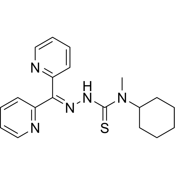
DpC is a selective, orally active iron chelator with anticancer activity. DpC acts on signaling pathway-related targets such as JNK, NF-κB, and its activity is competitively inhibited by another iron chelator Dp44mT (HY-18973). By chelating intracellular iron and copper ions in tumor cells to form redox-active complexes, DpC induces oxidative stress, activates the JNK, NF-κB pathways and downregulates IκBα, upregulates the expressions of neuroglobin and cytoglobin, activates caspase 3/9 to induce tumor cell apoptosis. It also overcomes P-glycoprotein-mediated multidrug resistance through a lysosome-targeting mechanism, and exhibits broad-spectrum synergistic effects when combined with various chemotherapeutic agents. DpC inhibits tumor metastasis and increases TNF-α levels in the tumor microenvironment to enhance endogenous immune responses. DpC is applicable to the research of various malignancies including neuroblastoma, pancreatic cancer, prostate cancer, lung cancer, and breast cancer.
연구목적의 판매만을 진행합니다. 환자를 대상으로 한 판매는 하지 않습니다.
- Purity : 98.73%
- CAS No.: 1382469-39-7
- 화학식: C19H23N5S
- 분자량:353.48
-
보관:Powder -20°C, 3 years ; In solvent -80°C, 6 months , -20°C, 1 month

To place orders, for customer services and technical support, please contact: MedChemExpress USA
Tel: 609-228-6898 E-mail: [email protected] [email protected]


-
 Biological Activity
-
Chemical Information
-
용액&용해도
- Protocol
- 순도&문서
- References
-
 Help & FAQs


All Caspase Isoforms
More

Biological Activity
제품 설명
IC50 & Target
[2]|
Caspase 3 |
Caspase-9 |
IL-10 |
Cellular Effect
|
Cell Line

|
Type | Value | Description | References |
|---|---|---|---|---|
| A549 | IC50 |
<0.03125 μM
Compound: DpC
|
Antiproliferative activity against human A549 cells assessed as reduction in cell growth incubated for 72 hrs by MTT assay
Antiproliferative activity against human A549 cells assessed as reduction in cell growth incubated for 72 hrs by MTT assay
|
[PMID: 32438199] |
| A549 | IC50 |
0.004 μM
Compound: DpC
|
Antiproliferative activity at human A549 cells after 72 hrs by MTT assay
Antiproliferative activity at human A549 cells after 72 hrs by MTT assay
|
[PMID: 22861499] |
| BGC-823 | IC50 |
<0.03125 μM
Compound: DpC
|
Antiproliferative activity against human BGC-823 cells assessed as reduction in cell growth incubated for 72 hrs by MTT assay
Antiproliferative activity against human BGC-823 cells assessed as reduction in cell growth incubated for 72 hrs by MTT assay
|
[PMID: 32438199] |
| Capan-2 | IC50 |
0.02 μM
Compound: 11; DPC
|
Cytotoxicity against human Capan2 cells incubated for 24 hrs by MTT assay
Cytotoxicity against human Capan2 cells incubated for 24 hrs by MTT assay
|
[PMID: 30904782] |
| CFPAC-1 | IC50 |
0.203 μM
Compound: 11; DPC
|
Cytotoxicity against human CFPAC-1 cells incubated for 24 hrs by MTT assay
Cytotoxicity against human CFPAC-1 cells incubated for 24 hrs by MTT assay
|
[PMID: 30904782] |
| DMS-53 | IC50 |
0.008 μM
Compound: DpC
|
Antiproliferative activity at human DMS53 cells after 72 hrs by MTT assay
Antiproliferative activity at human DMS53 cells after 72 hrs by MTT assay
|
[PMID: 22861499] |
| EC9706 | IC50 |
<0.03125 μM
Compound: DpC
|
Antiproliferative activity against human EC9706 cells assessed as reduction in cell growth incubated for 72 hrs by MTT assay
Antiproliferative activity against human EC9706 cells assessed as reduction in cell growth incubated for 72 hrs by MTT assay
|
[PMID: 32438199] |
| GES1 | IC50 |
0.44 μM
Compound: DPC
|
Antiproliferative activity against human GES-1 cells assessed as reduction in cell proliferation incubated for 72 hrs by MTT assay
Antiproliferative activity against human GES-1 cells assessed as reduction in cell proliferation incubated for 72 hrs by MTT assay
|
[PMID: 31614257] |
| GES1 | IC50 |
0.498 μM
Compound: DpC
|
Cytotoxicity against human GES1 cells assessed as reduction in cell viability incubated for 72 hrs by MTT assay
Cytotoxicity against human GES1 cells assessed as reduction in cell viability incubated for 72 hrs by MTT assay
|
[PMID: 32438199] |
| HGC-27 | IC50 |
0.07 μM
Compound: DpC
|
Antiproliferative activity against human HGC-27 cells assessed as reduction in cell growth incubated for 72 hrs by MTT assay
Antiproliferative activity against human HGC-27 cells assessed as reduction in cell growth incubated for 72 hrs by MTT assay
|
[PMID: 32438199] |
| MCF7 | IC50 |
<0.03125 μM
Compound: DpC
|
Antiproliferative activity against human MCF7 cells assessed as reduction in cell growth incubated for 72 hrs by MTT assay
Antiproliferative activity against human MCF7 cells assessed as reduction in cell growth incubated for 72 hrs by MTT assay
|
[PMID: 32438199] |
| MGC-803 | IC50 |
0.166 μM
Compound: DpC
|
Antiproliferative activity against human MGC-803 cells assessed as reduction in cell growth incubated for 72 hrs by MTT assay
Antiproliferative activity against human MGC-803 cells assessed as reduction in cell growth incubated for 72 hrs by MTT assay
|
[PMID: 32438199] |
| MIA PaCa-2 | IC50 |
0.005 μM
Compound: 11; DPC
|
Cytotoxicity against human MIAPaCa2 cells incubated for 24 hrs by MTT assay
Cytotoxicity against human MIAPaCa2 cells incubated for 24 hrs by MTT assay
|
[PMID: 30904782] |
| MRC5 | IC50 |
>10 μM
Compound: DpC
|
Antiproliferative activity at human MRC5 cells after 72 hrs by MTT assay
Antiproliferative activity at human MRC5 cells after 72 hrs by MTT assay
|
[PMID: 22861499] |
| PANC-1 | IC50 |
0.03 μM
Compound: 11; DPC
|
Cytotoxicity against human PANC1 cells incubated for 24 hrs by MTT assay
Cytotoxicity against human PANC1 cells incubated for 24 hrs by MTT assay
|
[PMID: 30904782] |
| PC-3 | IC50 |
0.12 μM
Compound: DPC
|
Antiproliferative activity against human PC3 cells assessed as reduction in cell proliferation incubated for 72 hrs by MTT assay
Antiproliferative activity against human PC3 cells assessed as reduction in cell proliferation incubated for 72 hrs by MTT assay
|
[PMID: 31614257] |
| PC-3 | IC50 |
0.122 μM
Compound: DPC
|
Antiproliferative activity against human PC-3 cells assessed as inhibition of cell proliferation measured after 72 hrs by MTT assay
Antiproliferative activity against human PC-3 cells assessed as inhibition of cell proliferation measured after 72 hrs by MTT assay
|
[PMID: 33153765] |
| SGC-7901 | IC50 |
<0.03125 μM
Compound: DpC
|
Antiproliferative activity against human SGC-7901 cells assessed as reduction in cell growth incubated for 72 hrs by MTT assay
Antiproliferative activity against human SGC-7901 cells assessed as reduction in cell growth incubated for 72 hrs by MTT assay
|
[PMID: 32438199] |
| SK-N-MC | IC50 |
0.013 μM
Compound: DpC
|
Antiproliferative activity at human SK-N-MC cells after 72 hrs by MTT assay
Antiproliferative activity at human SK-N-MC cells after 72 hrs by MTT assay
|
[PMID: 22861499] |
In Vitro
DpC (24 h; 72 h) exhibits potent and selective antiproliferative activity against PANC-1, PC3, DMS-53, DU-145 and MDA-MB-231 tumor cells. Meanwhile, it produces synergistic effects in combination with 9 clinical chemotherapeutic agents including Abiraterone (HY-70013), Carboplatin (HY-17393), Cisplatin (HY-17394) and Doxorubicin (HY-15142), but shows antagonistic effects when combined with Dp44mT[1].
Uptake of 14C-DpC (25 μM; 2 h) by SK-N-MC neuroepithelioma cells depends on temperature- and energy-dependent regulatory mechanisms, and DpC competes with Dp44mT for the same cellular uptake carrier/receptor[1].
DpC (25 μM; 24 h) exhibits antiproliferative activity against MSC, H9C2, MIHA, HK2 and SK-N-LP cells, with significantly higher activity in SK-N-LP cells than Dp44mT and L1. It also significantly upregulates the expressions of neuroglobin (Ngb) and cytoglobin (Cygb) in SK-N-LP and HK2 cells, increases the levels of phosphorylated JNK, cleaved caspase 3 and cleaved caspase 9, and decreases the level of IkBα[2].
MedChemExpress (MCE) has not independently confirmed the accuracy of these methods. They are for reference only.
-
Cell Line:PANC-1 pancreatic cancer cells, PC3 prostate cancer cells, DMS-53 lung cancer cells, DU-145 prostate cancer cells, and MDA- MB-231 breast cancer cell
-
Concentration:0.007-0.096 μM (72 h); 6.50-17.10 μM (24 h)
-
Incubation Time:24 h, 72 h
-
Result:Showed significant antiproliferative activity against five tumor cell lines, with specific IC50 values (24 h/72 h) for: PANC-1 cells (11.40 μM / 0.096 μM), PC3 cells (17.10 μM / 0.007 μM), DMS-53 cells (6.50 μM / 0.020 μM), DU-145 cells (7.90 μM / 0.007 μM), and MDA-MB-231 cells (9.40 μM/0.008 μM).
-
Cell Line:SK-N-LP
-
Concentration:25 μM
-
Incubation Time:24 h
-
Result:Significantly increased the expression levels of phosphorylated JNK, neuroglobin (Ngb), cytoglobin (Cygb), and cleaved caspase 3 and 9, while significantly decreasing the expression level of IkBα.
The ratio of phosphorylated JNK to total JNK was also significantly elevated, and total JNK levels remained unchanged.
In Vivo
MedChemExpress (MCE) has not independently confirmed the accuracy of these methods. They are for reference only.
-
Animal Model:BALB/c nu/nu male nude mice (4 weeks old) with orthotopic SK-N-LP/Luciferase neuroblastoma xenograft in the left adrenal gland fat pad[2]
-
Dosage:4 mg/kg
-
Administration:Tail vein injection; once daily; for 3 weeks
-
Result:After 3 weeks of daily intravenous administration via tail vein, the tumor imaging ROI value and tumor volume were significantly reduced compared with the vehicle control group.
The levels of Annexin V(+)/PI(+) cells, caspase 3, neuroglobin, cytoglobin, and tumor necrosis factor-α (TNFα) in tumor tissues were significantly increased, while interleukin-10 (IL-10) levels were slightly decreased.
The body weight gain of mice in the DpC-treated group was slightly but significantly reduced, and no obvious toxicity was observed in major organs such as the spleen, heart, kidney, and brain, but exudative inflammation was induced in the lung tissue.
- Multiplex immunofluorescence IHC
- Detection of Apoptosis
- TUNEL staining for apoptotic DNA fragmentation
- Annexin V plus membrane-impermeant dye apoptosis staining
- ROS/oxidative-stress fluorescent staining
- Multiplex immunohistochemistry
- Apoptosis Solutions
- Research Protocol for Inflammation-related Diseases
- Lysosome and acidic-vesicle live-cell staining
- Breast Cancer Modeling
- Orthotopic Cell-Line Xenograft
- Research Protocol for Cancer Immunology


Chemical Information
-
CAS No. 1382469-39-7
-
Appearance Solid
-
분자량 353.48
-
화학식 C19H23N5S
-
Color Light yellow to yellow
-
SMILES
S=C(N/N=C(C1=NC=CC=C1)\C2=NC=CC=C2)N(C3CCCCC3)C
-
선적
Room temperature in continental US; may vary elsewhere.
-
보관
Powder -20°C 3 years In solvent -80°C 6 months -20°C 1 month


용액&용해도
In Vitro:
DMSO : 62.5 mg/mL (176.81 mM; Need ultrasonic; Hygroscopic DMSO has a significant impact on the solubility of product, please use newly opened DMSO)
Please refer to the solubility information to select the appropriate solvent. Once prepared, please aliquot and store the solution to prevent product inactivation from repeated freeze-thaw cycles.
Storage method and period of stock solution: -80°C, 6 months; -20°C, 1 month. When stored at -80°C, please use it within 6 months. When stored at -20°C, please use it within 1 month.
Please refer to the solubility information to select the appropriate solvent. Once prepared, please aliquot and store the solution to prevent product inactivation from repeated freeze-thaw cycles.
Storage method and period of stock solution: -80°C, 6 months; -20°C, 1 month. When stored at -80°C, please use it within 6 months. When stored at -20°C, please use it within 1 month.
Concentration (start) × Volume (start) = Concentration (final) × Volume (final)
In Vivo:
Select the appropriate dissolution method based on your experimental animal and administration route.
- For the following dissolution methods, please ensure to first prepare a clear stock solution using an In Vitro approach and then sequentially add co-solvents:
- To ensure reliable experimental results, the clarified stock solution can be appropriately stored based on storage conditions. As for the working solution for In Vivo experiments, it is recommended to prepare freshly and use it on the same day.
- The percentages shown for the solvents indicate their volumetric ratio in the final prepared solution. If precipitation or phase separation occurs during preparation, heat and/or sonication can be used to aid dissolution.
Add each solvent one by one: 10% DMSO 40% PEG300 5% Tween-80 45% Saline
Solubility: ≥ 2.08 mg/mL (5.88 mM); Clear solution
This protocol yields a clear solution of ≥ 2.08 mg/mL (saturation unknown).
Taking 1 mL working solution as an example, add 100 μL DMSO stock solution (20.8 mg/mL) to 400 μL PEG300, and mix evenly; then add 50 μL Tween-80 and mix evenly; then add 450 μL Saline to adjust the volume to 1 mL.
Preparation of Saline: Dissolve 0.9 g sodium chloride in ddH₂O and dilute to 100 mL to obtain a clear Saline solution.
Add each solvent one by one: 10% DMSO 90% (20% SBE-β-CD in Saline)
Solubility: ≥ 2.08 mg/mL (5.88 mM); Clear solution
This protocol yields a clear solution of ≥ 2.08 mg/mL (saturation unknown).
Taking 1 mL working solution as an example, add 100 μL DMSO stock solution (20.8 mg/mL) to 900 μL 20% SBE-β-CD in Saline, and mix evenly.
Preparation of 20% SBE-β-CD in Saline (4°C, storage for one week): 2 g SBE-β-CD powder is dissolved in 10 mL Saline, completely dissolve until clear.
In Vivo Dissolution Calculator
Please enter the basic information of animal experiments:
-
-
-
-
Recommended: Prepare an additional quantity of animals to account for potential losses during experiments.
Please enter your animal formula composition:
-
%DMSO +
Recommended: Keep the proportion of DMSO in working solution below 2% if your animal is weak.
-
%+
-
+%Tween-80 + +
-
%Saline +
The co-solvents required include: DMSO, . All of co-solvents are available by MedChemExpress (MCE). , Tween 80. All of co-solvents are available by MedChemExpress (MCE).
Working solution concentration: 0.22 mg/mL
Method for preparing stock solution: mg drug dissolved in μL DMSO. Stock solution concentration: mg/mL.
1. Take μL DMSO stock solution;
2. Add μL .
μL , mix evenly;
3. Then add μL Tween 80, mix evenly;
4. Then add μL
Please ensure that the stock solution in the first step is dissolved to a clear state, and add co-solvents in sequence. You can use ultrasonic heating (ultrasonic cleaner, recommended frequency 20-40 kHz), vortexing, etc. to assist dissolution.


순도&문서
-
Data Sheet (289 KB)
-
SDS (480 KB)
- English - EN (480 KB)
- Français - FR (480 KB)
- Deutsch - DE (480 KB)
- Norwegian - NO (480 KB)
- Español - ES (480 KB)
- Swedish - SV (480 KB)
- Italian - IT (480 KB)
- Korean - KR (480 KB)
- Portuguese - PT (480 KB)
-
Handling Instructions (2659 KB)
References
[1]. Guo ZL, et al. The novel thiosemicarbazone, di-2-pyridylketone 4-cyclohexyl-4-methyl-3-thiosemicarbazone (DpC), inhibits neuroblastoma growth in vitro and in vivo via multiple mechanisms. J Hematol Oncol. 2016 Sep 27;9(1):98. [Content Brief]
[3]. Dharmasivam M, et al. The thiosemicarbazone, DpC, broadly synergizes with multiple anti-cancer therapeutics and demonstrates temperature- and energy-dependent uptake by tumor cells. Biochim Biophys Acta Gen Subj. 2022;1866(8):130152. [Content Brief]


Complete Stock Solution Preparation Table
Please refer to the solubility information to select the appropriate solvent. Once prepared, please aliquot and store the solution to prevent product inactivation from repeated freeze-thaw cycles.
Storage method and period of stock solution: -80°C, 6 months; -20°C, 1 month. When stored at -80°C, please use it within 6 months. When stored at -20°C, please use it within 1 month.
| Optional Solvent | Concentration Solvent Mass | 1 mg | 5 mg | 10 mg | 25 mg |
|---|---|---|---|---|---|
| DMSO | 1 mM | 2.8290 mL | 14.1451 mL | 28.2901 mL | 70.7254 mL |
| 5 mM | 0.5658 mL | 2.8290 mL | 5.6580 mL | 14.1451 mL | |
| 10 mM | 0.2829 mL | 1.4145 mL | 2.8290 mL | 7.0725 mL | |
| 15 mM | 0.1886 mL | 0.9430 mL | 1.8860 mL | 4.7150 mL | |
| 20 mM | 0.1415 mL | 0.7073 mL | 1.4145 mL | 3.5363 mL | |
| 25 mM | 0.1132 mL | 0.5658 mL | 1.1316 mL | 2.8290 mL | |
| 30 mM | 0.0943 mL | 0.4715 mL | 0.9430 mL | 2.3575 mL | |
| 40 mM | 0.0707 mL | 0.3536 mL | 0.7073 mL | 1.7681 mL | |
| 50 mM | 0.0566 mL | 0.2829 mL | 0.5658 mL | 1.4145 mL | |
| 60 mM | 0.0472 mL | 0.2358 mL | 0.4715 mL | 1.1788 mL | |
| 80 mM | 0.0354 mL | 0.1768 mL | 0.3536 mL | 0.8841 mL | |
| 100 mM | 0.0283 mL | 0.1415 mL | 0.2829 mL | 0.7073 mL |


DpC Related Classifications
HY-114243 Related Classifications
 Powered by Bioz