



L-Leucyl-L-Leucine methyl ester
Based on 23 publication(s) in Google Scholar
L-Leucyl-L-Leucine methyl ester (LLOMe; Leu-Leu methyl ester; H-Leu-Leu-OMe) is a lysosomal disrupting agent and apoptosis inducer that, through receptor-mediated endocytosis and lysosomal dipeptidyl peptidase I conversion, induces caspase activation, lysosomal membrane permeabilization, and DNA fragmentation, and requires cathepsin C to exert membrane-lytic activity, thereby selectively killing natural killer cells. L-Leucyl-L-Leucine methyl ester can be used for research related to lysosomal function and apoptosis.
For research use only. We do not sell to patients.
- CAS No.: 13022-42-9
- Formula: C13H26N2O3
- Molecular Weight:258.36
-
Storage:
Please store the product under the recommended conditions in the Certificate of Analysis.


To place orders, for customer services and technical support, please contact: MedChemExpress USA
Tel: 609-228-6898 E-mail: [email protected] [email protected]


-
 Biological Activity
-
Chemical Information
- Protocols
- Purity & Documentation
- References
-
 Help & FAQs


Publications Citing Use of MedChemExpress (MCE) L-Leucyl-L-Leucine methyl ester
More- Cell Res. 2026 Jan;36(1):51-71. [Abstract]
- Autophagy. 2026 Jul 10:1-21.
- Autophagy. 2024 Dec;20(12):2697-2718. [Abstract]
- Adv Sci (Weinh). 2026 May;13(25):e21681. [Abstract]
- Biomaterials. 2026 Jun 27:335:124405. [Abstract]
- Biomaterials. 2025 Jun:317:123091. [Abstract]
- Environ Chem Ecotoxicol. 2025 Nov 19;8:267-280.
- Pharmacol Res. 2026 Nov:233:108478.
- J Neuroinflammation. 2025 Oct 29;22(1):252. [Abstract]
- EMBO J. 2025 May;44(9):2596-2619. [Abstract]
- World J Gastroenterol. 2026 Feb 28;32(8):113299. [Abstract]
- J Cell Biol. 2025 Nov 3;224(11):e202404018. [Abstract]
- PLoS Pathog. 2025 Sep 16;21(9):e1013513. [Abstract]
- PLoS Pathog. 2024 Feb 14;20(2):e1011981. [Abstract]
- Toxicol Appl Pharmacol. 2026 Jan:506:117632. [Abstract]
- Int Immunol. 2021 Aug 23;33(9):491-504. [Abstract]
- FEBS Lett. 2026 Apr 24. [Abstract]
- Traffic. 2022 May;23(5):238-269. [Abstract]
- bioRxiv. 2026 Apr 21.
- SSRN. 2025 Aug 13.
- Preprints. 2025 Jul 8.
- bioRxiv. 2024 July 07.
- bioRxiv. 2024 Mar 13:2024.03.12.584718. [Abstract]
 Customer Validation & Images

-
IP
-
IF
-
Cell Imaging/Staining
-
IP
-
IP
All Caspase Isoforms
More

Biological Activity
Description
In Vitro
L-Leucyl-L-Leucine methyl ester (1-7 mM; 18 h) induces HeLa cell death only at concentrations above 4 mM after 18 h of continuous exposure[2].
L-Leucyl-L-Leucine methyl ester (0.5-7 mM; 5 min-1 h) causes rapid concentration- and time-dependent loss of catB/L activity in HeLa cells without releasing active cathepsins into the cytosol[2].
L-Leucyl-L-Leucine methyl ester (1-5 mM; 15 min-1 h) reduces mature catB, catL, and catC protein levels in HeLa cells within 15 min[2].
L-Leucyl-L-Leucine methyl ester (1 mM; up to 48 h) enables HeLa cells to reacquire catB/L activity through de novo protein synthesis between 12 and 24 h[2].
L-Leucyl-L-Leucine methyl ester (1-4 h) activates caspase-3-like proteases in HL-60 cells from 1 h onward, reaching a specific activity of 611 pmole AMC/min/mg protein at 4 h (approximately 10 times the control of 57 pmole AMC/min/mg protein)[1].
L-Leucyl-L-Leucine methyl ester (5 mM; 3-10 h) activates caspase-3-like activity in HeLa cells at 3 h, with maximal activity at 10 h[2].
L-Leucyl-L-Leucine methyl ester (0.25 mM; 10 min) causes complete loss of the lysosomal proton gradient in HeLa cells within 10 min[2].
L-Leucyl-L-Leucine methyl ester (1-5 mM; 30 min) permeabilizes HeLa lysosomes to molecules with a molecular weight of up to at least 4.4K, but does not permeabilize molecules with a molecular weight of 10K or greater[2].
L-Leucyl-L-Leucine methyl ester (1-5 mM; 15 min-1 h) retains cathepsin in HeLa lysosomes but inactivates it[2].
L-Leucyl-L-Leucine methyl ester (1-5 mM; 6 h) reduces lysosomal glycosidase activity in HeLa cells, with β-D-galactosidase being the most affected[2].
L-Leucyl-L-Leucine methyl ester (1 mM; up to 24 h) causes a transient loss of lysosomal acidity in HeLa cells, with reacidification beginning at 30 min and exceeding control levels at 4 h[2].
L-Leucyl-L-Leucine methyl ester (1 mM; up to 48 h) causes transient enlargement and perinuclear aggregation of LAMP-1-positive lysosomes in HeLa cells[2].
L-Leucyl-L-Leucine methyl ester (1 mM; 30 min-48 h) transiently enlarges HeLa lysosomes, increasing their volume 3-fold at 24 h and returning to normal at 48 h[2].
MedChemExpress (MCE) has not independently confirmed the accuracy of these methods. They are for reference only.
-
Cell Line:human HL-60 cells
-
Concentration:up to 150 µM; 200 µM
-
Incubation Time:20 min (room temperature, then further incubation); 1 h (200 µM)
-
Result:Induced apoptosis in a concentration-dependent manner, with maximum level at 150 µM.
Decrease in cell size was detected as early as 1 h after incubation with 200 µM, with 40% apoptotic cells at this time point.
With longer incubation, the percentage of apoptotic cells gradually increased.
-
Cell Line:human HL-60 cells
-
Concentration:200 µM; 500 µM
-
Incubation Time:4 h (37°C)
-
Result:DNA fragmentation was observed in HL-60 cells.
Lane 4 showed DNA fragmentation in cells incubated with 500 µM, and lane 2 showed DNA fragmentation with 200 µM.
The DPPI inhibitor (GFCHN2) suppressed DNA fragmentation.
-
Cell Line:HeLa cells
-
Concentration:1, 2, 3, 4, 5, 6, 7 mM
-
Incubation Time:18 h
-
Result:Both crystal violet and LDH assays agreed that cell death was induced only at concentrations above 4 mM after 18 h of continuous exposure.
-
Cell Line:HeLa cells
-
Concentration:1 or 5 mM (15 min after BMV109 pre-labeling; or 1 h BMV109 after prior exposure)
-
Incubation Time:15 min; 1 h
-
Result:Cells labeled with BMV109 for 1 h then exposed to 1 or 5 mM for 15 min showed preserved puncta representing cysteine cathepsin-positive lysosomes, indicating that cysteine cathepsins remained in lysosomes.
When cells were exposed first to 1 or 5 mM and then incubated with BMV109 for another hour, they showed no labeling, indicating that cysteine cathepsins were no longer active.
-
Cell Line:HeLa cells
-
Concentration:1, 3, 5 mM
-
Incubation Time:15 min; 1 h
-
Result:Compared to control cells, protein levels for the mature forms of catB, catL, and catC were visibly reduced already 15 min after exposure to 1, 3, or 5 mM, and even more so after 1 h.
This indicates that exposure-mediated loss of cysteine cathepsin activities was due to a decrease in total enzyme amount rather than only changes in enzyme kinetics.
-
Cell Line:HeLa cells
-
Concentration:1 mM
-
Incubation Time:1, 2, 3, 6, 12, 24, 36, 48 h
-
Result:Following continuous culture with 1 mM, LAMP-1-positive vesicles became enlarged and localized to the perinuclear region.
By 48 h after exposure, the pattern of LAMP-1 labeling reversed and resembled the appearance of lysosomes in untreated cells.


Chemical Information
-
CAS No. 13022-42-9
-
Molecular Weight 258.36
-
Formula C13H26N2O3
-
SMILES
CC(C)C[C@H](N)C(N[C@@H](CC(C)C)C(OC)=O)=O
-
Synonyms
LLOMe; Leu-Leu methyl ester; H-Leu-Leu-OMe
-
Shipping
Room temperature in continental US; may vary elsewhere.
-
Storage
Please store the product under the recommended conditions in the Certificate of Analysis.


Publications (23)
-
Journal Impact Factor
-
Most Recent
-
Cell Res
2026 Jan;36(1):51-71. PMID: 41478856 -
-
Autophagy
2024 Dec;20(12):2697-2718. PMID: 39081059 -
Adv Sci (Weinh)
Loss of VMP1 Impairs Tight Junction Recycling and Aggravates Intestinal Barrier Dysfunction in Inflammatory Bowel Disease. [Abstract]2026 May;13(25):e21681. PMID: 41757559 -
Biomaterials
In-situ cascade assembled peptide-drug conjugate for the treatment of bladder cancer by enhancing membrane-entry and lysosome destabilization. [Abstract]2026 Jun 27:335:124405. PMID: 42372507 -
Biomaterials
Engineered cell membrane vesicles loaded with lysosomophilic drug for acute myeloid leukemia therapy via organ-cell-organelle cascade-targeting. [Abstract]2025 Jun:317:123091. PMID: 39778270 -
-
-
J Neuroinflammation
Immune subversion by Leishmania infantum parasites suppresses NLRP3-driven inflammatory responses in amyloid-β-activated microglia. [Abstract]2025 Oct 29;22(1):252. PMID: 41162989 -
EMBO J
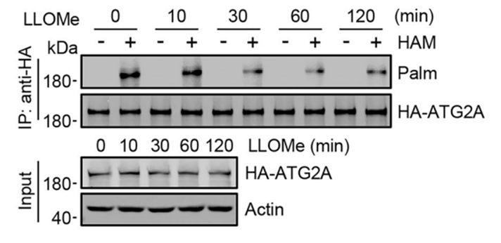
S-palmitoylation modulates ATG2-dependent non-vesicular lipid transport during starvation-induced autophagy. [Abstract]2025 May;44(9):2596-2619. PMID: 40128367
L-Leucyl-L-Leucine methyl ester purchased from MedChemExpress. Usage Cited in: EMBO J. 2025 May;44(9):2596-2619. [Abstract]
S-palmitoylation of HA-ATG2A detected by ABE assay in HEK293T cells treated with 1 mM LLOMe for indicated times.
-
World J Gastroenterol
Wogonin derivative V8 enhances bortezomib efficacy in gastric carcinoma by disrupting lysosome-mediated drug resistance. [Abstract]2026 Feb 28;32(8):113299. PMID: 41809875 -
J Cell Biol
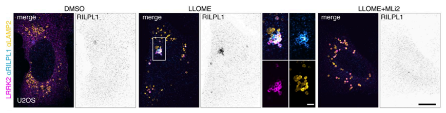
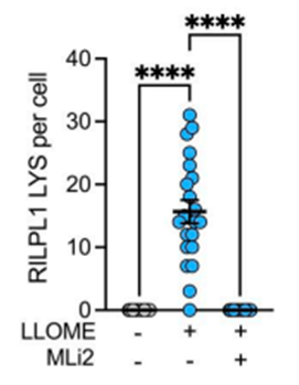
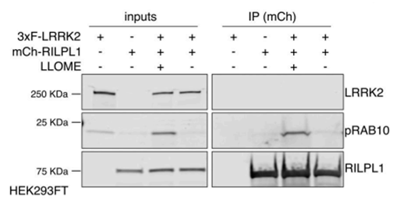
JIP4 and RILPL1 utilize opposing motor force to dynamically regulate lysosomal tubulation. [Abstract]2025 Nov 3;224(11):e202404018. PMID: 40990465
L-Leucyl-L-Leucine methyl ester purchased from MedChemExpress. Usage Cited in: J Cell Biol. 2025 Nov 3;224(11):e202404018. [Abstract]
U2OS cells were transfected with HaloTag-LRRK2 for 36 h. Cells were then treated with DMSO, LLOME (1 mM), or LLOME+MLi2 for 2 h before fixation. Cells were then stained for RILPL1 and LAMP2.
L-Leucyl-L-Leucine methyl ester purchased from MedChemExpress. Usage Cited in: J Cell Biol. 2025 Nov 3;224(11):e202404018. [Abstract]
Quantification of the RILPL1 lysosomes per cell. Cells were treated with DMSO, LLOME (1 mM), or LLOME+MLi2 for 2 h.
L-Leucyl-L-Leucine methyl ester purchased from MedChemExpress. Usage Cited in: J Cell Biol. 2025 Nov 3;224(11):e202404018. [Abstract]
HEK293FT cells were transfected with 3xflag-LRRK2 and mCherry-RILPL1 for 24 h. Cells were treated or not with LLOME (1 mM) for 2 h, and lysates were subjected to immunoprecipitation with anti-RFP antibodies. RFP, LRRK2, and pT73-RAB10 were blotted.
L-Leucyl-L-Leucine methyl ester purchased from MedChemExpress. Usage Cited in: J Cell Biol. 2025 Nov 3;224(11):e202404018. [Abstract]
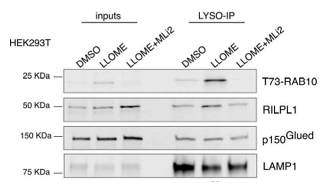
HEK293T cells stably expressing GFP-LRRK2 and TMEM192-3xHA were seeded and treated with DMSO, LLOME (1 mM), or LLOME + MLi2 24 h later. Lysosomes were purified with anti-HA beads following the LYSO-IP technique. Lysosomes were then lysed, and their content was analyzed via immunoblotting.
-
PLoS Pathog
A non-canonical activation of the host's ESCRT machinery is required for the scission of parasitophorous vacuoles and the replication of Leishmania donovani. [Abstract]2025 Sep 16;21(9):e1013513. PMID: 40956840 -
PLoS Pathog
The HN protein of Newcastle disease virus induces cell apoptosis through the induction of lysosomal membrane permeabilization. [Abstract]2024 Feb 14;20(2):e1011981. PMID: 38354122 -
Toxicol Appl Pharmacol
Honokiol ameliorates hepatic fibrosis by inducing lysosomal membrane permeabilization and impairing lipophagy in hepatic stellate cells. [Abstract]2026 Jan:506:117632. PMID: 41207513 -
Int Immunol
Helicobacter urease suppresses cytotoxic CD8+ T-cell responses through activating Myh9-dependent induction of PD-L1. [Abstract]2021 Aug 23;33(9):491-504. PMID: 34297096 -
FEBS Lett
2026 Apr 24. PMID: 42032884 -
Traffic
2022 May;23(5):238-269. PMID: 35343629 -
-
-
-
-
bioRxiv
Autophagosome turnover requires Arp2/3 complex-mediated maintenance of lysosomal integrity. [Abstract]2024 Mar 13:2024.03.12.584718. PMID: 38559247


Purity & Documentation
References

Calculators
Concentration (start) × Volume (start) = Concentration (final) × Volume (final)


L-Leucyl-L-Leucine methyl ester Related Classifications
HY-165398 Related Classifications
 Powered by Bioz
Keywords
- L-Leucyl-L-Leucine methyl ester
- 13022-42-9
- LLOMe
- Leu-Leu methyl ester
- H-Leu-Leu-OMe
- Apoptosis
- Caspase
- DNA fragmentation
- apoptosis inducer
- caspase-3-like activation
- cathepsin C
- de novo protein synthesis
- dipeptidyl peptidase I
- lysosomal disruptor
- lysosomal membrane permeabilization
- natural killer cells
- receptor-mediated endocytosis
- Inhibitor
- inhibitor
- inhibit