



Lidocaine-d10

Based on 1 Customer Validation
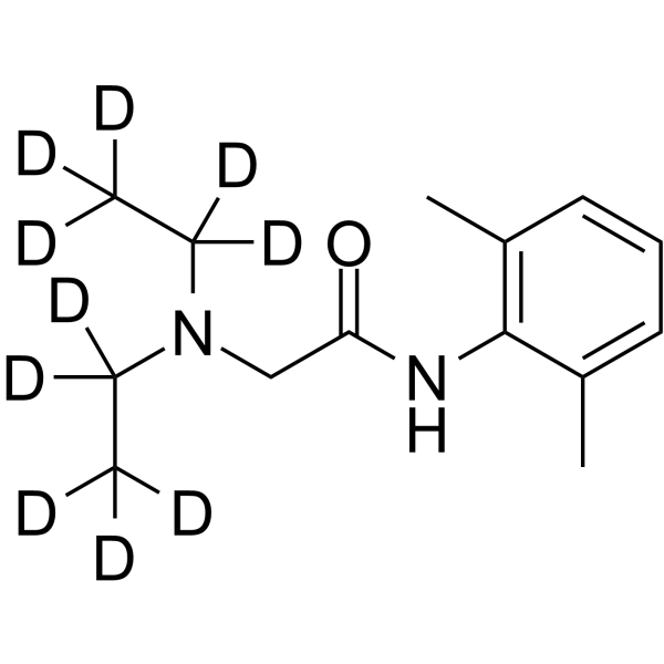
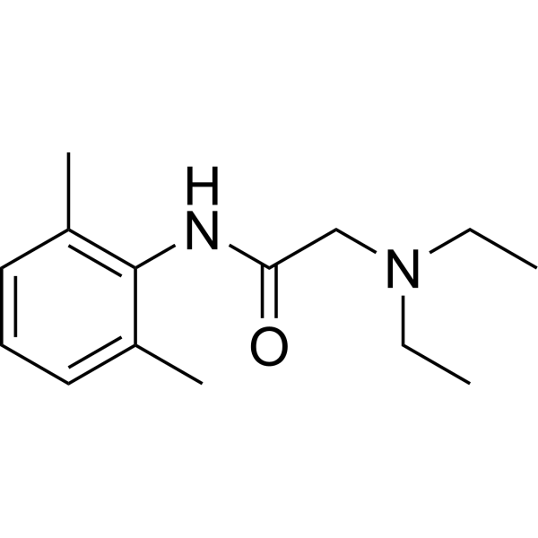
Lidocaine-d10 is the deuterium labeled Lidocaine. Lidocaine (Lignocaine) inhibits sodium channels involving complex voltage and using dependence. Lidocaine decreases growth, migration and invasion of gastric carcinoma cells via up-regulating miR-145 expression and further inactivation of MEK/ERK and NF-κB signaling pathways. Lidocaine is an amide derivative and has potential for the research of ventricular arrhythmia.
For research use only. We do not sell to patients.
- Purity : 98.89%
- CAS No.: 851528-09-1
- Formula: C14H12D10N2O
- Molecular Weight:244.40
-
Storage:Powder -20°C, 3 years , 4°C, 2 years ; In solvent -80°C, 6 months , -20°C, 1 month

To place orders, for customer services and technical support, please contact: MedChemExpress USA
Tel: 609-228-6898 E-mail: [email protected] [email protected]


-
 Biological Activity
-
Chemical Information
-
Solvent & Solubility
- Protocols
- Purity & Documentation
- References
-
 Help & FAQs


All MEK Isoforms
More

Biological Activity
Description
In Vitro
Stable heavy isotopes of hydrogen, carbon, and other elements have been incorporated into drug molecules, largely as tracers for quantitation during the drug development process. Deuteration has gained attention because of its potential to affect the pharmacokinetic and metabolic profiles of drugs[1].
MedChemExpress (MCE) has not independently confirmed the accuracy of these methods. They are for reference only. Further protocols information, click here.
Application
1. This compound can be used as a tracer
2. This compound can be used as an internal standard for quantitative analysis by NMR, GC-MS, or LC-MS.


Chemical Information
-
CAS No. 851528-09-1
-
Unlabeled CAS 73-78-9
-
Appearance Solid
-
Molecular Weight 244.40
-
Formula C14H12D10N2O
-
Color Off-white to light yellow
-
SMILES
[2H]C([2H])([2H])C([2H])([2H])N(C([2H])([2H])C([2H])([2H])[2H])CC(NC1=C(C=CC=C1C)C)=O
-
Shipping
Room temperature in continental US; may vary elsewhere.
-
Storage
Powder -20°C 3 years 4°C 2 years In solvent -80°C 6 months -20°C 1 month


Solvent & Solubility
In Vitro:
DMSO : 100 mg/mL (409.17 mM; Need ultrasonic and warming; Hygroscopic DMSO has a significant impact on the solubility of product, please use newly opened DMSO)
Please refer to the solubility information to select the appropriate solvent. Once prepared, please aliquot and store the solution to prevent product inactivation from repeated freeze-thaw cycles.
Storage method and period of stock solution: -80°C, 6 months; -20°C, 1 month. When stored at -80°C, please use it within 6 months. When stored at -20°C, please use it within 1 month.
Please refer to the solubility information to select the appropriate solvent. Once prepared, please aliquot and store the solution to prevent product inactivation from repeated freeze-thaw cycles.
Storage method and period of stock solution: -80°C, 6 months; -20°C, 1 month. When stored at -80°C, please use it within 6 months. When stored at -20°C, please use it within 1 month.
Concentration (start) × Volume (start) = Concentration (final) × Volume (final)

Protocols
-
RNA extraction experimental
By lysing cells, releasing RNA, and removing impurities such as proteins and DNA, high-purity RNA products are finally obtained. The commonly used traditional method is the guanidine isothiocyanate/phenol/chloroform method (Trizol), which is suitable for a variety of animal materials including animal tissues, microorganisms, cultured cells, etc., and most plant materials.
-
Cardiac voltage-sensitive optical mapping
Cardiac voltage-sensitive optical mapping records changes in transmembrane potential from cardiac tissue by staining the preparation with a voltage-sensitive dye and imaging fluorescence changes during electrical activation; the resulting optical action potentials can be used to map activation time, action potential duration, conduction velocity, wavefront propagation, and arrhythmia dynamics. The optical signal represents a relative fluorescence change from a tissue volume rather than a single-cell intracellular recording, so spatial resolution, sampling rate, voltage resolution, optical magnification, light penetration, and motion control must be considered together when interpreting optical action potentials.
-
Cell migration
Cell migration is a method that plays an important role in wound healing, cell differentiation, embryonic development, etc.
-
Cell invasion
Cell invasion is the ability of cells to migrate from one area to another via the extracellular matrix. Cell invasion is the response of normal and cancer cells to chemical and mechanical stimuli. Before migrating to a new region, the extracellular matrix is degraded by proteases within the cell. Cell invasion often occurs during wound repair, vascularization and inflammation, abnormal tissue invasion, and tumor cell metastasis.
-
Research Protocol for Inflammation-related Diseases
The NLRP3 inflammasome is a cytosolic innate immune signaling platform that integrates priming signals and danger-signal activation to promote caspase-1 activation, maturation of IL-1β and IL-18, and gasdermin D-mediated pyroptotic cell death. The core experimental logic is to determine whether inflammatory disease phenotypes are driven by increased NLRP3 expression, ASC-containing inflammasome assembly, caspase-1 cleavage, GSDMD cleavage, and extracellular release of IL-1β/IL-18 rather than by nonspecific cell injury alone. The pathway is strongly linked to inflammation-related disease phenotypes because monosodium urate crystals activate NALP3/NLRP3 inflammasome signaling in gout-like crystal inflammation, cholesterol crystals activate NLRP3 inflammasomes in atherogenesis models, and DSS-induced intestinal inflammation has been reported to involve NLRP3 inflammasome activity. However, experimental colitis studies also show context-dependent protective effects of NLRP3 inflammasome co


Purity & Documentation
-
Data Sheet (277 KB)
-
SDS (392 KB)
- English - EN (392 KB)
- Français - FR (392 KB)
- Deutsch - DE (392 KB)
- Norwegian - NO (392 KB)
- Español - ES (392 KB)
- Swedish - SV (392 KB)
- Italian - IT (392 KB)
- Korean - KR (392 KB)
- Portuguese - PT (392 KB)
-
Handling Instructions (2659 KB)
References
[1]. Russak EM, et al. Impact of Deuterium Substitution on the Pharmacokinetics of Pharmaceuticals. Ann Pharmacother. 2019;53(2):211-216. [Content Brief]
[2]. Cummins TR, et al. Setting up for the block: the mechanism underlying lidocaine's use-dependent inhibition of sodium channels. J Physiol. 2007 Jul 1;582(Pt 1):11. [Content Brief]
[3]. Sui H, et al. Lidocaine inhibits growth, migration and invasion of gastric carcinoma cells by up-regulation of miR-145. BMC Cancer. 2019 Mar 15;19(1):233. [Content Brief]
[4]. Li Z, et al. Evaluation of the antinociceptive effects of lidocaine and bupivacaine on the tail nerves of healthy rats. Basic Clin Pharmacol Toxicol. 2013 Jul;113(1):31-6. [Content Brief]


Complete Stock Solution Preparation Table
Please refer to the solubility information to select the appropriate solvent. Once prepared, please aliquot and store the solution to prevent product inactivation from repeated freeze-thaw cycles.
Storage method and period of stock solution: -80°C, 6 months; -20°C, 1 month. When stored at -80°C, please use it within 6 months. When stored at -20°C, please use it within 1 month.
| Optional Solvent | Concentration Solvent Mass | 1 mg | 5 mg | 10 mg | 25 mg |
|---|---|---|---|---|---|
| DMSO | 1 mM | 4.0917 mL | 20.4583 mL | 40.9165 mL | 102.2913 mL |
| 5 mM | 0.8183 mL | 4.0917 mL | 8.1833 mL | 20.4583 mL | |
| 10 mM | 0.4092 mL | 2.0458 mL | 4.0917 mL | 10.2291 mL | |
| 15 mM | 0.2728 mL | 1.3639 mL | 2.7278 mL | 6.8194 mL | |
| 20 mM | 0.2046 mL | 1.0229 mL | 2.0458 mL | 5.1146 mL | |
| 25 mM | 0.1637 mL | 0.8183 mL | 1.6367 mL | 4.0917 mL | |
| 30 mM | 0.1364 mL | 0.6819 mL | 1.3639 mL | 3.4097 mL | |
| 40 mM | 0.1023 mL | 0.5115 mL | 1.0229 mL | 2.5573 mL | |
| 50 mM | 0.0818 mL | 0.4092 mL | 0.8183 mL | 2.0458 mL | |
| 60 mM | 0.0682 mL | 0.3410 mL | 0.6819 mL | 1.7049 mL | |
| 80 mM | 0.0511 mL | 0.2557 mL | 0.5115 mL | 1.2786 mL | |
| 100 mM | 0.0409 mL | 0.2046 mL | 0.4092 mL | 1.0229 mL |


Lidocaine-d10 Related Classifications
HY-B0185S1 Related Classifications
 Powered by Bioz