



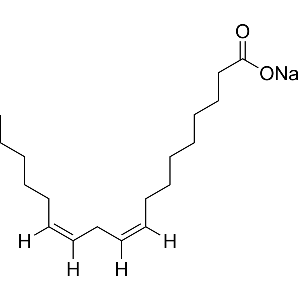
Linoleate sodium

Based on 1 Customer Validation
Linoleate sodium is an orally active IL8 regulator via the JNK and NF-κB pathway. Linoleate sodium can change the composition of fatty acids and the production of metabolites in cells. Linoleate sodium has anti-inflammatory, immune-regulating, and tumor cell growth-affecting activities.
For research use only. We do not sell to patients.
- Purity : 99.88%
- CAS No.: 822-17-3
- Formula: C18H31NaO2
- Molecular Weight:302.43
-
Storage:
-20°C, stored under nitrogen, away from moisture
* In solvent : -80°C, 6 months; -20°C, 1 month (stored under nitrogen, away from moisture)

To place orders, for customer services and technical support, please contact: MedChemExpress USA
Tel: 609-228-6898 E-mail: [email protected] [email protected]


-
 Biological Activity
-
Chemical Information
-
Solvent & Solubility
- Protocols
- Purity & Documentation
- References
-
 Help & FAQs


-
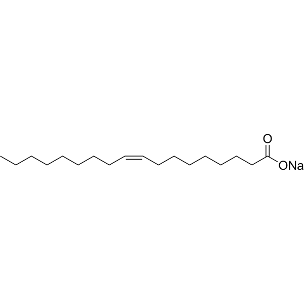
HY-N1446B
Oleic acid sodiumNa+/K+ ATPase Activator -
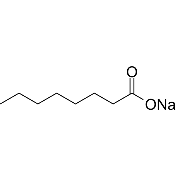
HY-W015339
Octanoate, 99% sodiumBiochemical Assay Reagent -
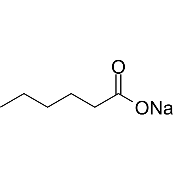
HY-W099696
N-Caproicacid sodiumBiochemical Assay Reagent -
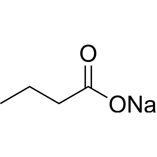
HY-B0350A
Sodium butyrateHDAC Inhibitor -
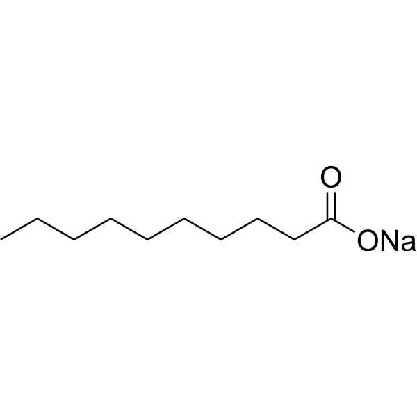
HY-115340
Decanoic acid sodiumBiochemical Assay Reagent -
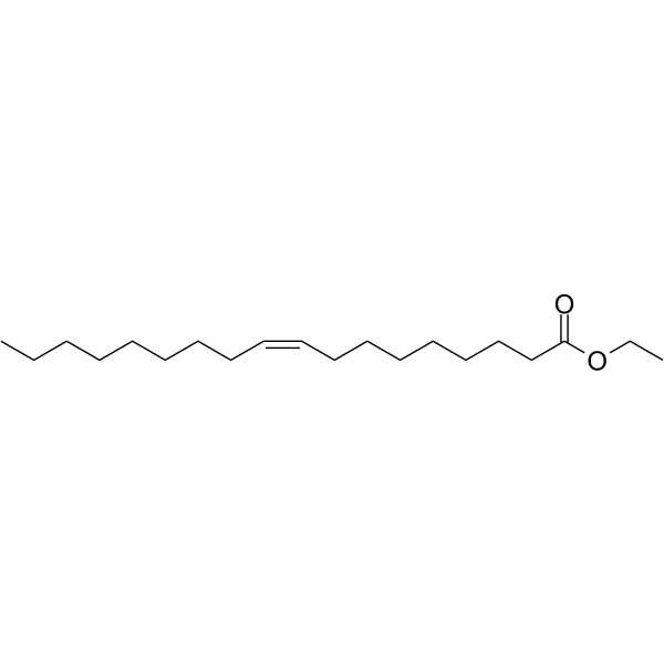
HY-N7103
Ethyl oleateFatty Acid Ester -
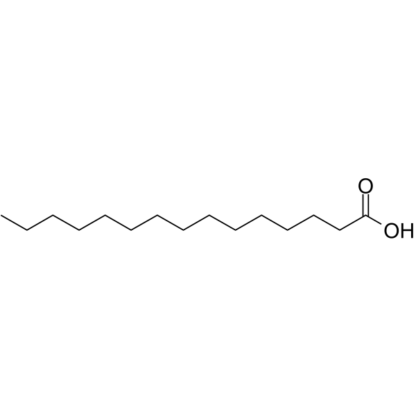
HY-W004283
Pentadecanoic acidSTAT3 Inhibitor -
HY-W004260
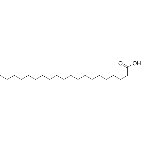
Arachidic acidReceptor/Ion Channel Effector -
HY-W004284
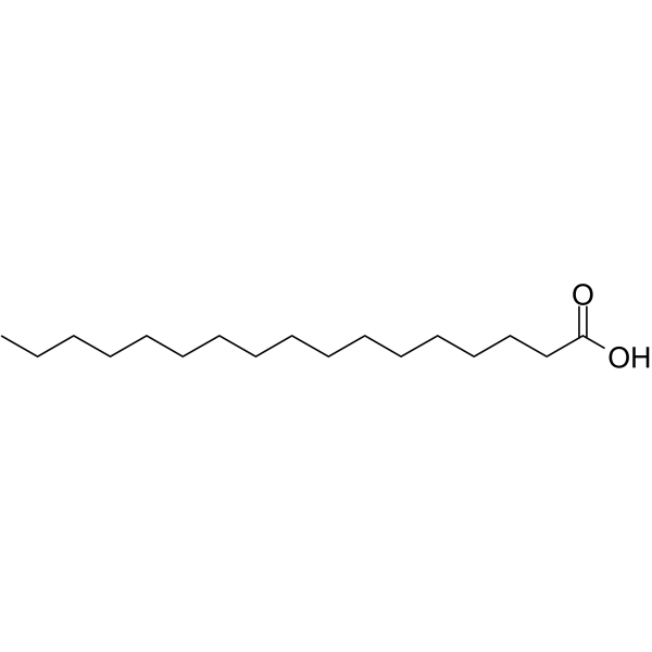
Heptadecanoic acidEndogenous Metabolite -

HY-B1773A
Sodium propionateShort-chain Fatty Acid -
HY-42935A
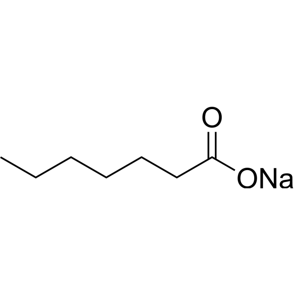
Heptanoate sodiumBiochemical Assay Reagent
All Parasite Isoforms
More

Biological Activity
Description
|
IL-8 |
In Vitro
Linoleate (0.125-1.0 mM, 24 h (for viability test)) sodium increases viability of hepatocytes and regulates TG accumulation in goose primary hepatocytes[1].
Linoleate (100-1000 μM, 9 h) sodium combined with palmitate (500 μM) significantly inhibits IL8 (mRNA and protein) production in Huh7 and HepG2 cells, below that of palmitate alone[2].
MedChemExpress (MCE) has not independently confirmed the accuracy of these methods. They are for reference only. Further protocols information, click here.
-
Cell Line:goose primary hepatocytes
-
Concentration:0.125, 0.25, 0.5, 1.0 mM
-
Incubation Time:24 h
-
Result:Increased PLIN mRNA and reduced PPARγ mRNA.
Increased mRNA expression of PPARα and CPT-1.
Increased FoxO1 mRNA expression.
Increased MTP mRNA.
Significantly elevated DGAT2 mRNA.
In Vivo
Linoleate (in the form of a diet containing 15% (w/w) safflower oil; p.o., through diet) sodium reduces the induction of fatty acid synthetase activity in rat liver[4].
Linoleate (in the form of linoleate-enriched safflower oil, 100 ppm, dietary) sodium increases arachidonic acid (20:4n-6) levels in the serum of mice with antigen-induced antibodies and allergic reactions[5].
Linoleate (in the form of linoleate-enriched safflower oil, corresponding to an 18:2n-6/18:3n-3 ratio of 127; dietary) sodium attenuates gastric mucosal damage induced by ethanol, ischemia/reperfusion, and water immersion stress in a rat experimental gastric ulcer model[6].
Linoleate sodium (1 mg/mouse; ip; 1 time) significantly increases the median survival of mice inoculated with EAT cells from 18 days to 48 days, and completely inhibited tumor growth in more than 40% of mice[7].
Linoleate (in the form of linoleate-rich safflower oil, 10% of the semi-purified diet; fed) sodium makes ICR mice more sensitive to pentobarbital, as manifested by a shorter onset of anesthetic effect and a longer duration of anesthetic[8].
Linoleate (in the form of linoleate-rich safflower oil corresponding to an 18:2n-6/18:3n-3 ratio of less than 0.1; fed) can reduce urine protein levels, plasma urea nitrogen levels, glomerular crescent formation, and fibrinoid necrosis in rats with crescent-type anti-glomerular basement membrane nephritis, while increasing the proportion of arachidonic acid (20:4n-6) in glomerular phospholipids[9].
MedChemExpress (MCE) has not independently confirmed the accuracy of these methods. They are for reference only.
-
Animal Model:Male lean SHHF rats; spontaneously hypertensive heart failure model[3]
-
Dosage:High-linoleate safflower oil (HLSO), 10% (w/w)
-
Administration:ad libitum for 4 weeks
-
Result:Preserved L4CL and total CL to 90% of non-failing levels.
Attenuated 17-22% decreases in state 3 mitochondrial respiration.
Increased left ventricular fractional shortening significantly, and decreased plasma insulin levels.
Augmented the production of several eicosanoid species in serum compared with the control and lard groups.


Chemical Information
-
CAS No. 822-17-3
-
Appearance Solid
-
Molecular Weight 302.43
-
Formula C18H31NaO2
-
Color White to light yellow
-
SMILES
CCCCC/C=C\C/C=C\CCCCCCCC(O[Na])=O
-
Shipping
Room temperature in continental US; may vary elsewhere.
-
Storage
-20°C, stored under nitrogen, away from moisture
* In solvent : -80°C, 6 months; -20°C, 1 month (stored under nitrogen, away from moisture)


Solvent & Solubility
In Vitro:
Ethanol : 4 mg/mL (13.23 mM; ultrasonic and warming and heat to 60°C)
Please refer to the solubility information to select the appropriate solvent. Once prepared, please aliquot and store the solution to prevent product inactivation from repeated freeze-thaw cycles.
Storage method and period of stock solution: -80°C, 6 months; -20°C, 1 month (stored under nitrogen, away from moisture). When stored at -80°C, please use it within 6 months. When stored at -20°C, please use it within 1 month.
Please refer to the solubility information to select the appropriate solvent. Once prepared, please aliquot and store the solution to prevent product inactivation from repeated freeze-thaw cycles.
Storage method and period of stock solution: -80°C, 6 months; -20°C, 1 month (stored under nitrogen, away from moisture). When stored at -80°C, please use it within 6 months. When stored at -20°C, please use it within 1 month.
Concentration (start) × Volume (start) = Concentration (final) × Volume (final)

Protocols
-
Protocol for Cell Counting and Cell Density Analysis
Cell counting and cell-density analysis estimate the number of cells in a known volume or field area. Manual hemocytometer counting uses a chamber of defined geometry to convert counted cells into cells/mL, while automated counters and image-analysis workflows detect cell objects from optical, brightfield, fluorescence, impedance, or digital-image features. Trypan blue viability counting is based on dye exclusion: viable cells with intact membranes exclude dye, while non-viable cells with compromised membranes stain blue. The readout is total cell density, viable-cell density, dead-cell density, and percent viability. Cell density can also be estimated from microscopy images by counting objects per image area, from flow cytometry using calibrated volume or reference particles, or from in situ microscopy in bioreactors after calibration against reference methods such as hemocytometer or flow cytometry.
-
Somatic Cell Culture
A method of simulating the in vivo environment in vitro to maintain the cell growth, differentation and main functions.
-
CCK-8/WST-8 Cell Proliferation Assay
The CCK-8/WST-8 assay is based on the reduction of the water-soluble tetrazolium salt WST-8 to a water-soluble formazan product by cellular dehydrogenases in metabolically active cells, where the generated formazan amount is proportional to the number of living cells and is quantified by measuring absorbance in the visible range, providing a colorimetric readout for cell viability and proliferation assessment. This class of tetrazolium-based assays improves upon earlier MTT-based systems by producing a water-soluble formazan, eliminating the need for organic solubilization steps and enabling direct spectrophotometric measurement in culture medium.
-
Research Protocol for Inflammation-related Diseases
The NLRP3 inflammasome is a cytosolic innate immune signaling platform that integrates priming signals and danger-signal activation to promote caspase-1 activation, maturation of IL-1β and IL-18, and gasdermin D-mediated pyroptotic cell death. The core experimental logic is to determine whether inflammatory disease phenotypes are driven by increased NLRP3 expression, ASC-containing inflammasome assembly, caspase-1 cleavage, GSDMD cleavage, and extracellular release of IL-1β/IL-18 rather than by nonspecific cell injury alone. The pathway is strongly linked to inflammation-related disease phenotypes because monosodium urate crystals activate NALP3/NLRP3 inflammasome signaling in gout-like crystal inflammation, cholesterol crystals activate NLRP3 inflammasomes in atherogenesis models, and DSS-induced intestinal inflammation has been reported to involve NLRP3 inflammasome activity. However, experimental colitis studies also show context-dependent protective effects of NLRP3 inflammasome co
-
Cell Counting-Based Growth Curve Assay
Cell counting-based growth curve assays quantify cell proliferation by directly measuring changes in viable cell number over time using manual or automated counting methods such as hemocytometer-based counting or instrument-assisted cell enumeration, enabling construction of growth curves that reflect population expansion dynamics in response to culture conditions. A widely used approach is trypan blue exclusion with hemocytometer counting, where membrane-compromised (non-viable) cells take up the dye, allowing discrimination between viable and non-viable cells while simultaneously enabling total cell number quantification. Repeated sampling across time points allows estimation of proliferation rate, growth phases, and comparative growth kinetics between experimental conditions.
-
MTT Cell Proliferation Assay
The MTT assay is a colorimetric endpoint assay for estimating viable cell number, cell growth, cytotoxicity, or cell activation in cultured mammalian cells. Living cells reduce the yellow tetrazolium salt MTT into purple/blue formazan, while dead cells do not generate the same signal; the resulting color can be quantified with a multiwell spectrophotometer. MTT reduction is commonly interpreted as a readout of metabolic activity that often correlates with viable cell number, but it should not be treated as a direct cell-counting method unless the assay is optimized for the cell type and experimental condition. Studies show that MTT reduction can involve mitochondrial and non-mitochondrial reducing systems, and formazan may accumulate in intracellular lipid droplets rather than simply marking mitochondria.
-
Cell Viability Determination by MTT Colorimetric Assay
The following protocol uses the MTT colorimetric assay as a classic literature-established method for assessing cell viability/metabolic activity in cultured mammalian cells. MTT[3-(4,5-dimethylthiazol-2-yl)-2,5-diphenyltetrazolium bromide] is reduced by metabolically active cells to a colored formazan product; the amount of formazan is quantified spectrophotometrically and provides an indirect measure of metabolically active viable cells. Importantly, MTT reduction reflects cellular oxidoreductase/metabolic activity rather than an absolute direct count of living cells, so changes in cellular metabolism can alter the signal independently of cell number.


Purity & Documentation
-
Data Sheet (273 KB)
-
SDS (393 KB)
- English - EN (393 KB)
- Français - FR (393 KB)
- Deutsch - DE (393 KB)
- Norwegian - NO (393 KB)
- Español - ES (393 KB)
- Swedish - SV (393 KB)
- Italian - IT (393 KB)
- Korean - KR (393 KB)
- Portuguese - PT (393 KB)
-
Handling Instructions (2659 KB)
References
[1]. Pan Z, et al. Effects of linoleate on cell viability and lipid metabolic homeostasis in goose primary hepatocytes. Comp Biochem Physiol A Mol Integr Physiol. 2011 Jun;159(2):113-8. [Content Brief]
[2]. Maruyama H, et al. Linoleate appears to protect against palmitate-induced inflammation in Huh7 cells. Lipids Health Dis. 2014 May 13;13:78. [Content Brief]
[3]. Mulligan CM, et al. Dietary linoleate preserves cardiolipin and attenuates mitochondrial dysfunction in the failing rat heart. Cardiovasc Res. 2012 Jun 1;94(3):460-8. [Content Brief]
[4]. Flick PK, et al. Effect of dietary linoleate on synthesis and degradation of fatty acid synthetase from rat liver. J Biol Chem. 1977 Jun 25;252(12):4242-9. [Content Brief]
[5]. Oh-hashi K, et al. Reevaluation of the effect of a high alpha-linolenate and a high linoleate diet on antigen-induced antibody and anaphylactic responses in mice. Biol Pharm Bull. 1997 Mar;20(3):217-23. [Content Brief]
[6]. Matsuba S, et al. Effect of dietary linoleate/alpha-linolenate balance on experimentally induced gastric injury in rats. Prostaglandins Leukot Essent Fatty Acids. 1998 Nov;59(5):317-23. [Content Brief]
[7]. Norman A, et al. Antitumor activity of sodium linoleate. Nutr Cancer. 1988;11(2):107-15. [Content Brief]
[8]. Nakashima Y, et al. Effect of a high linoleate and a high alpha-linolenate diet on general behavior and drug sensitivity in mice. J Lipid Res. 1993 Feb;34(2):239-47. [Content Brief]
[9]. Watanabe S, et al. Effect of dietary alpha-linolenate/linoleate balance on crescent type-anti-glomerular basement membrane nephritis in rats. Lipids. 1990 May;25(5):267-72. [Content Brief]


Complete Stock Solution Preparation Table
Please refer to the solubility information to select the appropriate solvent. Once prepared, please aliquot and store the solution to prevent product inactivation from repeated freeze-thaw cycles.
Storage method and period of stock solution: -80°C, 6 months; -20°C, 1 month (stored under nitrogen, away from moisture). When stored at -80°C, please use it within 6 months. When stored at -20°C, please use it within 1 month.
| Optional Solvent | Concentration Solvent Mass | 1 mg | 5 mg | 10 mg | 25 mg |
|---|---|---|---|---|---|
| Ethanol | 1 mM | 3.3066 mL | 16.5328 mL | 33.0655 mL | 82.6638 mL |
| 5 mM | 0.6613 mL | 3.3066 mL | 6.6131 mL | 16.5328 mL | |
| 10 mM | 0.3307 mL | 1.6533 mL | 3.3066 mL | 8.2664 mL |


Linoleate sodium Related Classifications
HY-W011398 Related Classifications
 Powered by Bioz