



Malvidin-3-glucoside chloride

Based on 1 Customer Validation
Malvidin-3-glucoside (Malvidin-3-O-glucoside; Oenin) chloride is an orally active inhibitor of the NF-κB pathway, which blocks inflammatory responses induced by TNF-α, reduces IκB-α degradation and p65 nuclear translocation, and upregulates endothelial nitric oxide synthase eNOS to increase NO production. Malvidin-3-glucoside chloride exerts anti-inflammatory and antioxidant effects by inhibiting pro-inflammatory molecules such as MCP-1, ICAM-1, and IL-6, and regulating intestinal microorganisms and metabolites, while protecting endothelial cells and improving intestinal microecological dysbiosis under inflammatory conditions. Malvidin-3-glucoside chloride can be used to study chronic inflammatory-related diseases such as atherosclerosis and inflammatory bowel disease, and has the potential to prevent vascular inflammation and improve intestinal health.
For research use only. We do not sell to patients.
- Purity : 99.87%
- CAS No.: 7228-78-6
- Formula: C23H25ClO12
- Molecular Weight:528.89
-
Storage:
4°C, sealed storage, away from moisture and light
* In solvent : -80°C, 6 months; -20°C, 1 month (sealed storage, away from moisture and light)

To place orders, for customer services and technical support, please contact: MedChemExpress USA
Tel: 609-228-6898 E-mail: [email protected] [email protected]


-
 Biological Activity
-
Chemical Information
- Protocols
- Purity & Documentation
- References
-
 Help & FAQs


-
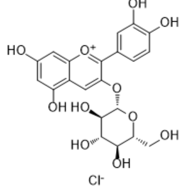
HY-N0640
Kuromanin chlorideBlood glucose regulator -
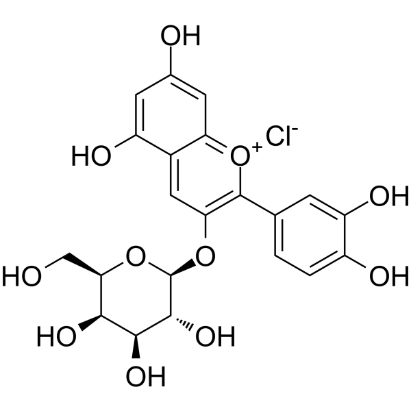
HY-N4142
Cyanidin-3-O-galactoside chlorideAntioxidant Agent -
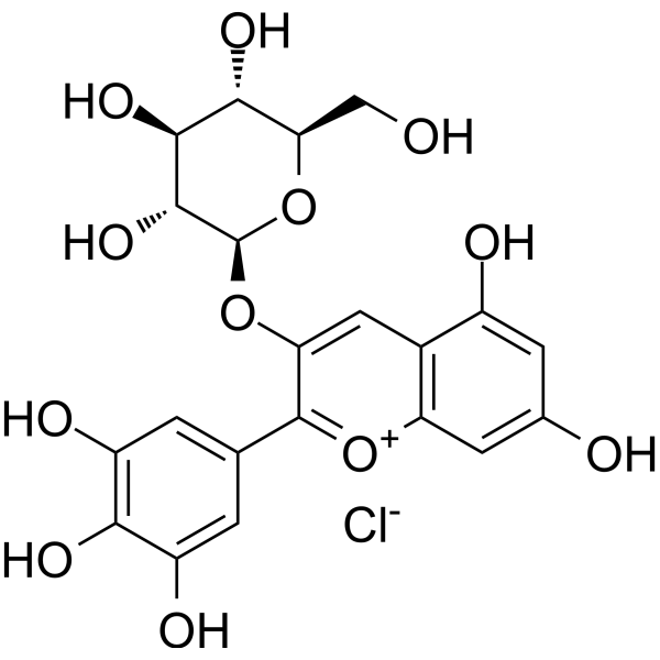
HY-108052
Delphinidin 3-glucoside chlorideEGFR Inhibitor -
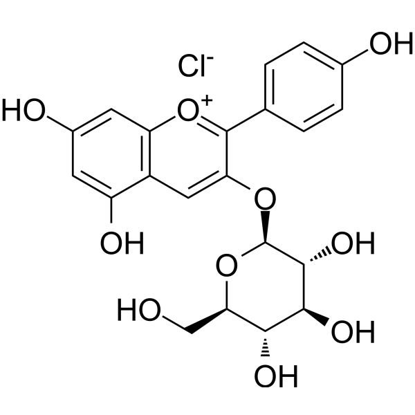
HY-W040045
Callistephin chlorideROS/iNOS/TNF-α/COX-2 Inhibitor -
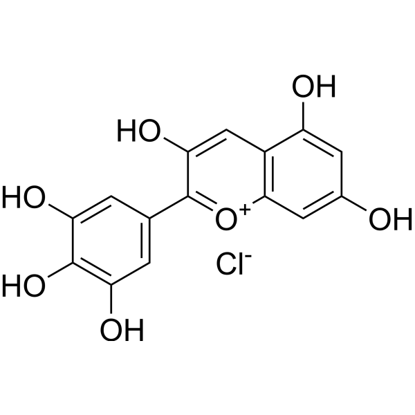
HY-N2409
Delphinidin chlorideAnthocyanidin -
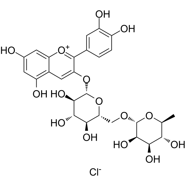
HY-105935
Keracyanin chlorideAntioxidant Agent -
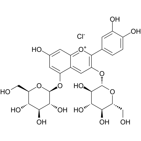
HY-129138
Cyanidin 3,5-diglucoside chlorideAnthocyanin -
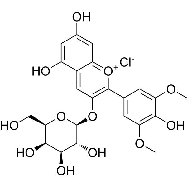
HY-N6623
Malvidin-3-galactoside chlorideROS Inhibitor -
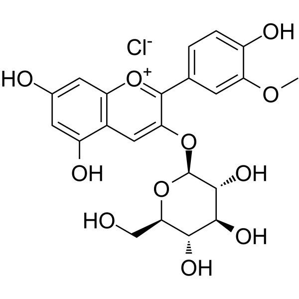
HY-W040127
Peonidin 3-O-glucoside chlorideInsulin Secretagogue -
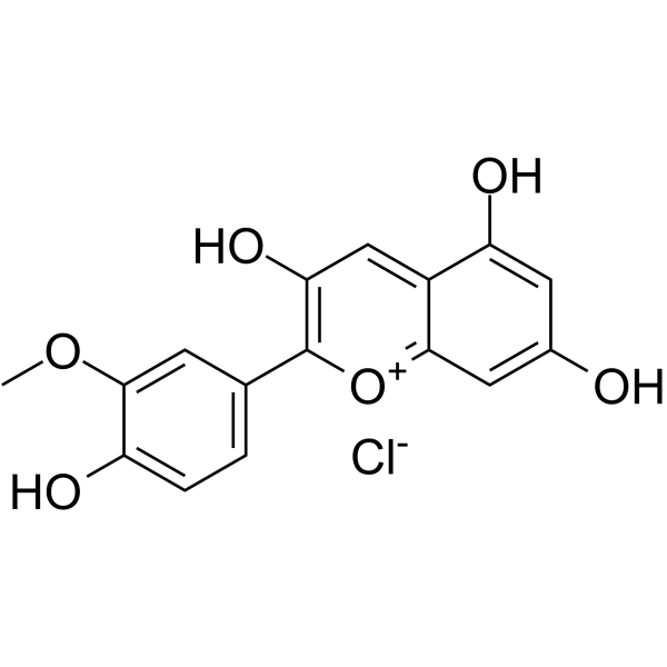
HY-N2459
Peonidin chlorideAnthocyanin Compound -

HY-125740R
Malvidin-3-glucoside chloride (Standard)NF-κB Inhibitor -
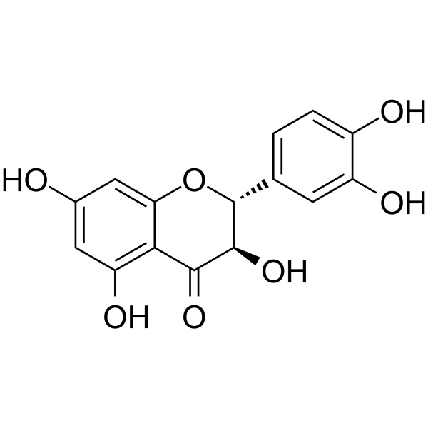
HY-N0136
TaxifolinAntifibrotic Agent


Biological Activity
Description
IC50 & Target
|
IL-6 |
eNOS |
In Vitro
Malvidin-3-glucoside (1, 10, 50, 100 μM; pretreatment for 18 h + TNF-α stimulation for 6 h) inhibits TNF-α-induced MCP-1, ICAM-1, VCAM-1 protein and mRNA expressions in human umbilical vein endothelial cells (HUVECs) in a concentration-dependent manner, reduces IκB-α degradation and inhibits NF-κB p65 subunit nuclear translocation[1].
Malvidin-3-glucoside (25 μM; pretreatment 14 h+ ONOO- aggression) upregulates eNOS mRNA and activity in bovine aortic endothelial cells (BAECs), increases NO production, while inhibiting peroxynitrite-induced iNOS, COX-2 expression and IL-6 production, and blocking NF-κB activation[2].
MedChemExpress (MCE) has not independently confirmed the accuracy of these methods. They are for reference only. Further protocols information, click here.
-
Cell Line:Human umbilical vein endothelial cells (HUVECs)
-
Concentration:1 μM, 10 μM, 50 μM, 100 μM
-
Incubation Time:18 h pretreatment with malvidin-3-glucoside, followed by 6 h stimulation with TNF-α
-
Result:Significantly reduced the protein levels of MCP-1, ICAM-1, and VCAM-1 in cell supernatants in a concentration-dependent manner.
Resulted 35.9% MCP-1, 54.4% ICAM-1, and 44.7% VCAM-1 protein increases inhibition at 1 μM; and suppressed over 90% mRNA expressions of MCP-1 and ICAM-1 as well, with 100 μM.
-
Cell Line:Human umbilical vein endothelial cells (HUVECs)
-
Concentration:1 μM, 10 μM, 50 μM, 100 μM
-
Incubation Time:18 h pretreatment with malvidin-3-glucoside, followed by 6 h stimulation with TNF-α
-
Result:Decreased IκB-α degradation (inhibition rates: 84.8% at 50 μM).
-
Cell Line:Human umbilical vein endothelial cells (HUVECs)
-
Concentration:1 μM, 10 μM, 50 μM, 100 μM
-
Incubation Time:18 h pretreatment with malvidin-3-glucoside, followed by 6 h stimulation with TNF-α
-
Result:Reduced nuclear translocation of p65 in immunofluorescence (IF) assay.
In Vivo
MedChemExpress (MCE) has not independently confirmed the accuracy of these methods. They are for reference only.
-
Animal Model:Male C57BL/6J mice (4-5 weeks old), Dextran sulfate sodium (HY-116282C)-induced colitis model[3]Dosage:24 mg/kg Malvidin-3-glucoside in dietAdministration:Dietary supplementation ad libitum, daily for 50 days (including 8 days pre-Dextran sulfate sodium (DSS) induction and two DSS cycles)Result:Significantly reduced histopathological scores by improving crypt dilation, ulceration, and smooth muscle thickness in colonic tissues.
Increased IL-10 mRNA expression in colon mucosa.
Decreased microbial evenness but enhanced microbial interactions, with Firmicutes/Bacteroidetes ratio restored from 0.38 (DSS group) to 0.56.


Chemical Information
-
CAS No. 7228-78-6
-
Appearance Solid
-
Molecular Weight 528.89
-
Formula C23H25ClO12
-
Color Brown to black
-
SMILES
OC1=CC2=C(C=C(C(C3=CC(OC)=C(C(OC)=C3)O)=[O+]2)O[C@H]4[C@@H]([C@H]([C@@H]([C@H](O4)CO)O)O)O)C(O)=C1.[Cl-]
-
Synonyms
Malvidin-3-O-glucoside chloride; Oenin chloride
-
Structure Classification
-
Initial Source
-
Shipping
Room temperature in continental US; may vary elsewhere.
-
Storage
4°C, sealed storage, away from moisture and light
* In solvent : -80°C, 6 months; -20°C, 1 month (sealed storage, away from moisture and light)

Protocols
-
Cytoplasmic-Nuclear Fractionated Protein Extraction
Cytoplasmic-nuclear fractionated protein extraction separates soluble cytoplasmic proteins from nuclear-enriched proteins by mild plasma-membrane permeabilization, differential centrifugation, washing of nuclei, and extraction of nuclear proteins for downstream immunoblotting or related molecular analysis. The readout is the relative abundance of a protein in cytoplasmic and nuclear fractions, commonly assessed by western blotting together with compartment markers such as tubulin or pyruvate kinase for cytoplasm and lamin, nucleoporin, hnRNP, H2AX, or Lamin B for nuclear fractions.
-
DSS-Induced Colitis
Dextran sulfate sodium (DSS)-induced colitis is generated by administering DSS in mouse drinking water, producing epithelial injury, barrier disruption, weight loss, diarrhea, fecal blood, colon shortening, histologic mucosal damage, and inflammatory mediator changes; the model is mainly used to study acute or chronic intestinal inflammation resembling selected features of ulcerative colitis. DSS injury is interpreted through clinical and tissue readouts rather than a single molecular endpoint: daily body weight, stool consistency, and bleeding are combined into a disease activity index, while colon length, histology, cytokines, myeloperoxidase activity, intestinal permeability, and tight-junction markers provide complementary measures of inflammation and barrier damage.
-
TNBS-Induced Colitis
TNBS-induced colitis is produced by intrarectal delivery of 2,4,6-trinitrobenzene sulfonic acid in ethanol, where ethanol disrupts the mucosal barrier and TNBS haptenates colonic proteins, generating immune-mediated colonic inflammation with weight loss, diarrhea, ulceration, transmural injury, inflammatory-cell infiltration, and cytokine responses. The model is used as an experimental intestinal inflammation model with Crohn’s disease–like features, especially when Th1-type responses, IL-12–dependent inflammation, chronic relapsing inflammation, or fibrosis-related endpoints are studied.
-
Endothelial Tube Formation Assay
Endothelial tube formation assay evaluates the ability of endothelial cells to attach, migrate, align, and organize into capillary-like networks when cultured on gelled basement membrane extract or Matrigel; the readout is the morphology and quantity of tube-like networks, which reflects an in vitro endothelial morphogenesis step related to angiogenesis. Basement membrane extract/Matrigel provides laminin-rich extracellular matrix cues that support endothelial differentiation into capillary-like structures, but it can contain biologically active growth factors, so growth-factor-reduced matrix is preferred when testing defined angiogenic stimulators or inhibitors.
-
Research Protocol for Cardiovascular Diseases
Cardiovascular disease can be modeled as maladaptive cardiac remodeling, where ischemic injury or pressure overload activates inflammatory signaling, fibroblast activation, extracellular-matrix deposition, cardiomyocyte hypertrophy, vascular remodeling, and progressive ventricular dysfunction. The TGF-β/SMAD axis is a central profibrotic pathway after myocardial injury and pressure overload, while innate immune and cytokine pathways regulate leukocyte recruitment, scar formation, and adverse remodeling. Key unresolved questions include which inflammatory signals are reparative versus harmful, when fibrosis is protective versus maladaptive, and whether pathway inhibition improves function without weakening necessary infarct healing or compensatory remodeling.
-
Research Protocol for Inflammation-related Diseases
The NLRP3 inflammasome is a cytosolic innate immune signaling platform that integrates priming signals and danger-signal activation to promote caspase-1 activation, maturation of IL-1β and IL-18, and gasdermin D-mediated pyroptotic cell death. The core experimental logic is to determine whether inflammatory disease phenotypes are driven by increased NLRP3 expression, ASC-containing inflammasome assembly, caspase-1 cleavage, GSDMD cleavage, and extracellular release of IL-1β/IL-18 rather than by nonspecific cell injury alone. The pathway is strongly linked to inflammation-related disease phenotypes because monosodium urate crystals activate NALP3/NLRP3 inflammasome signaling in gout-like crystal inflammation, cholesterol crystals activate NLRP3 inflammasomes in atherogenesis models, and DSS-induced intestinal inflammation has been reported to involve NLRP3 inflammasome activity. However, experimental colitis studies also show context-dependent protective effects of NLRP3 inflammasome co


Purity & Documentation
-
Data Sheet (287 KB)
-
SDS (393 KB)
- English - EN (393 KB)
- Français - FR (393 KB)
- Deutsch - DE (393 KB)
- Norwegian - NO (393 KB)
- Español - ES (393 KB)
- Swedish - SV (393 KB)
- Italian - IT (393 KB)
- Korean - KR (393 KB)
- Portuguese - PT (393 KB)
-
Handling Instructions (2659 KB)
References
[1]. Huang WY, et al. Anti-inflammatory effect of the blueberry anthocyanins malvidin-3-glucoside and malvidin-3-galactoside in endothelial cells. Molecules. 2014 Aug 21;19(8):12827-41. [Content Brief]
[2]. Paixão J, et al. Malvidin-3-glucoside protects endothelial cells up-regulating endothelial NO synthase and inhibiting peroxynitrite-induced NF-kB activation. Chem Biol Interact. 2012 Sep 30;199(3):192-200. [Content Brief]
[3]. Liu F, et al. Malvidin 3-Glucoside Modulated Gut Microbial Dysbiosis and Global Metabolome Disrupted in a Murine Colitis Model Induced by Dextran Sulfate Sodium. Mol Nutr Food Res. 2019 Nov;63(21):e1900455. [Content Brief]

Calculators
Concentration (start) × Volume (start) = Concentration (final) × Volume (final)


Malvidin-3-glucoside chloride Related Classifications
HY-125740 Related Classifications
 Powered by Bioz
Keywords