



Nonactin

Based on 1 Customer Validation
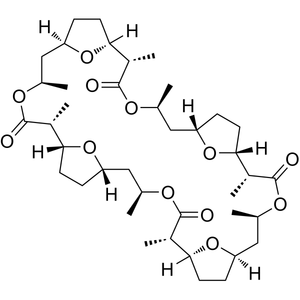
Nonactin is a macrotetrolide antibiotic and mitochondrial uncoupler with antibacterial, insecticidal, and acaricidal activities. Nonactin acts as an ionophore for monovalent cations, including K+, and NH4+, and it can also inhibit the surface expression of endogenous HSP60. In addition, Nonactin can induce apoptosis in β-catenin mutant tumor cells and has anti-tumor activity.
For research use only. We do not sell to patients.
- Purity : 99.0%
- CAS No.: 6833-84-7
- Formula: C40H64O12
- Molecular Weight:736.93
-
Storage:Powder -20°C, 3 years ; In solvent -80°C, 6 months , -20°C, 1 month

To place orders, for customer services and technical support, please contact: MedChemExpress USA
Tel: 609-228-6898 E-mail: [email protected] [email protected]


-
 Biological Activity
-
Chemical Information
-
Solvent & Solubility
- Protocols
- Purity & Documentation
- References
-
 Help & FAQs


-
HY-N6693
ValinomycinAntibiotic -
HY-B0401
TolbutamideKATP Inhibitor -

HY-B0401R
Tolbutamide (Standard)KATP Inhibitor -
HY-15209
RepaglinideInsulin Secretagogue -
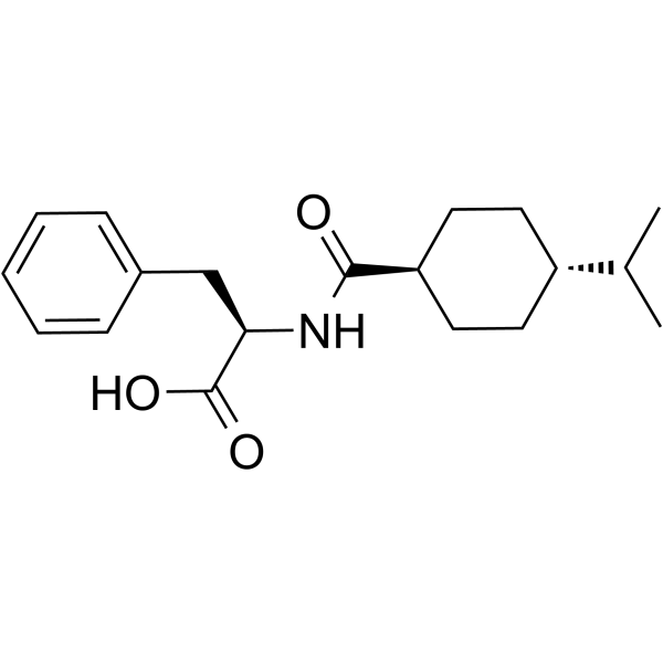
HY-B0422
NateglinideK+ Channel Inhibitor -
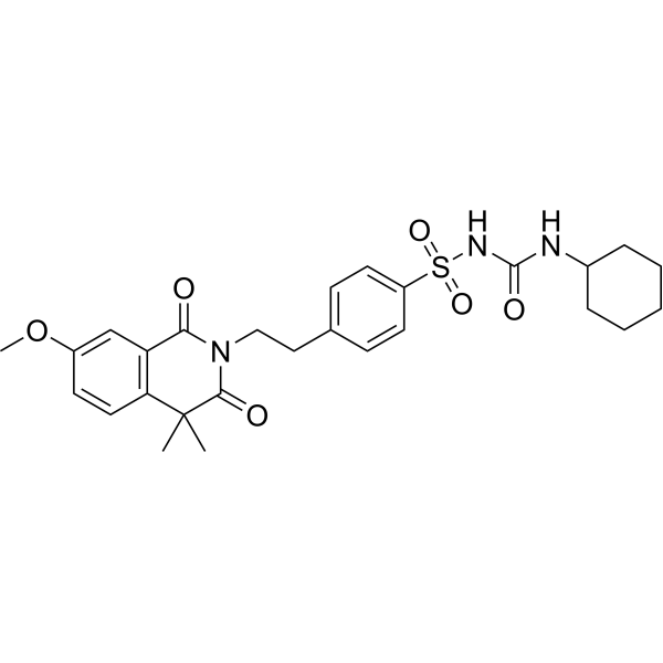
HY-B1114
GliquidoneAnti-Diabetic Drug -
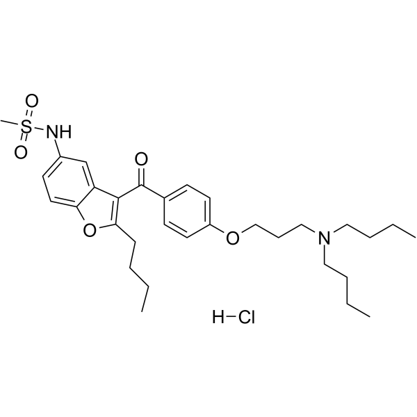
HY-75839
Dronedarone HydrochloridePotassium Channel Inhibitor -
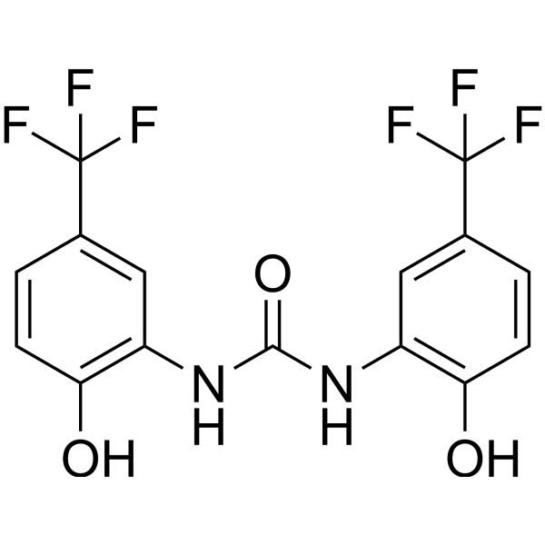
HY-16916
NS1643hERG Channels Agonist -
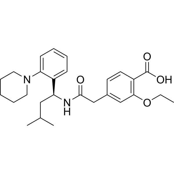
HY-15209R
Repaglinide (Standard)Insulin Secretagogue -
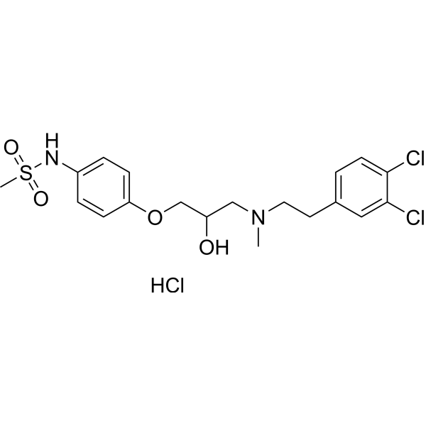
HY-101253
AM-92016 hydrochloridePotassium Channel Blocker -
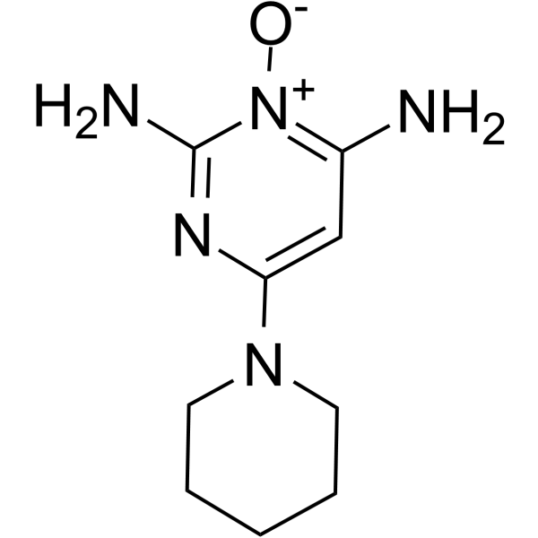
HY-B0112
MinoxidilEndogenous Metabolite
All Antibiotic Isoforms
More

Biological Activity
Description
In Vitro
Nonactin (0.01-10 μM; 24 h) induces apoptosis in β-catenin-mutated HCT 116 cells, and the mechanism is independent of ERK[2].
MedChemExpress (MCE) has not independently confirmed the accuracy of these methods. They are for reference only.
-
Cell Line:β-catenin mutant HCT 116 cells
-
Concentration:0.01, 0.1, 1 and 10 μM
-
Incubation Time:24 h
-
Result:Significantly increased the expression levels of cleaved-PARP.
Did not inhibit ERK1/2 phosphorylation.
In Vivo


Chemical Information
-
CAS No. 6833-84-7
-
Appearance Solid
-
Molecular Weight 736.93
-
Formula C40H64O12
-
Color White to off-white
-
SMILES
O=C(O[C@H](C)C[C@@]1([H])CC[C@@](O1)([H])[C@H](C)C(O[C@@H](C)C[C@]2([H])CC[C@](O2)([H])[C@@H](C)C(O[C@H](C)C[C@@]3([H])CC[C@@](O3)([H])[C@@H]4C)=O)=O)[C@H](C)[C@@](O5)([H])CC[C@@]5([H])C[C@H](C)OC4=O
-
Synonyms
Ammonium ionophore I
-
Structure Classification
-
Initial Source
Streptomyces griseus ETH A7796
-
Shipping
Room temperature in continental US; may vary elsewhere.
-
Storage
Powder -20°C 3 years In solvent -80°C 6 months -20°C 1 month


Solvent & Solubility
In Vitro:
DMF : 50 mg/mL (67.85 mM; ultrasonic and warming and heat to 60°C)
DMSO : 6.67 mg/mL (9.05 mM; Need ultrasonic; Hygroscopic DMSO has a significant impact on the solubility of product, please use newly opened DMSO)
Please refer to the solubility information to select the appropriate solvent. Once prepared, please aliquot and store the solution to prevent product inactivation from repeated freeze-thaw cycles.
Storage method and period of stock solution: -80°C, 6 months; -20°C, 1 month. When stored at -80°C, please use it within 6 months. When stored at -20°C, please use it within 1 month.
Please refer to the solubility information to select the appropriate solvent. Once prepared, please aliquot and store the solution to prevent product inactivation from repeated freeze-thaw cycles.
Storage method and period of stock solution: -80°C, 6 months; -20°C, 1 month. When stored at -80°C, please use it within 6 months. When stored at -20°C, please use it within 1 month.
Concentration (start) × Volume (start) = Concentration (final) × Volume (final)


Purity & Documentation
-
Data Sheet (283 KB)
-
SDS (394 KB)
- English - EN (394 KB)
- Français - FR (394 KB)
- Deutsch - DE (394 KB)
- Norwegian - NO (394 KB)
- Español - ES (394 KB)
- Swedish - SV (394 KB)
- Italian - IT (394 KB)
- Korean - KR (394 KB)
- Portuguese - PT (394 KB)
-
Handling Instructions (2659 KB)
References
[1]. Martínez-Haya B, et al. On the ionophoric selectivity of nonactin and related macrotetrolide derivatives. Phys Chem Chem Phys. 2017 Jan 4;19(2):1288-1297. [Content Brief]
[2]. Shikata Y, et al. Mitochondrial uncoupler exerts a synthetic lethal effect against β-catenin mutant tumor cells. Cancer Sci. 2017 Apr;108(4):772-784. [Content Brief]
[3]. Barazi HO, et al. Identification of heat shock protein 60 as a molecular mediator of alpha 3 beta 1 integrin activation. Cancer Res. 2002 Mar 1;62(5):1541-8. [Content Brief]
[4]. Boykova I, et al. Strain Streptomyces sp. P-56 Produces Nonactin and Possesses Insecticidal, Acaricidal, Antimicrobial and Plant Growth-Promoting Traits. Microorganisms. 2023 Mar 16;11(3):764. [Content Brief]


Complete Stock Solution Preparation Table
Please refer to the solubility information to select the appropriate solvent. Once prepared, please aliquot and store the solution to prevent product inactivation from repeated freeze-thaw cycles.
Storage method and period of stock solution: -80°C, 6 months; -20°C, 1 month. When stored at -80°C, please use it within 6 months. When stored at -20°C, please use it within 1 month.
| Optional Solvent | Concentration Solvent Mass | 1 mg | 5 mg | 10 mg | 25 mg |
|---|---|---|---|---|---|
| DMSO / DMF | 1 mM | 1.3570 mL | 6.7849 mL | 13.5698 mL | 33.9245 mL |
| 5 mM | 0.2714 mL | 1.3570 mL | 2.7140 mL | 6.7849 mL | |
| DMF | 10 mM | 0.1357 mL | 0.6785 mL | 1.3570 mL | 3.3925 mL |
| 15 mM | 0.0905 mL | 0.4523 mL | 0.9047 mL | 2.2616 mL | |
| 20 mM | 0.0678 mL | 0.3392 mL | 0.6785 mL | 1.6962 mL | |
| 25 mM | 0.0543 mL | 0.2714 mL | 0.5428 mL | 1.3570 mL | |
| 30 mM | 0.0452 mL | 0.2262 mL | 0.4523 mL | 1.1308 mL | |
| 40 mM | 0.0339 mL | 0.1696 mL | 0.3392 mL | 0.8481 mL | |
| 50 mM | 0.0271 mL | 0.1357 mL | 0.2714 mL | 0.6785 mL | |
| 60 mM | 0.0226 mL | 0.1131 mL | 0.2262 mL | 0.5654 mL |


Nonactin Related Classifications
HY-N6790 Related Classifications
 Powered by Bioz