



Prostaglandin A1

Based on 1 Customer Validation
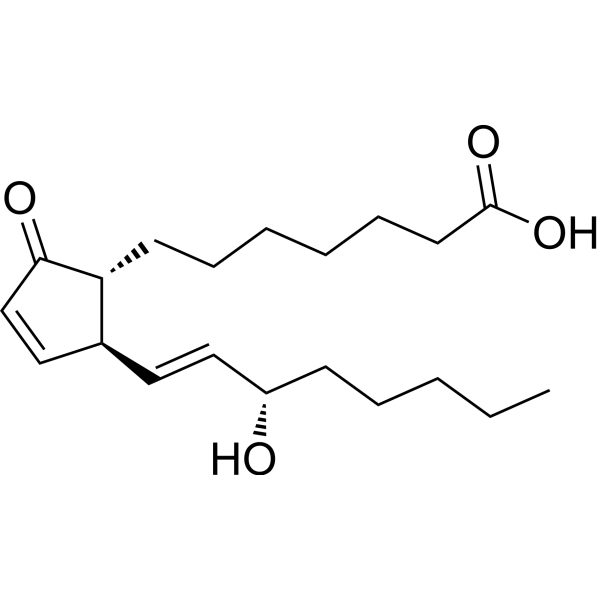
Prostaglandin A1 is a dehydrated derivative of Prostaglandin E1. Prostaglandin A1 exhibits antiviral, anti-inflammatory, antitumor, neuroprotective, and antihypertensive activities. Prostaglandin A1 covalently binds to cysteine residues of target proteins through Michael addition, or activates pathways such as PPARγ and HSPs via transcriptional regulation. Prostaglandin A1 is used in research on Mayaro virus infection, stroke, Alzheimer's disease, neuroblastoma, melanoma, and severe gestational hypertension.
For research use only. We do not sell to patients.
- Purity : 99.40%
- CAS No.: 14152-28-4
- Formula: C20H32O4
- Molecular Weight:336.47
-
Storage:
-20°C, sealed storage, away from moisture
* In solvent : -80°C, 6 months; -20°C, 1 month (sealed storage, away from moisture)

To place orders, for customer services and technical support, please contact: MedChemExpress USA
Tel: 609-228-6898 E-mail: [email protected] [email protected]


-
 Biological Activity
-
Chemical Information
- Protocols
- Purity & Documentation
- References
-
 Help & FAQs


-
HY-113369
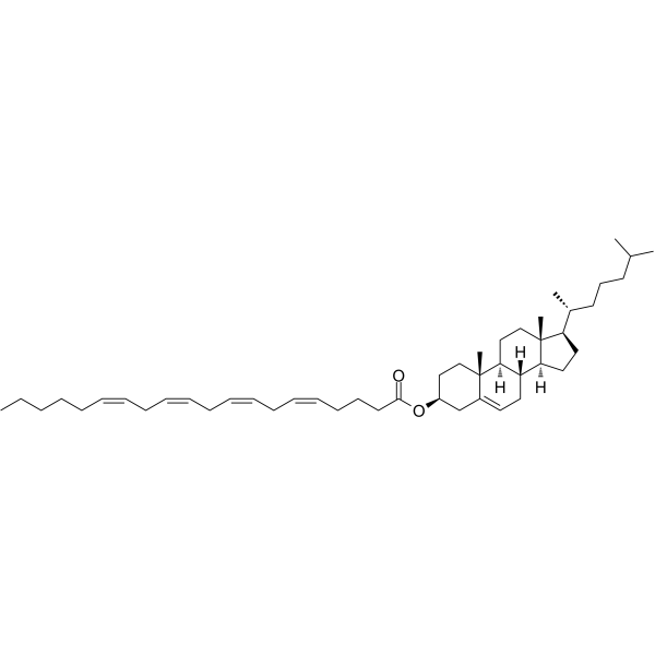
Cholesteryl arachidonateEndogenous Metabolite -
HY-B2130A
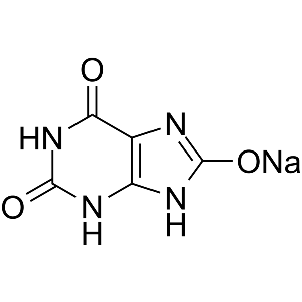
Uric acid sodiumOxygen Radical Scavenger -
HY-B0813
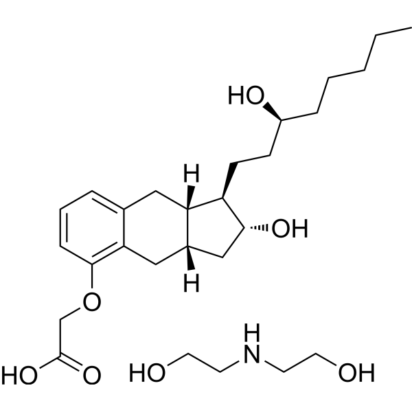
Treprostinil diethanolamineEP2/DP1/IP Agonist -
HY-113042
Prostaglandin B2Prostaglandin -
HY-118827
VedaprofenAnti-inflammatory Agent -
HY-139414
LysophosphatidylcholinesHemolytic Agent -
HY-P1540
Angiotensin III, human, mouseAT2R Agonist -
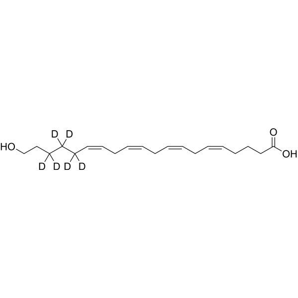
HY-15598S
20-HETE-d6Stable Isotope -

HY-B2130AR
Uric acid sodium (Standard)Reference Standard -
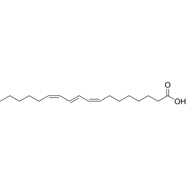
HY-121619
Jacaric acidAnticancer Agent -
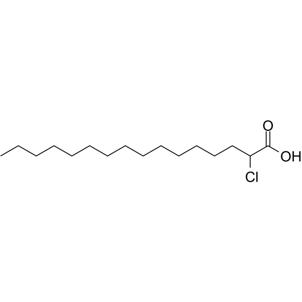
HY-131688
2-Chlorohexadecanoic acidInflammatory Lipid Mediator -
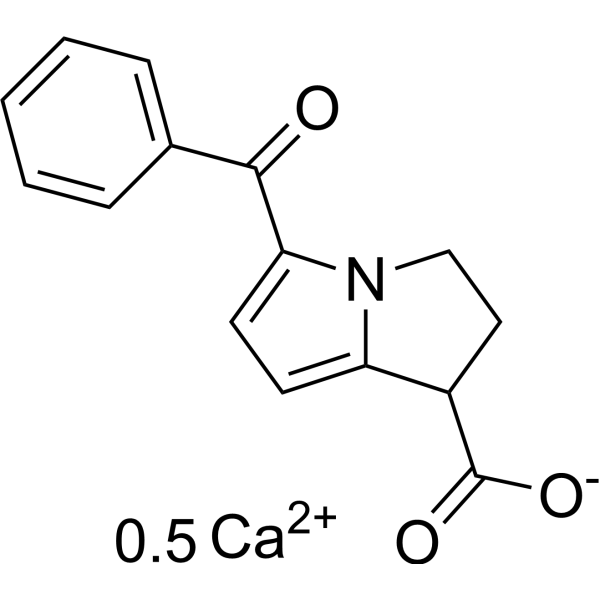
HY-B0580C
Ketorolac hemicalciumCOX Inhibitor


Biological Activity
Description
|
PPAR-γ |
HSP70 |
Cellular Effect
|
Cell Line

|
Type | Value | Description | References |
|---|---|---|---|---|
| B104 | IC50 |
10 nM
|
Inhibition of growth of rat B104 neuroblastoma cells incubated in defined medium for 24 hrs by Coulter counter.
Inhibition of growth of rat B104 neuroblastoma cells incubated in defined medium for 24 hrs by Coulter counter.
|
6580494 |
| C6 | IC50 |
100 nM
|
Inhibition of growth of rat C6 glioma cells incubated in defined medium for 24 hrs by Coulter counter.
Inhibition of growth of rat C6 glioma cells incubated in defined medium for 24 hrs by Coulter counter.
|
6580494 |
In Vitro
Prostaglandin A1 (0.1-6 µg/mL; 6-48 h) inhibits Mayaro virus (MAYV) replication, induces HSP70 expression, but only slightly inhibits viral structural protein synthesis in Hep-2 cells[4].
Prostaglandin A1 (6-24 h) inhibits cell proliferation in rat B104 neuroblastoma and C6 glioma cells (IC50 values of 10 nM and 100 nM, respectively), and is itself converted to polar metabolites, but does not inhibit normal L929 or CEF fibroblasts[5].
Prostaglandin A1 (100 ng/mL-1 μg/mL; 10-14 days) inhibits tumor colony formation in soft agar in S91 CCL 53.1 mouse melanoma and C8146c human melanoma cells[6].
Prostaglandin A1 (10 µM; 48 h) promotes cholesterol efflux, decreases PEN-2 expression, and inhibits Aβ production through the PPARγ/ABCA1 pathway in APPsw cells and N2a cells[2].
Prostaglandin A1 (30 µg/mL; 2-24 h) induces heat shock response, increases IκBα gene and polypeptide expression, and inhibits TNF-α (HY-P70426A)-mediated IL-8 secretion and NF-κB activation in BEAS-2B cells[7].
Prostaglandin A1 (0.1-1.0 µg/mL) inhibits spontaneous motility in isolated myometrium[1].
Prostaglandin A1 (10 µM; 2-48 h) decreases Tau protein phosphorylation by binding to PPP2R1A to activate PP2A in N2a cells[3].
MedChemExpress (MCE) has not independently confirmed the accuracy of these methods. They are for reference only. Further protocols information, click here.
-
Cell Line:Hep-2
-
Concentration:1, 5 μg/mL
-
Incubation Time:24 h
-
Result:Increased HSP70 levels by 52% at 1 μg/mL and 130% at 5 μg/mL relative to the untreated control.
-
Cell Line:APPsw cells
-
Concentration:10 μM
-
Incubation Time:48 h
-
Result:Induced a marked accumulation of cholesterol in the supernatant.
DIDS reversed the effects on increasing the synthesis of ABCA1 and inducing cholesterol efflux.
DIDS completely abolished the effects on suppressing the expression of PEN-2 and decreasing the production of Aβ1-42.
ABCA1 knockout completely reversed the effects on downregulating PEN-2, inducing cholesterol efflux, and decreasing the levels of Aβ1-42.
-
Cell Line:BEAS-2B
-
Concentration:30 μg/mL
-
Incubation Time:2 h
-
Result:Inhibited secretion of immunoreactive IL-8.
Immunoreactive IL-8 was not detectable in the media of control cells or in cells treated with PGA1 alone.
-
Cell Line:N2a
-
Concentration:10 μM
-
Incubation Time:48 h
-
Result:Inhibited tau phosphorylation at Thr181, Ser202, and Ser404.
The inhibitory effects of PGA1 on tau phosphorylation were reversed by the addition of OA.
-
Cell Line:Murine S91 CCL 53.1 and human C8146c
-
Concentration:100, 500, 1000 ng/mL
-
Incubation Time:10, 14 days
-
Result:Significantly inhibited melanoma colony formation in soft agar.
In Vivo
Prostaglandin A1 (1 µg/µL, 5 µL total; i.c.v.; single administration; 48 h) decreases Aβ levels in cerebrospinal fluid and upregulates ABCA1 expression in APP/PS1 double transgenic mice[2].
Prostaglandin A1 (2.5 mg/kg/day; i.n.; every day; for 6 consecutive months) activates PP2A, reduces Tau phosphorylation, and ameliorates cognitive decline in tau P301S transgenic mice (a model of AD and tauopathies)[3].
Prostaglandin A1 (0.01-10 µg/µl, 5 µl total; i.c.v.; single administration; 48 h) activates PP2A and dose-dependently inhibits Tau phosphorylation in tau P301S transgenic mice[3].
Prostaglandin A1 (16.5-33 nM; i.c.v.; single administration; observation up to 24 h post-ischemia) enhances HSF-1, HO-1, and HSP90α expression, restores HSP90β expression, and decreases the expression of the pro-apoptotic factor Apaf-1 in a permanent middle cerebral artery occlusion focal cerebral ischemia model in Sprague-Dawley rats[9].
MedChemExpress (MCE) has not independently confirmed the accuracy of these methods. They are for reference only.
-
Animal Model:Sprague-Dawley (Male, 280-300 g, pMCAO)[9]
-
Dosage:16.5 nM; 33.3 nM
-
Administration:icv; single injection; 15 min prior to pMCAO
-
Result:Elevated ischemia-induced levels of HSF-1, HO-1, and HSP90α, significantly recovered downregulated HSP90β expression, and significantly inhibited Apaf-1 induction in the ischemic striatum.
-
Animal Model:C57BL/6 mice (male, wild-type, 9 months old); APP/PS1 transgenic mice [B6C3-Tg (APPswe, PSEN1dE9) 85Dbo/J] (male, 9 months old)[2]
-
Dosage:5 μL of 1 μg/μL
-
Administration:i.c.v.; single injection; 48 h before assessment
-
Result:Significantly decreased the increased levels of PEN-2 mRNA and protein in APP/PS1 Tg mice.
Markedly increased the extent of sAPPα, while sAPPβ showed the opposite trend.
Obviously decreased Aβ1-42 levels in PGA1-treated mice.
Significantly reduced the CSF levels of low molecular weight Aβ oligomers.
Did not affect the levels of high molecular weight Aβ aggregates or fibrils in the hippocampus and cortex.
Substantially reduced cholesterol levels in PGA1 treatment groups compared to the untreated group.
Significantly increased the mRNA and protein levels of ABCA1 in APP/PS1 Tg mice.
-
Animal Model:TauP301S transgenic (Tg) mice (stock No. 008169)[3]
-
Dosage:5 μl of 1 μg/μl (PGA1); 2 ng/μl (OA)
-
Administration:i.c.v.; single administration; 48 h
-
Result:Suppressed tau phosphorylation at Thr181, Ser202, and Ser404 by activating PP2A, an effect that was blocked by okadaic acid.


Chemical Information
-
CAS No. 14152-28-4
-
Appearance Solid
-
Molecular Weight 336.47
-
Formula C20H32O4
-
Color White to off-white
-
SMILES
CCCCC[C@H](O)/C=C/[C@@H]1[C@H](C(C=C1)=O)CCCCCCC(O)=O
-
Structure Classification
-
Initial Source
Human semen
-
Shipping
Room temperature in continental US; may vary elsewhere.
-
Storage
-20°C, sealed storage, away from moisture
* In solvent : -80°C, 6 months; -20°C, 1 month (sealed storage, away from moisture)

Protocols
-
Research Protocol for Infectious Diseases
Infectious-disease experiments test how pathogens interact with host barriers, innate immune receptors, inflammatory signaling, pathogen replication, and tissue injury; pattern-recognition receptors such as TLRs, RIG-I-like receptors, NOD-like receptors, and inflammasomes detect microbial molecules and activate NF-κB, interferon, and cytokine responses. The central hypothesis is that infection severity reflects the balance between pathogen burden and host response: protective inflammation restricts pathogen growth, whereas excessive or mislocalized inflammation contributes to tissue damage and disease phenotype. Unresolved questions include which host pathways are protective versus pathogenic, why some infection models fail to translate to human disease, and which combined readouts best predict clinically relevant infection outcomes.
-
Research Protocol for Cardiovascular Diseases
Cardiovascular disease can be modeled as maladaptive cardiac remodeling, where ischemic injury or pressure overload activates inflammatory signaling, fibroblast activation, extracellular-matrix deposition, cardiomyocyte hypertrophy, vascular remodeling, and progressive ventricular dysfunction. The TGF-β/SMAD axis is a central profibrotic pathway after myocardial injury and pressure overload, while innate immune and cytokine pathways regulate leukocyte recruitment, scar formation, and adverse remodeling. Key unresolved questions include which inflammatory signals are reparative versus harmful, when fibrosis is protective versus maladaptive, and whether pathway inhibition improves function without weakening necessary infarct healing or compensatory remodeling.
-
Research Protocol for Inflammation-related Diseases
The NLRP3 inflammasome is a cytosolic innate immune signaling platform that integrates priming signals and danger-signal activation to promote caspase-1 activation, maturation of IL-1β and IL-18, and gasdermin D-mediated pyroptotic cell death. The core experimental logic is to determine whether inflammatory disease phenotypes are driven by increased NLRP3 expression, ASC-containing inflammasome assembly, caspase-1 cleavage, GSDMD cleavage, and extracellular release of IL-1β/IL-18 rather than by nonspecific cell injury alone. The pathway is strongly linked to inflammation-related disease phenotypes because monosodium urate crystals activate NALP3/NLRP3 inflammasome signaling in gout-like crystal inflammation, cholesterol crystals activate NLRP3 inflammasomes in atherogenesis models, and DSS-induced intestinal inflammation has been reported to involve NLRP3 inflammasome activity. However, experimental colitis studies also show context-dependent protective effects of NLRP3 inflammasome co
-
Alzheimer’s Disease Modeling
Alzheimer’s Disease (AD) is a neurodegenerative disorder characterized by a progressive decline in cognitive functions and loss of specific types of neurons and synapses. Alzheimer's symptoms can be simulated in mice by injecting drugs (such as Aβ) or genetically modified.


Purity & Documentation
-
Data Sheet (286 KB)
-
SDS (557 KB)
- English - EN (557 KB)
- Français - FR (557 KB)
- Deutsch - DE (557 KB)
- Norwegian - NO (557 KB)
- Español - ES (557 KB)
- Swedish - SV (557 KB)
- Italian - IT (557 KB)
- Korean - KR (557 KB)
- Portuguese - PT (557 KB)
-
Handling Instructions (2659 KB)
References

Calculators
Concentration (start) × Volume (start) = Concentration (final) × Volume (final)


Prostaglandin A1 Related Classifications
HY-113053 Related Classifications
 Powered by Bioz