1431 Results for "

fluorometric imaging plate reader

" in MedChemExpress (MCE) Product Catalog:
Products (1431)

1431 Results for "fluorometric imaging plate reader" in MCE Product Catalog:

Cat. No.: HY-NP163A
Synonyms: WGA-AF488
Wheat germ agglutinin-AF488 (WGA-AF488) is a cell membrane-specific staining agent prepared by conjugating wheat germ agglutinin with the Alexa Fluor 488 (HY-D1304) fluorescent dye, and it binds to cell surface glycoproteins with high affinity. Wheat germ agglutinin-AF488 is applied in fluorescence microscopy and confocal imaging techniques, and it can clearly label the membrane structures of various cells including breast cancer cells, enabling high-resolution visual observation. Wheat germ agglutinin-AF488 is used in studies of breast cancer and triple-negative breast cancer to observe cell morphology and membrane dynamic changes .
loading...
    loading...
Cat. No.: HY-P11287A
DOTA-Pep-1L TFA is a polypeptide formed by the conjugation of DOTA and Pep-1L. DOTA-Pep-1L TFA specifically binds to IL13RA2 and can be used for the synthesis of targeted polypeptides. DOTA-Pep-1L TFA yields the isotopically labeled product [ 225Ac]DOTA-Pep-1L, which exerts α-radiation killing effects on orthotopic glioma cells and extends the median survival time of mice bearing orthotopic glioma models after stereotactic injection. DOTA-Pep-1L TFA can be used for PET imaging, tumor targeting and glioma research .
loading...
    loading...
Cat. No.: HY-P5362A
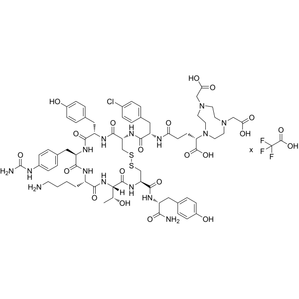
NODAGA-LM3 TFA is a ligand that can cross the blood-brain barrier and targets somatostatin receptor SSTR2 with high affinity (IC50 = 1.3 nM). NODAGA-LM3 TFA does not trigger the internalization of SSTR2 and can inhibit agonist-induced internalization processes. NODAGA-LM3 TFA shows low uptake in normal tissues such as the liver and spleen, but high uptake in the lungs and blood pool. 68Ga-labeled NODAGA-LM3 TFA can serve as a PET imaging agent for well-differentiated neuroendocrine tumors, and is applied in studies related to small cell lung cancer and well-differentiated neuroendocrine tumors .
loading...
    loading...
Cat. No.: HY-P992340

Research Areas:  

Cancer

CTB006 is a monoclonal antibody targeting a humanized chimeric recombinant anti-DR5. CTB006 specifically binds to and activates DR5, thereby inducing tumor cell apoptosis, inhibiting tumor growth and reducing tumor drug resistance. 177Lu-radiolabeled CTB006 can deliver targeted radiotherapy to tumor cells; while 89Zr- or 177Lu-labeled CTB006 can serve as a PET/CT imaging agent for detecting DR5 expression levels in preclinical tumor models and screening cancers with DR5 overexpression. CTB006 can be applied to research related to gastrointestinal cancer, colorectal cancer and other solid tumors .
loading...
    loading...
Cat. No.: HY-113553
CAS No.: 102185-28-4
Synonyms: (p-Aminophenyl)phosphocholine; p-Aminophenylphosphorylcholine; 4-APPC
Research Areas:  

Others

4-Aminophenylphosphorylcholine ((p-Aminophenyl) phosphocholine; p-Aminophenylphosphorylcholine; 4-APPC) is a phosphatidylcholine derivative that serves as a coupling ligand and antigen probe. When conjugated with gold nanoclusters, the phosphocholine head epitope of 4-Aminophenylphosphorylcholine is specifically recognized by anti-Pc IgM and CRP. It can be covalently coupled to human serum albumin (HSA) or glutathione-protected gold nanoclusters (Au-GSH) via covalent chemistry, respectively. The 4-Aminophenylphosphorylcholine-HSA complex acts as an ELISA coating antigen for detecting endogenous anti-Pc IgM antibodies in plasma; 4-Aminophenylphosphorylcholine-modified Au-GSH can be used to construct an ultra-stealthy NIR-II fluorescent gold nanocluster probe for imaging-related studies in mouse breast cancer and colon cancer models .
loading...
    loading...
Cat. No.: HY-131025
CAS No.: 1811539-88-4
Synonyms: JF585, SE; JF585, NHS
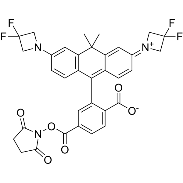
Janelia Fluor 585, SE (JF585, SE) is an orange fluorescent dye containing an NHS ester that can be conjugated with primary amine groups. Janelia Fluor 585, SE can be used immediately for structured illumination (SIM) and stimulated emission depletion (STED) imaging and could be converted to photoactivatable derivative for single-molecule localization microscopy (SMLM) experiments . Janelia Fluor fluorescent dyes can be chemically conjugated to the specific HaloTag substrate (a chloroalkane ligand) to form a fluorescent ligand, rather than binding directly to the HaloTag protein itself. Janelia Fluor products are licensed under U.S. Pat. Nos. 9,933,417, 10,018,624 and 10,161,932 and other patents from Howard Hughes Medical Institute (Ex/Em = 585/609 nm).
loading...
    loading...
Cat. No.: HY-138659
CAS No.: 2042192-00-5
Purity:  95.29%
Synonyms: JF646, Tetrazine
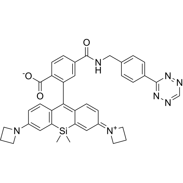
Janelia Fluor 646, Tetrazine (JF646, Tetrazine) a red fluorescent dye that contains a tetrazine group. JF646, Tetrazine can be used in cellular imaging . Janelia Fluor fluorescent dyes can be chemically conjugated to the specific HaloTag substrate (a chloroalkane ligand) to form a fluorescent ligand, rather than binding directly to the HaloTag protein itself. Janelia Fluor products are licensed under U.S. Pat. Nos. 9,933,417, 10,018,624 and 10,161,932 and other patents from Howard Hughes Medical Institute. Janelia Fluor 646, Tetrazine is a click chemistry reagent, it contains a Tetrazine group that can undergo an inverse electron demand Diels-Alder reaction (iEDDA) with molecules containing TCO groups (Ex/Em = 646/664 nm).
loading...
    loading...
Cat. No.: HY-141637S
CAS No.: 478518-95-5
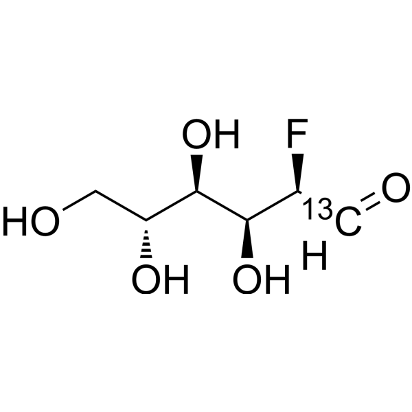
2-Deoxy-2-fluoro-D-glucose- 13C is the 13C labeled 2-Deoxy-2-fluoro-D-glucose (HY-141637). 2-Deoxy-2-fluoro-D-glucose (2-Fluoro-2-deoxy-D-glucose) is a glucose analog that can be absorbed by cells. 2-Deoxy-2-fluoro-D-glucose is capable of being labelled with 18F for positron emission tomography (PET) imaging. 18F-2-Deoxy-2-fluoro-D-glucose can be used to detect breast cancer and gastric cancer .
loading...
    loading...
Cat. No.: HY-160184
CAS No.: 524699-85-2
Target:  

COX

COX-1-IN-5 (example 13, compound PS13) is a potent and selective COX-1 inhibitor (COX-1 IC50 = 1 nM, COX-2 IC50 > 0.1 μM) exhibiting >1000-fold selectivity over COX-2. COX-1-IN-5 possesses strong anti-inflammatory, antipyretic, analgesic, antithrombotic, anti-cancer activities. COX-1-IN-5 can be used for COX-mediated diseases research, such as inflammatory conditions and pain. COX-1-IN-5 radiolabeled with 11C can be used as a selective PET tracer for whole‑body imaging of COX‑1 distribution and target engagement in vivo .
loading...
    loading...
Cat. No.: HY-165424
CAS No.: 3036456-18-2
NIR-BG2 is a fluorescent indicator targeting senescence-associated β-galactosidase (SA-β-Gal). NIR-BG2 contains a hemicyanine fluorophore caged by SA-β-Gal; cleavage of its β-galactosyl group by SA-β-Gal generates an electrophilic quinone methide, which can form covalent bonds with adjacent proteins to achieve long-term signal retention. NIR-BG2 activates a near-infrared fluorescent signal upon recognition of SA-β-Gal. NIR-BG2 can be used for non-invasive in vivo imaging of cellular senescence in mouse xenograft models. The excitation/emission wavelengths are 675/708 nm .
loading...
    loading...
Cat. No.: HY-165426
CAS No.: 3031522-14-9
Research Areas:  

Inflammation/Immunology

HB-230 is a Transglutaminase-2 (TG2) (human IC50 = 4.9 μM) inhibitor and a selective fluorescent imaging probe for active TG2 localization. HB-230 modifies TG2 Cys277 to sustain the enzyme open conformation. HB-230 forms a ternary complex with TG2 and α2-macroglobulin, undergoes receptor-mediated endocytosis, and trafficks directly to lysosomes without early/late endosome fusion. HB-230 exhibits TG2 activity-dependent and α2-macroglobulin-dependent uptake. HB-230 can be used for the research of celiac disease, (Ex/Em = 649/665 nm) .
loading...
    loading...
Cat. No.: HY-174322
Target:  

SHP2 ERK Fluorescent Dye

Research Areas:  

Cancer

SHP2-IN-38 is a novel green-fluorescent SHP2 inhibitor with IC50 values of 2.89 μM (SHP2), 8.73 μM (SHP1), 11.08 μM (PTP1B), 33.07 μM (TCPTP). SHP2-IN-38 blocks the SHP2-mediated ERK signaling pathway and inhibits MV4-11 cells proliferation in vitro with IC50 of 7.90 μM. SHP2-IN-38 has an excitation wavelength of 360 nm, with a maximum emission wavelength of 550 nm in DMSO and 540 nm in DMF. SHP2-IN-38 shows green fluorescence imaging in HeLa cells and zebrafish.
loading...
    loading...
Cat. No.: HY-19888
CAS No.: 1001389-72-5
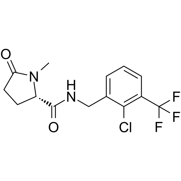
GSK-1482160 is an orally active and blood-brain barrier penetrant P2X7 receptor (P2X7R) negative allosteric modulator with pIC50s of 8.5 (human) and 6.5 (rat). GSK-1482160 reduces the efficacy of ATP at the P2X7 receptor without affecting its affinity, thereby inhibiting the release of IL-1β. GSK-1482160 is an effective radioligand and can be labeled with radioactive isotopes like 11C or 18F to image P2X7R. GSK-1482160 can be used for the studies of chronic joint pain and chronic constriction injury (CCI) .
loading...
    loading...
Cat. No.: HY-400882
CAS No.: 958233-17-5
Tosylate-DPA-714 is a GMP-grade precursor for the radiosynthesis of DPA-714 (HY-122607). It crosses the blood-brain barrier and exhibits steroidogenic activity. DPA-714 is a specific ligand for the 18 kDa translocator protein (TSPO) with a Ki of 7.0 nM. Tosylate-DPA-714 is safe and does not bind to central benzodiazepine receptors (benzodiazepine receptor). As a radiolabeling precursor, Tosylate-DPA-714 reacts with 18F via nucleophilic aliphatic substitution to form 18F-DPA-714, enabling specific targeted binding to TSPO. 18F-DPA-714 accurately reflects the level of neuroinflammation mediated by microglial activation, and is used for PET molecular imaging of neurological diseases such as Alzheimer's disease and multiple sclerosis .
loading...
    loading...
Cat. No.: HY-D0920
CAS No.: 166196-17-4
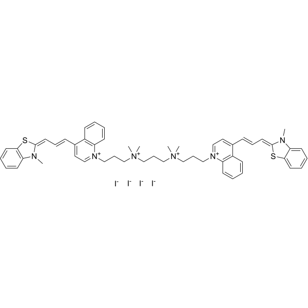
TOTO-3 is a cyanine nucleic acid intercalating fluorescent probe that binds to both RNA and DNA (Ex=642 nm, Em=660 nm). TOTO-3 cannot enter intact cells, but can penetrate cells with permeabilized plasma membranes, staining cytoplasmic RNA and nucleoli to distinguish intact cells from damaged ones. TOTO-3 is taken up by macrophages in an ultrasound-independent manner. TOTO-3 accumulates in tumor cells via ultrasound-mediated enhancement of plasma membrane permeability. TOTO-3 supports optical imaging for real-time assessment of plasma membrane integrity, nucleic acid detection, and monitoring of ultrasound-mediated intracellular delivery processes. TOTO-3 is applicable to studies related to rectal cancer .
loading...
    loading...
Cat. No.: HY-D1991
CAS No.: 906664-68-4
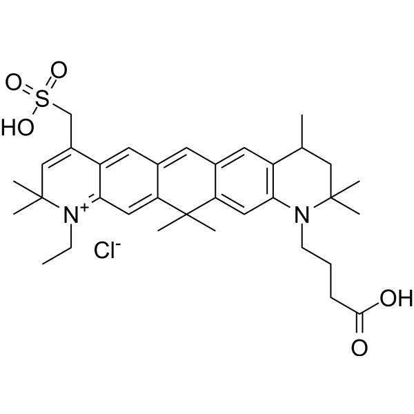
ATTO 647 is a carborhodamine fluorophore and imaging tracer with photostable properties. ATTO 647 serves as a fluorescent probe to investigate cell membrane structure and diffusion characteristics. When conjugated with wheat germ agglutinin, ATTO 647 specifically binds to N-acetyl-β-D-glucosamine and sialic acid residues on membrane glycoproteins, enabling single-molecule tracing of glycoprotein diffusion. ATTO 647 exhibits highly stable fluorescence properties with significantly reduced blinking in mounting media such as ROXS (AA/MV) and ROXS (TX/TQ), whereas its brightness properties vary in Ibidi-MM and Vectashield. ATTO 647 can also be used to label histone H2B-GFP in fixed cells for confocal microscopy photobleaching experiments .
loading...
    loading...
Cat. No.: HY-D3108
CAS No.: 2569032-08-0
Target:  

Fluorescent Dye

Research Areas:  

Others

BV-1 is a fluorescent probe for viscosity detection and imaging in living cells. BV-1 can be used to detect changes in intracellular viscosity and mitochondrial viscosity in living cells. BV-1 responds to environmental viscosity via a molecular rotor mechanism. In a low-viscosity environment, the centrally located unsaturated structure induces a twisted intramolecular charge transfer (TICT) state and promotes non-radiative transitions, resulting in fluorescence quenching; when viscosity increases, molecular rotation is restricted, the TICT state is attenuated, and fluorescence is enhanced with a red shift. BV-1 can be used for real-time monitoring of changes in cytoplasmic viscosity and the viscosity increase during cell apoptosis. Ex/Em = 480/570 nm .
loading...
    loading...
Cat. No.: HY-D3189
CAS No.: 2370825-44-6
5GluAF-2MeTG is an activatable fluorescent probe targeting the glutamate carboxypeptidase (CP) activity of PSMA (Ex/Em=490/500-600 nm). After being hydrolyzed by PSMA, 5GluAF-2MeTG releases a cell membrane-permeable fluorescent product, and achieves fluorescence activation by disrupting donor-excited photoinduced electron transfer (d-PeT). 5GluAF-2MeTG enables fluorescence imaging of live PSMA-expressing prostate cancer cells in vitro and visualizes the carboxypeptidase activity of PSMA. 5GluAF-2MeTG can be used to detect prostate cancer regions in preclinical excised tissue specimens .
loading...
    loading...
Cat. No.: HY-D3197
CDg16 is a selective fluorescent dye targeting SLC18B1 (λabs/λem=458/544 nm) that is actively transported into lysosomal vesicles of activated macrophages independent of the endocytic pathway. CDg16 enables highly specific vesicle localization in live cells. CDg16 exhibits no cytotoxicity and accurately distinguishes activated M1 and M2 subsets from different origins. CDg16 shows low background staining in non-activated cells and normal organs, making it suitable for time-lapse imaging. In preclinical animal models of inflammatory sites, atherosclerotic plaques and liver inflammation, CDg16 allows visualization of activated macrophages. CDg16 can be used to study inflammation-related diseases and atherosclerosis .
loading...
    loading...
Cat. No.: HY-D3351
CAS No.: 2668963-61-7
Target:  

Fluorescent Dye

Research Areas:  

Inflammation/Immunology

PNO1 is a fluorescent probe for pulmonary fibrosis detection. The detection mechanism of PNO1 relies on sensing upregulated nitric oxide (NO) in the pulmonary fibrosis (PF) microenvironment. PNO1 contains an aniline group that reacts with NO or its derivatives under acidic conditions to form an N-nitrosylated intermediate, which is subsequently converted into a diazonium salt and undergoes deamination, eliminating the intramolecular electron transfer quenching effect and triggering a fluorescence turn-on response. The excitation wavelength of PNO1 includes 545 nm in in vitro solution studies, while an excitation range of 520-550 nm is used for cell, tissue and in vivo imaging; its emission wavelength centers at 572 nm in the unreacted state, and shifts to 559 nm with enhanced fluorescence intensity upon binding to NO. PNO1 can be used for pulmonary fibrosis research .
loading...
    loading...

