



Xanthoangelol
Based on 2 publication(s) in Google Scholar
Xanthoangelol, extracted from Angelica keiskei, suppresses obesity-induced inflammatory responses. Xanthoangelol possesses antibacterial activity. Xanthoangelol inhibits monoamine oxidases. Xanthoangelol induces apoptosis in neuroblastoma and leukemia cells.
For research use only. We do not sell to patients.
- Purity : 99.88%
- CAS No.: 62949-76-2
- Formula: C25H28O4
- Molecular Weight:392.49
-
Storage:Powder -20°C, 3 years , 4°C, 2 years ; In solvent -80°C, 6 months , -20°C, 1 month

To place orders, for customer services and technical support, please contact: MedChemExpress USA
Tel: 609-228-6898 E-mail: [email protected] [email protected]


-
 Biological Activity
-
Chemical Information
-
Solvent & Solubility
- Protocols
- Purity & Documentation
- References
-
 Help & FAQs


-
HY-N7204
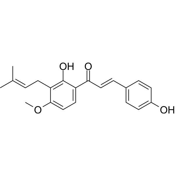
4-HydroxyderricinMonoamine Oxidase Inhibitor -
HY-101392
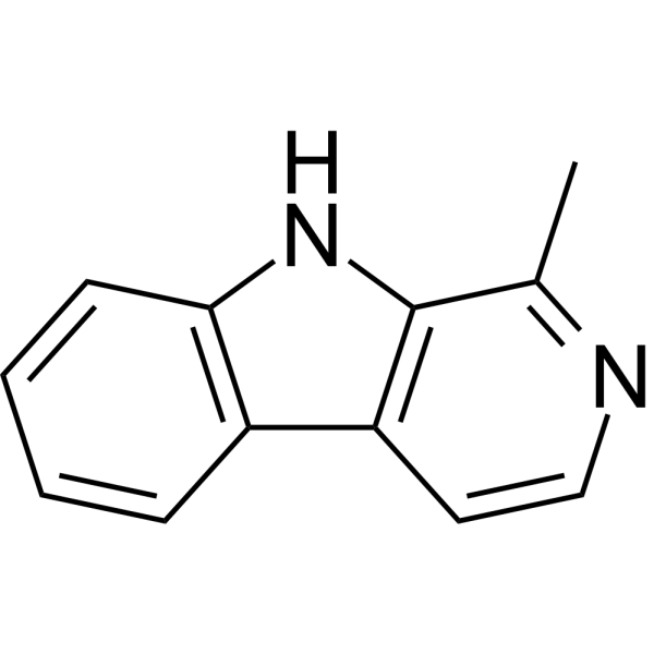
HarmaneMAO Inhibitor -
HY-A0091A
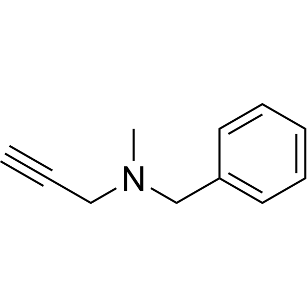
PargylineMonoamine Oxidase Inhibitor -
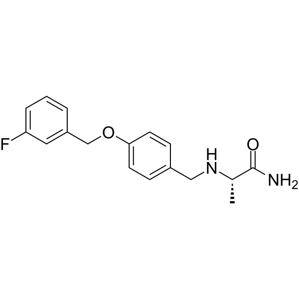
HY-70057
SafinamideMAO-B Inhibitor -
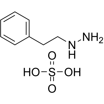
HY-B1018A
Phenelzine sulfateMAO Inhibitor -
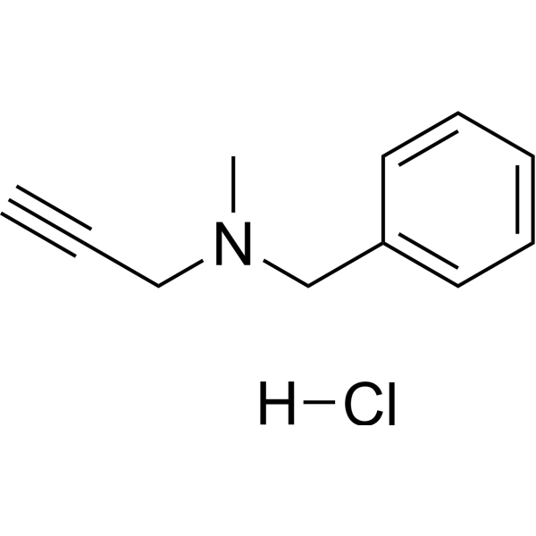
HY-A0091
Pargyline hydrochlorideMonoamine Oxidase Inhibitor -
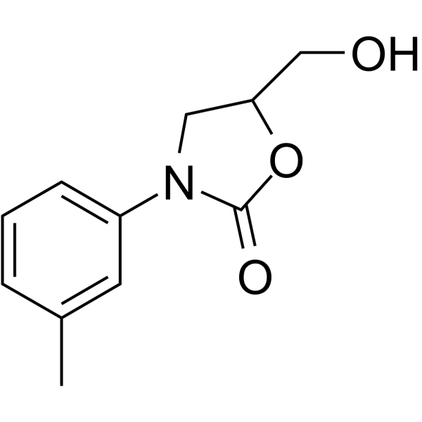
HY-14196
ToloxatoneMAO-A Inhibitor -
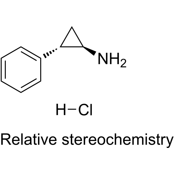
HY-17447A
Tranylcypromine hydrochlorideLSD1/MAO Inhibitor -
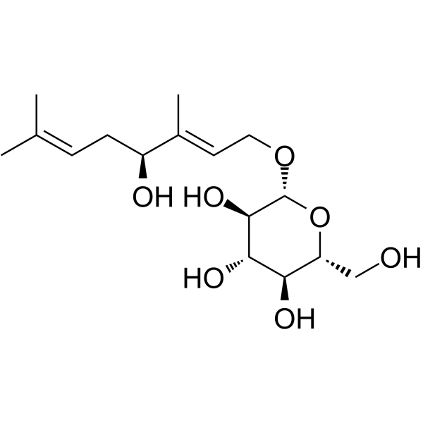
HY-N0505
RosiridinMAO A/B Inhibitor -
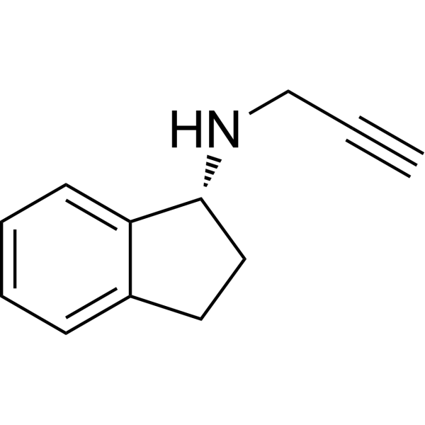
HY-14605A
RasagilineMAO Inhibitor -

HY-Y0882
Hydroxylamine hydrochlorideMAO Inhibitor -
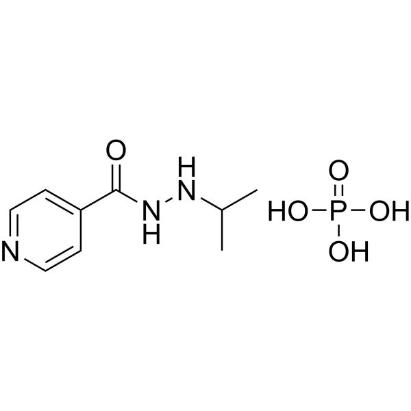
HY-B0886
Iproniazid phosphateMAO Inhibitor
Publications Citing Use of MedChemExpress (MCE) Xanthoangelol
MoreAll AMPK Isoforms
MoreAll Caspase Isoforms
More

Biological Activity
Description
Cellular Effect
|
Cell Line

|
Type | Value | Description | References |
|---|---|---|---|---|
| BHK-21 | CC50 |
11.8 μM
Compound: 30
|
Cytotoxicity against hamster BHK-21 cells assessed as reduction in cell viability measured after 48 hrs by MTT assay
Cytotoxicity against hamster BHK-21 cells assessed as reduction in cell viability measured after 48 hrs by MTT assay
|
[PMID: 36940609] |
| HEK293 | IC50 |
21.5 μM
Compound: Xanthoangelol
|
Cytotoxicity against human HEK293 cells assessed as reduction in cell viability incubated for 48 hrs by resazurin reagent based assay
Cytotoxicity against human HEK293 cells assessed as reduction in cell viability incubated for 48 hrs by resazurin reagent based assay
|
[PMID: 31648878] |
| HeLa | IC50 |
1.03 μM
Compound: 11
|
Inhibition of TNF-alpha-induced NF-kappaB activation in human HeLa cells by SEAP reporter gene assay
Inhibition of TNF-alpha-induced NF-kappaB activation in human HeLa cells by SEAP reporter gene assay
|
[PMID: 18841906] |
| HepG2 | IC50 |
13.7 μM
Compound: Xanthoangelol
|
Cytotoxicity against human HepG2 cells assessed as reduction in cell viability incubated for 48 hrs by resazurin reagent based assay
Cytotoxicity against human HepG2 cells assessed as reduction in cell viability incubated for 48 hrs by resazurin reagent based assay
|
[PMID: 31648878] |
| HUVEC | IC50 |
>100 μM
Compound: 1b
|
Toxicity against HUVEC incubated for 48 hrs by MTT assay
Toxicity against HUVEC incubated for 48 hrs by MTT assay
|
[PMID: 25590864] |
| K562 | IC50 |
3.98 μM
Compound: 1b
|
Antitumor activity against human K562 cells incubated for 48 hrs by MTT assay
Antitumor activity against human K562 cells incubated for 48 hrs by MTT assay
|
[PMID: 25590864] |
| MRC5 | IC50 |
21.7 μM
Compound: Xanthoangelol
|
Cytotoxicity against human MRC5 cells assessed as reduction in cell viability incubated for 48 hrs by resazurin reagent based assay
Cytotoxicity against human MRC5 cells assessed as reduction in cell viability incubated for 48 hrs by resazurin reagent based assay
|
[PMID: 31648878] |
| MRC5 | IC50 |
25 μM
Compound: 23
|
Cytotoxicity against human MRC5 cells after 48 hrs by resazurin dye-based fluorescence analysis
Cytotoxicity against human MRC5 cells after 48 hrs by resazurin dye-based fluorescence analysis
|
[PMID: 28075580] |
| Raji | IC50 |
273 molar ratio
Compound: 1, xanthoangelol
|
Inhibition of TPA-induced EBV-early antigen activation in human Raji cells
Inhibition of TPA-induced EBV-early antigen activation in human Raji cells
|
[PMID: 16441065] |
| RAW264.7 | IC50 |
1.83 μM
Compound: 11
|
Inhibition of LPS-induced NF-kappaB activation in mouse RAW264.7 cells by SEAP reporter gene assay
Inhibition of LPS-induced NF-kappaB activation in mouse RAW264.7 cells by SEAP reporter gene assay
|
[PMID: 18841906] |
| THP-1 | IC50 |
23.6 μM
Compound: Xanthoangelol
|
Cytotoxicity against human THP1 cells assessed as reduction in cell viability incubated for 48 hrs by resazurin reagent based assay
Cytotoxicity against human THP1 cells assessed as reduction in cell viability incubated for 48 hrs by resazurin reagent based assay
|
[PMID: 31648878] |
| THP-1 | IC50 |
25 μM
Compound: 23
|
Cytotoxicity against human THP1 cells after 48 hrs by resazurin dye-based fluorescence analysis
Cytotoxicity against human THP1 cells after 48 hrs by resazurin dye-based fluorescence analysis
|
[PMID: 28075580] |
In Vitro
Xanthoangelol (1 and 5 μM, 24 h) combined with 4-HD suppresses proinflammatory factors, such as NO, TNF-a, and MCP-1 through inhibition of the JNK pathway in the co-culture system of fully differentiated 3T3-L1 adipocytes and RAW264.7 macrophages[1].
Xanthoangelol (1-100 μM, 48 h) reduces the neuroblastoma and leukemia cells survival with cytotoxicity[4].
Xanthoangelol (1-10 μM, 4 h) induces concentration-dependent early apoptosis followed by secondary cell death through activation of caspase-3 in Jurkat cells[4].
MedChemExpress (MCE) has not independently confirmed the accuracy of these methods. They are for reference only. Further protocols information, click here.
-
Cell Line:LPS-stimulated RAW264.7 macrophages
-
Concentration:1 and 5 μM
-
Incubation Time:24 h
-
Result:Decreased NO release, TNF-a and MCP-1 secretion, as well as the mRNA expression in LPS-stimulated RAW264.7 macrophages.
-
Cell Line:LPS-stimulated RAW264.7 macrophages
-
Concentration:5 μM
-
Incubation Time:1 h
-
Result:Alleviated JNK phosphorylation (by 38% and 49%, respectively), but had no effect on IjB degradation and p38 phosphorylation in LPS-stimulated RAW264.7 macrophages.
-
Cell Line:IMR-32 and Jurkat cells
-
Concentration:1-100 μM
-
Incubation Time:48 h
-
Result:Markedly reduced IMR-32 and Jurkat cells viability in a concentration-dependent manner.
-
Cell Line:Jurkat cells
-
Concentration:1-10 μM
-
Incubation Time:4 h
-
Result:Significantly increased the ratio of apoptotic IMR-32 cells.
In Vivo
MedChemExpress (MCE) has not independently confirmed the accuracy of these methods. They are for reference only.
-
Animal Model:High-fat diet-induced obese (DIO) mice[1]
-
Dosage:0.1% or 0.15% w/w
-
Administration:mixed in a high-fat diet, daily for 14 weeks
-
Result:Showed fewer positively stained macrophages, suppressed the gene expression of inflammatory marker MCP-1 and TNF-a, increased oxygen consumption in DIO mice.


Chemical Information
-
CAS No. 62949-76-2
-
Appearance Solid
-
Molecular Weight 392.49
-
Formula C25H28O4
-
Color Light yellow to yellow
-
SMILES
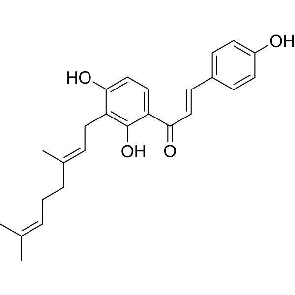
O=C(/C=C/C1=CC=C(O)C=C1)C2=CC=C(O)C(C/C=C(C)/CC/C=C(C)/C)=C2O
-
Structure Classification
-
Shipping
Room temperature in continental US; may vary elsewhere.
-
Storage
Powder -20°C 3 years 4°C 2 years In solvent -80°C 6 months -20°C 1 month


Publications (2)
-
Journal Impact Factor
-
Most Recent
-
Reprod Biomed Online
Xanthoangelol promotes early embryonic development of porcine embryos by relieving endoplasmic reticulum stress and enhancing mitochondrial function. [Abstract]2023 Aug;47(2):103211. PMID: 37246104 -


Solvent & Solubility
In Vitro:
DMSO : 100 mg/mL (254.78 mM; Need ultrasonic; Hygroscopic DMSO has a significant impact on the solubility of product, please use newly opened DMSO)
Please refer to the solubility information to select the appropriate solvent. Once prepared, please aliquot and store the solution to prevent product inactivation from repeated freeze-thaw cycles.
Storage method and period of stock solution: -80°C, 6 months; -20°C, 1 month. When stored at -80°C, please use it within 6 months. When stored at -20°C, please use it within 1 month.
Please refer to the solubility information to select the appropriate solvent. Once prepared, please aliquot and store the solution to prevent product inactivation from repeated freeze-thaw cycles.
Storage method and period of stock solution: -80°C, 6 months; -20°C, 1 month. When stored at -80°C, please use it within 6 months. When stored at -20°C, please use it within 1 month.
Concentration (start) × Volume (start) = Concentration (final) × Volume (final)

Protocols
-
Research Protocol for Infectious Diseases
Infectious-disease experiments test how pathogens interact with host barriers, innate immune receptors, inflammatory signaling, pathogen replication, and tissue injury; pattern-recognition receptors such as TLRs, RIG-I-like receptors, NOD-like receptors, and inflammasomes detect microbial molecules and activate NF-κB, interferon, and cytokine responses. The central hypothesis is that infection severity reflects the balance between pathogen burden and host response: protective inflammation restricts pathogen growth, whereas excessive or mislocalized inflammation contributes to tissue damage and disease phenotype. Unresolved questions include which host pathways are protective versus pathogenic, why some infection models fail to translate to human disease, and which combined readouts best predict clinically relevant infection outcomes.
-
Apoptosis
Apoptosis, also called programmed cell death, is generally characterized by distinct morphological characteristics.
-
TUNEL staining for apoptotic DNA fragmentation
TUNEL staining detects DNA strand breaks by using terminal deoxynucleotidyl transferase to add labeled nucleotides to exposed 3′-OH DNA termini, generating either microscopic staining in fixed cells or tissue sections, or fluorescence/cytometric signal in cell suspensions. TUNEL positivity reflects DNA fragmentation but should not be interpreted alone as definitive apoptosis, because TUNEL can also label necrotic, autolytic, mechanically damaged, or DNA-repair-associated DNA breaks.
-
Annexin V plus membrane-impermeant dye apoptosis staining
Annexin V-based apoptosis assays rely on the detection of phosphatidylserine (PS) externalization from the inner leaflet of the plasma membrane to the outer leaflet, an early biochemical hallmark of apoptosis. Fluorescently labeled Annexin V binds PS in a calcium-dependent manner, enabling identification of early apoptotic cells by flow cytometry or fluorescence microscopy. When combined with a membrane-impermeant DNA-binding dye (e. g. , propidium iodide), this approach allows discrimination between viable (Annexin V−/dye−), early apoptotic (Annexin V+/dye−), and late apoptotic or necrotic (Annexin V+/dye+) cell populations by assessing membrane integrity and PS exposure.
-
Apoptosis Solutions
Apoptosis is a regulated, generally non-lytic cell-death pathway that removes unwanted, damaged, infected, or abnormal cells through coordinated morphological changes, caspase activation, DNA fragmentation, and membrane remodeling. The intrinsic apoptosis pathway is controlled mainly by mitochondrial outer membrane permeabilization, BCL-2 family proteins, cytochrome c release, apoptosome formation, caspase-9 activation, and downstream executioner caspase-3/7 activation. The extrinsic apoptosis pathway is initiated by death receptors such as Fas, TNFR, and TRAIL receptors, which recruit adaptor proteins and activate caspase-8 before engaging executioner caspases or mitochondrial amplification through BID cleavage. Apoptosis is linked to many phenotypes, including cancer cell killing, tissue homeostasis, immune regulation, neurodegeneration, infection response, and treatment-induced cytotoxicity; unresolved questions include how apoptosis interacts with necroptosis, pyroptosis, ferroptos
-
Research Protocol for Metabolic Diseases
AMP-activated protein kinase, AMPK, is a conserved cellular energy sensor that responds to reduced cellular energy status and coordinates metabolism by increasing ATP-generating catabolic pathways while suppressing ATP-consuming anabolic processes. In metabolic disease research, the AMPK pathway is experimentally relevant because it regulates hepatic lipid synthesis, fatty acid oxidation, glucose production, skeletal-muscle glucose disposal, mTORC1-linked biosynthesis, autophagy, mitochondrial homeostasis, and whole-body energy balance. The central pathway logic is that energy stress, metformin, exercise-like stimulation, or direct AMPK activators increase AMPKα Thr172 phosphorylation and downstream substrate phosphorylation, including ACC and RAPTOR. Phosphorylation of ACC suppresses lipogenesis and supports fatty acid oxidation, whereas phosphorylation of RAPTOR suppresses mTORC1 signaling and links cellular energy status to growth and protein synthesis control. The pathway is linked
-
Bacterial live/dead nucleic-acid viability staining
The LIVE/DEAD bacterial viability staining method is based on differential permeability of nucleic-acid-binding fluorescent dyes, most commonly SYTO 9 and propidium iodide (PI), which enables discrimination of bacterial populations with intact versus compromised cytoplasmic membranes. SYTO 9 penetrates both intact and damaged bacterial membranes and binds nucleic acids to produce green fluorescence, whereas propidium iodide penetrates only cells with compromised membranes and fluoresces red while also reducing SYTO 9 signal through competitive binding and fluorescence interactions. The resulting fluorescence pattern is interpreted as a proxy for membrane integrity, which is widely used as an indicator of bacterial viability in microscopy, flow cytometry, and spectroscopic platforms. However, mechanistic studies show that SYTO 9 and PI interactions involve displacement and fluorescence resonance energy transfer effects, which can influence signal interpretation depending on dye ratios a


Purity & Documentation
-
Data Sheet (277 KB)
-
SDS (393 KB)
- English - EN (393 KB)
- Français - FR (393 KB)
- Deutsch - DE (393 KB)
- Norwegian - NO (393 KB)
- Español - ES (393 KB)
- Swedish - SV (393 KB)
- Italian - IT (393 KB)
- Korean - KR (393 KB)
- Portuguese - PT (393 KB)
-
Handling Instructions (2659 KB)
References
[1]. Li Y, et al. Xanthoangelol and 4-hydroxyderrcin suppress obesity-induced inflammatory responses. Obesity (Silver Spring). 2016 Nov;24(11):2351-2360. [Content Brief]
[2]. Inamori Y, et al. Antibacterial activity of two chalcones, xanthoangelol and 4-hydroxyderricin, isolated from the root of Angelica keiskei KOIDZUMI. Chem Pharm Bull (Tokyo). 1991 Jun;39(6):1604-5. [Content Brief]
[3]. Kim JH, et al. Xanthoangelol and 4-Hydroxyderricin Are the Major Active Principles of the Inhibitory Activities against Monoamine Oxidases on Angelica keiskei K. Biomol Ther (Seoul). 2013 May 30;21(3):234-40. [Content Brief]


Complete Stock Solution Preparation Table
Please refer to the solubility information to select the appropriate solvent. Once prepared, please aliquot and store the solution to prevent product inactivation from repeated freeze-thaw cycles.
Storage method and period of stock solution: -80°C, 6 months; -20°C, 1 month. When stored at -80°C, please use it within 6 months. When stored at -20°C, please use it within 1 month.
| Optional Solvent | Concentration Solvent Mass | 1 mg | 5 mg | 10 mg | 25 mg |
|---|---|---|---|---|---|
| DMSO | 1 mM | 2.5478 mL | 12.7392 mL | 25.4784 mL | 63.6959 mL |
| 5 mM | 0.5096 mL | 2.5478 mL | 5.0957 mL | 12.7392 mL | |
| 10 mM | 0.2548 mL | 1.2739 mL | 2.5478 mL | 6.3696 mL | |
| 15 mM | 0.1699 mL | 0.8493 mL | 1.6986 mL | 4.2464 mL | |
| 20 mM | 0.1274 mL | 0.6370 mL | 1.2739 mL | 3.1848 mL | |
| 25 mM | 0.1019 mL | 0.5096 mL | 1.0191 mL | 2.5478 mL | |
| 30 mM | 0.0849 mL | 0.4246 mL | 0.8493 mL | 2.1232 mL | |
| 40 mM | 0.0637 mL | 0.3185 mL | 0.6370 mL | 1.5924 mL | |
| 50 mM | 0.0510 mL | 0.2548 mL | 0.5096 mL | 1.2739 mL | |
| 60 mM | 0.0425 mL | 0.2123 mL | 0.4246 mL | 1.0616 mL | |
| 80 mM | 0.0318 mL | 0.1592 mL | 0.3185 mL | 0.7962 mL | |
| 100 mM | 0.0255 mL | 0.1274 mL | 0.2548 mL | 0.6370 mL |


Xanthoangelol Related Classifications
HY-111588 Related Classifications
 Powered by Bioz