



Z-VAD
Based on 26 publication(s) in Google Scholar
Z-VAD is an irreversible, broad-spectrum pan-caspase inhibitor that can inhibit a variety of caspases including caspase-3, -6, -7, -8, -9, etc. (with a weaker inhibitory effect on caspase-2). Z-VAD can block apoptosis signaling pathways, induce autophagy and necrosis in tumor cells, and has anti-angiogenic activity. Z-VAD can enhance the sensitivity of breast cancer and lung cancer cells to radiotherapy in vitro and in vivo, and prolong the growth delay of tumor xenograft models. Z-VAD is well tolerated and is mainly used in research related to cancer radiosensitization and cell death pathway regulation.
For research use only. We do not sell to patients.
- Purity : 99.93%
- CAS No.: 162852-62-2
- Formula: C20H27N3O8
- Molecular Weight:437.44
-
Storage:
Sealed storage, away from moisture.
Powder -80°C, 2 years , -20°C, 1 year* In solvent : -80°C, 6 months; -20°C, 1 month (sealed storage, away from moisture)


To place orders, for customer services and technical support, please contact: MedChemExpress USA
Tel: 609-228-6898 E-mail: [email protected] [email protected]


-
 Biological Activity
-
Chemical Information
-
Solvent & Solubility
- Protocols
- Purity & Documentation
- References
-
 Help & FAQs


-

HY-100579
Ferrostatin-1Ferroptosis Inhibitor -
HY-15760
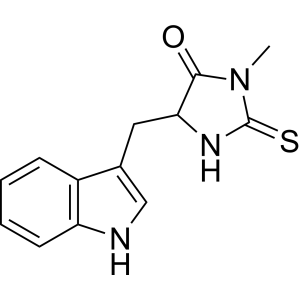
Necrostatin-1RIPK1 Inhibitor -
HY-19312
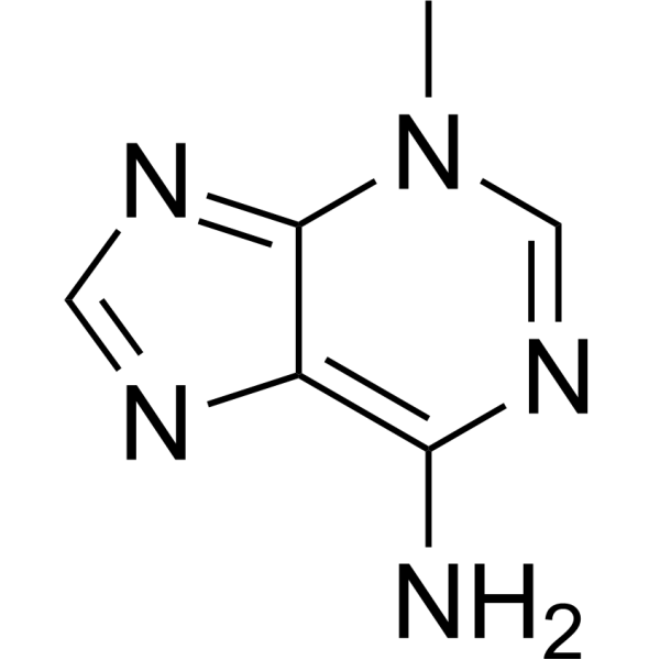
3-MethyladenineAutophagy/PI3K Inhibitor -
HY-100218A
RSL3GPX4 Inhibitor -

HY-B0215
AcetylcysteineMucolytic Agent -

HY-15763
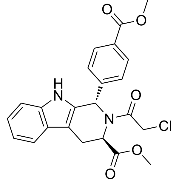
ErastinFerroptosis Inducer -
HY-17589A
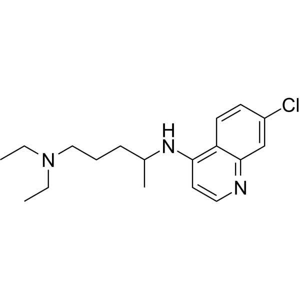
ChloroquineAntimalarial Agent -
HY-100573
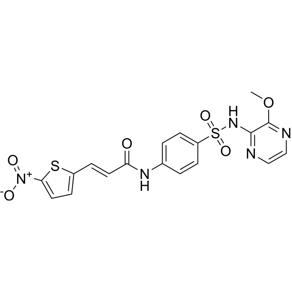
NecrosulfonamideMLKL/GSDMD Inhibitor -

HY-100558
Bafilomycin A1V-ATPase Inhibitor -
HY-13629
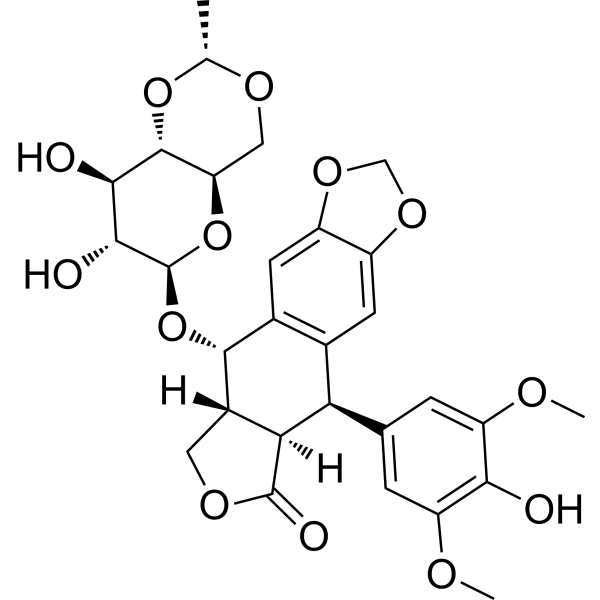
EtoposideTopoisomerase II Inhibitor -

HY-18739
Phorbol 12-myristate 13-acetatePKC Activator -

HY-15141
StaurosporinePKC/PKA Inhibitor
Publications Citing Use of MedChemExpress (MCE) Z-VAD
More- Cell. 2026 Sep 14:S0092-8674(26)01003-2.
- Nat Commun. 2026 Apr 17;17(1):5369. [Abstract]
- Nat Commun. 2025 Jan 31;16(1):1212. [Abstract]
- Adv Sci (Weinh). 2026 May;13(25):e19248. [Abstract]
- Adv Sci (Weinh). 2026 Mar 18:e74886. [Abstract]
- Cell Death Differ. 2025 Feb;32(2):242-255. [Abstract]
- Cell Death Dis. 2026 Jul 1.
- Phytomedicine. 2024 Dec:135:156110. [Abstract]
- Mater Today Bio. 2025 Sep 25:35:102341. [Abstract]
- Environ Int. 2026 May:211:110262. [Abstract]
- J Colloid Interface Sci. 2025 Jun 15:688:44-58. [Abstract]
- Emerg Microbes Infect. 2026 Apr 28;15(1):2667557. [Abstract]
- Biomater Adv. 2026 Jun:183:214770. [Abstract]
- Int Immunopharmacol. 2026 Mar 15:173:116250. [Abstract]
- PLoS Pathog. 2026 Apr 24;22(4):e1014174. [Abstract]
- FASEB J. 2025 Apr 15;39(7):e70488. [Abstract]
- Fish Shellfish Immunol. 2025 Aug:163:110416. [Abstract]
- J Virol. 2026 Mar 24;100(3):e0212125. [Abstract]
- Biochem Biophys Rep. 2026 Jan 15:45:102454. [Abstract]
- Exp Eye Res. 2025 Dec:261:110656. [Abstract]
- Vet Microbiol. 2025 Dec:311:110776. [Abstract]
- Vet Microbiol. 2025 Sep:308:110658. [Abstract]
- Albany College of Pharmacy and Health Sciences. 2026.
- Res Sq. 2026 Mar 10.
- Res Sq. 2025 Apr 20.
- bioRxiv. 2025 March 18.
 Customer Validation & Images

-
WB

-
Cell Imaging/Staining

-
RT-PCR
-
Cell Proliferation/Viability Assay
-
WB
All Caspase Isoforms
More

Biological Activity
Description
IC50 & Target
|
Caspase 3 |
Caspase-6 |
Caspase-7 |
Caspase-8 |
Caspase-9 |
In Vitro
Z-VAD (50 μM; 24 h) enhances the sensitivity of MDA-MB-231 breast cancer cells and H460 lung cancer cells to ionizing radiation[1].
Z-VAD (50 μM; 24h, 48h) combined with radiotherapy induces increasing autophagosome formation in breast cancer and lung cancer cells and upregulated the expression of autophagy-related proteins such as ATG5-ATG12 complex and Beclin-1[1].
Z-VAD (50 μM) combined with radiotherapy significantly reduces the formation of capillary-like tube structures in human umbilical vein endothelial cells (HUVECs)[1].
MedChemExpress (MCE) has not independently confirmed the accuracy of these methods. They are for reference only.
-
Cell Line:MDA-MB-231 (breast cancer), H460 (lung cancer)
-
Concentration:50 μM
-
Incubation Time:24 h; followed by irradiation (0-6 Gy)
-
Result:Significantly reduced the surviving fraction of both cell lines, with a dose enhancement ratio (DER) of 1.31, indicating increased radiosensitivity compared to radiation alone.
-
Cell Line:MDA-MB-231 (breast cancer), H460 (lung cancer)
-
Concentration:50 μM
-
Incubation Time:24 h (GFP-LC3 imaging), 24 h/48 h (Western blotting); combined with 5 Gy radiation
-
Result:Significantly increased punctate autophagosome formation, compared to single treatments.
Upregulated the expression of ATG5-ATG12 complex and Beclin-1 proteins at 24 h and 48 h after combined treatment, indicating induction of autophagy-related pathways.
-
Cell Line:HUVECs (human umbilical vein endothelial cells)
-
Concentration:50 μM
-
Incubation Time:Pre-treatment for 24 h before seeding on Matrigel
-
Result:Significantly decreased the number of capillary-like tubules formed by HUVECs on Matrigel, with an average reduction from 47.66 (control) to 6.0 tubules per microscopic field, demonstrating anti-angiogenic effects.
In Vivo
MedChemExpress (MCE) has not independently confirmed the accuracy of these methods. They are for reference only.
-
Animal Model:Female BALB/c nude mice (6-8 weeks old, 18-22 g) bearing MDA-MB-231 breast cancer or H460 lung cancer xenograft tumors[1]
-
Dosage:5 mg/kg
-
Administration:Intraperitoneal injection, three times a week, for 4 weeks
-
Result:Treatment with 5 mg/kg Z-VAD via intraperitoneal injection three times a week for 4 weeks, combined with radiotherapy, significantly reduced the tumor volume in both MDA-MB-231 and H460 xenograft models compared to radiotherapy alone.
The mice showed no significant signs of toxicity or adverse effects, indicating good tolerance to the treatment.
- Mammalian live/dead viability and cytotoxicity staining
- Annexin V plus membrane-impermeant dye apoptosis staining
- Breast Cancer Modeling
- Detection of Apoptosis
- TUNEL staining for apoptotic DNA fragmentation
- Cell Cytotoxicity Assay
- Autophagy
- Endothelial Tube Formation Assay
- Patient-Derived Xenograft (PDX)
- Apoptosis Solutions
- Splenic/Portal-Vein Liver Metastasis Xenograft
- Lysosome and acidic-vesicle live-cell staining
- Subcutaneous Cell-Line-Derived Xenograft
- Macroautophagy Solutions
- Orthotopic Cell-Line Xenograft
- Intraperitoneal/Peritoneal Dissemination Xenograft


Chemical Information
-
CAS No. 162852-62-2
-
Appearance Solid
-
Molecular Weight 437.44
-
Formula C20H27N3O8
-
Color White to off-white
-
Sequence
Z-Val-Ala-Asp
-
Sequence Shortening
Z-VAD
-
Shipping
Room temperature in continental US; may vary elsewhere.
-
Storage
Sealed storage, away from moisture
Powder -80°C 2 years -20°C 1 year * In solvent : -80°C, 6 months; -20°C, 1 month (sealed storage, away from moisture)


Publications (26)
-
Journal Impact Factor
-
Most Recent
-
Z-VAD purchased from MedChemExpress. Usage Cited in: Cell. 2026 Sep 14:S0092-8674(26)01003-2.
MYH9 polymerization upon treatment with cell death inhibitors. A375 cells were pretreated with different reagents (Z-VAD (40 μM), Necrostatin-1 (Nec-1, 40 μM), Liproxstatin-1 (Lip-1, 2 μM), CQ (20 μM) and NAC (5 mM)), MYH9 polymerization was detected.
Z-VAD purchased from MedChemExpress. Usage Cited in: Cell. 2026 Sep 14:S0092-8674(26)01003-2.
Effects of different inhibitors on cuproptotic cell death. Different inhibitors, including autophagy inhibitor CQ (20 μM), apoptosis inhibitor Z-VAD (40 μM), necroptosis inhibitors NSA (10 μM) and Necrostatin-1 (Nec-1, 40 μM), and ferroptosis inhibitors Liproxstatin-1 (Lip-1, 2 μM) and Fer-1 (10 μM), were used to pretreat A375 cells for 2 h, and then HThPA treatment, cuproptosis was analyzed.
-
Nat Commun
ELMO2 is a therapeutic vulnerability in mesenchymal-like and drug-resistant non-small cell lung cancer. [Abstract]2026 Apr 17;17(1):5369. PMID: 41997974 -
Nat Commun
Cellular senescence-associated gene IFI16 promotes HMOX1-dependent evasion of ferroptosis and radioresistance in glioblastoma. [Abstract]2025 Jan 31;16(1):1212. PMID: 39890789 -
Adv Sci (Weinh)
Elevated TRIM25 Impairs Poly (ADP-ribose) Metabolism via PARG Degradation and Mediates Compression-Induced Intervertebral Disc Degeneration. [Abstract]2026 May;13(25):e19248. PMID: 41700742 -
Adv Sci (Weinh)
2026 Mar 18:e74886. PMID: 41848091 -
Cell Death Differ
The IDH1-R132H mutation aggravates cisplatin-induced acute kidney injury by promoting ferroptosis through disrupting NDUFA1 and FSP1 interaction. [Abstract]2025 Feb;32(2):242-255. PMID: 39306640 -
-
Phytomedicine
2024 Dec:135:156110. PMID: 39369568 -
Mater Today Bio
Injectable magnetic hydrogel induces multi-programmed cell death and deep tumor regression in magnetic hyperthermia therapy in hepatocellular carcinoma. [Abstract]2025 Sep 25:35:102341. PMID: 41089724 -
Environ Int
Trimethyltin chloride triggers ferroptosis in myocardial injury: mitochondria-dependent protection by luteolin. [Abstract]2026 May:211:110262. PMID: 42013553 -
J Colloid Interface Sci
Nanozyme as tumor energy homeostasis disruptor mediated ferroptosis for high-efficiency radiotherapy. [Abstract]2025 Jun 15:688:44-58. PMID: 39987840 -
Emerg Microbes Infect
TRIM7 Inhibits Rabies Virus Replication by Promoting K48-Linked Ubiquitination and Degradation of RABV-M. [Abstract]2026 Apr 28;15(1):2667557. PMID: 42047619 -
Biomater Adv
Acoustic hydrogen delivery to treat PANoptosis induced by myocardial ischemia/reperfusion injury in rats. [Abstract]2026 Jun:183:214770. PMID: 41671925 -
Int Immunopharmacol
Trimethyltin chloride (TMT) - induced vascular injury through ubiquitination proteasome pathway - regulated GPX4 degradation. [Abstract]2026 Mar 15:173:116250. PMID: 41605050 -
PLoS Pathog
Duck plague virus LORF2 utilizes RNF34 to inhibit antiviral innate immunity by ubiquitination and degradation of IRF7. [Abstract]2026 Apr 24;22(4):e1014174. PMID: 42030364 -
FASEB J
2025 Apr 15;39(7):e70488. PMID: 40168090 -
Fish Shellfish Immunol
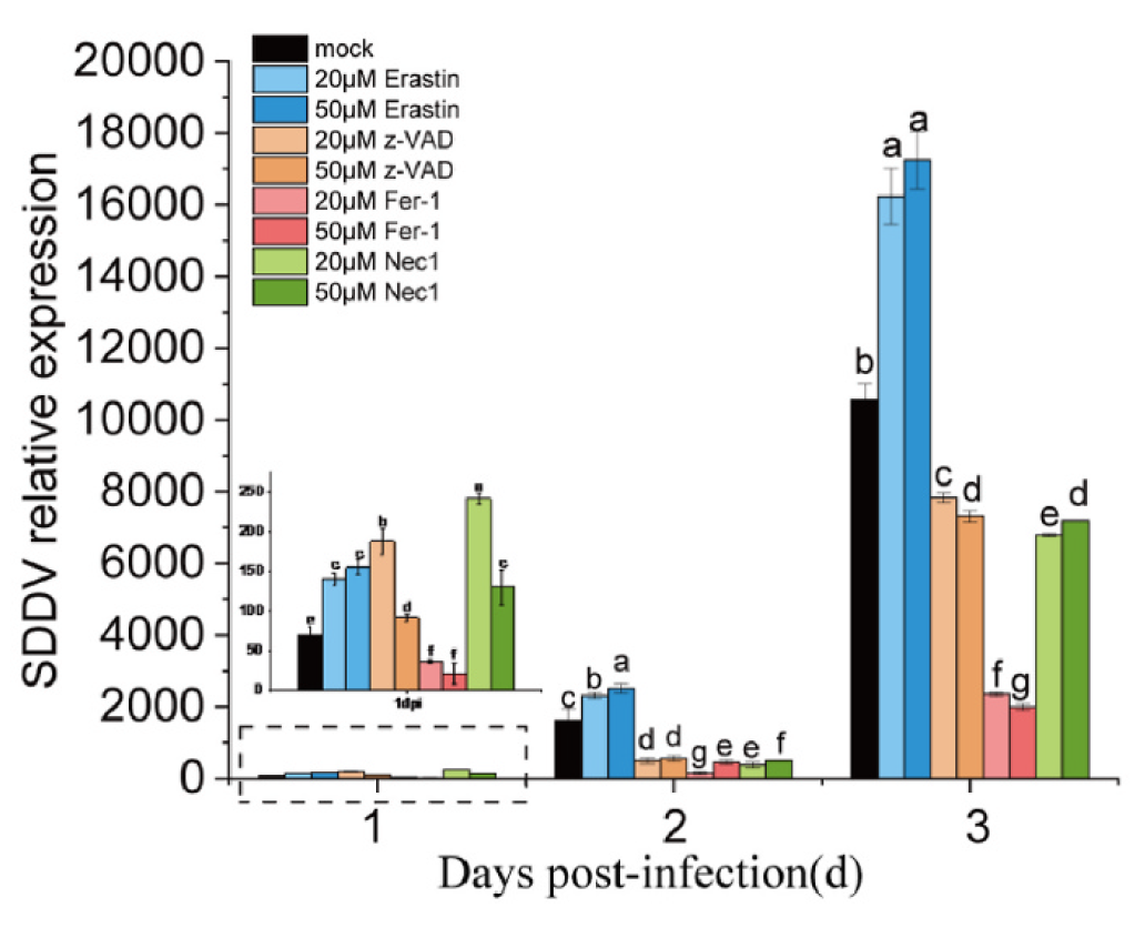
Scale drop disease virus (SDDV) triggers ferroptosis both in mandarin fish (Siniperca chuatsi) and MFF-1 cells to facilitate virus infection via linking to transferrin receptor 1 (TfR1). [Abstract]2025 Aug:163:110416. PMID: 40373888
Z-VAD purchased from MedChemExpress. Usage Cited in: Fish Shellfish Immunol. 2025 Aug:163:110416. [Abstract]
Relative SDDV expression levels in MFF-1 cells after treatment with Z-VAD (20-50 μM; 4 h), followed by SDDV infection.
-
J Virol
Small-molecule PS10 inhibits PRRSV replication by targeting HSP90 and multiple viral non-structural proteins. [Abstract]2026 Mar 24;100(3):e0212125. PMID: 41705837 -
Biochem Biophys Rep
2026 Jan 15:45:102454. PMID: 41584303 -
Exp Eye Res
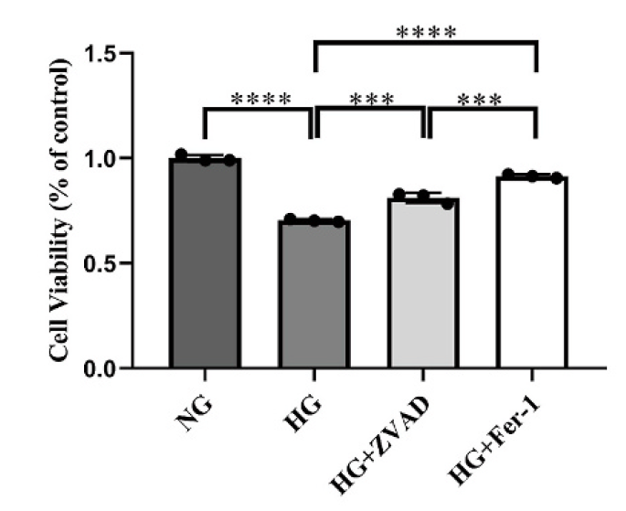
YAP1 aggravates hyperglycemia-induced retinal capillary endothelial cells injury via ferroptosis and inflammation regulation. [Abstract]2025 Dec:261:110656. PMID: 40992587
Z-VAD purchased from MedChemExpress. Usage Cited in: Exp Eye Res. 2025 Dec:261:110656. [Abstract]
Z-VAD (50 μM; 48 h) partially attenuated the reduction in viability of HRCECs after HG stimulation.
-
Vet Microbiol
Sanguinarine inhibits bovine parainfluenza virus type 3 replication through CD97 suppression. [Abstract]2025 Dec:311:110776. PMID: 41135374
Z-VAD purchased from MedChemExpress. Usage Cited in: Vet Microbiol. 2025 Dec:311:110776. [Abstract]
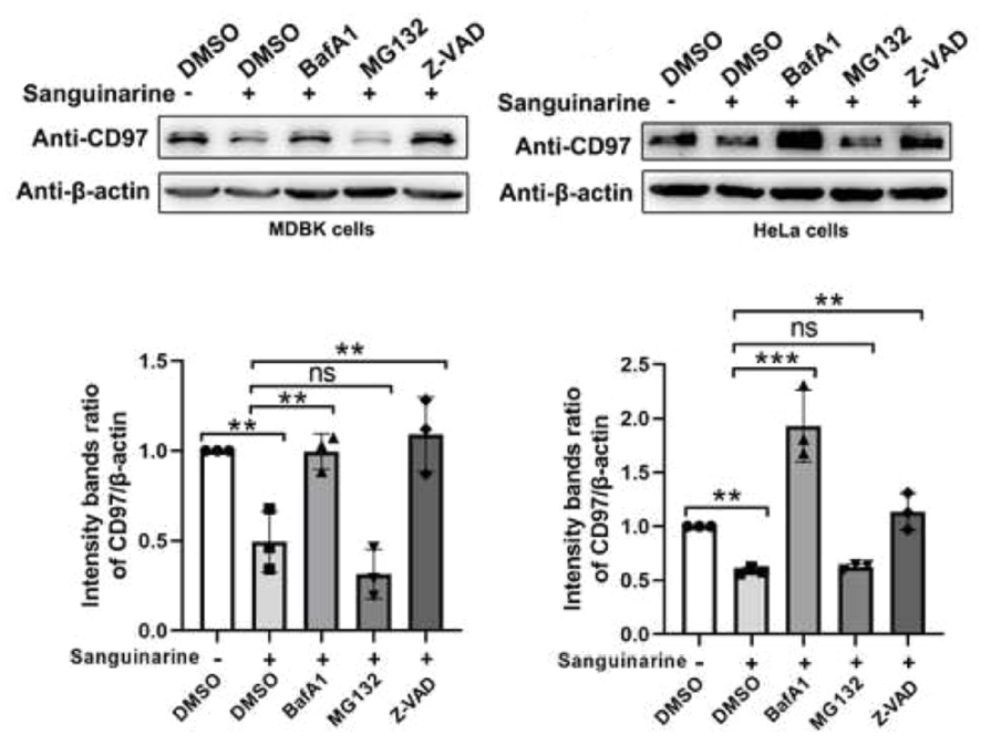
The caspase inhibitor Z-VAD (40 μM) specifically blocked the downregulation of CD97 in MDBK and HeLa cells by sanguinarine.
Z-VAD purchased from MedChemExpress. Usage Cited in: Vet Microbiol. 2025 Dec:311:110776. [Abstract]
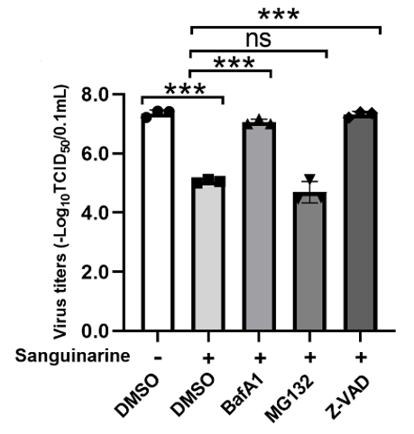
Z-VAD (40 μM) abolished the inhibition of BPIV3 replication in MDBK cells by sanguinarine.
-
Vet Microbiol
Sec6 suppresses BEFV-triggered type I IFN responses by promoting P62-mediated MAVS degradation. [Abstract]2025 Sep:308:110658. PMID: 40780029 -
-
-
-


Solvent & Solubility
In Vitro:
DMSO : 50 mg/mL (114.30 mM; Need ultrasonic; Hygroscopic DMSO has a significant impact on the solubility of product, please use newly opened DMSO)
Please refer to the solubility information to select the appropriate solvent. Once prepared, please aliquot and store the solution to prevent product inactivation from repeated freeze-thaw cycles.
Storage method and period of stock solution: -80°C, 6 months; -20°C, 1 month (sealed storage, away from moisture). When stored at -80°C, please use it within 6 months. When stored at -20°C, please use it within 1 month.
Please refer to the solubility information to select the appropriate solvent. Once prepared, please aliquot and store the solution to prevent product inactivation from repeated freeze-thaw cycles.
Storage method and period of stock solution: -80°C, 6 months; -20°C, 1 month (sealed storage, away from moisture). When stored at -80°C, please use it within 6 months. When stored at -20°C, please use it within 1 month.
Concentration (start) × Volume (start) = Concentration (final) × Volume (final)
In Vivo:
Select the appropriate dissolution method based on your experimental animal and administration route.
- For the following dissolution methods, please ensure to first prepare a clear stock solution using an In Vitro approach and then sequentially add co-solvents:
- To ensure reliable experimental results, the clarified stock solution can be appropriately stored based on storage conditions. As for the working solution for In Vivo experiments, it is recommended to prepare freshly and use it on the same day.
- The percentages shown for the solvents indicate their volumetric ratio in the final prepared solution. If precipitation or phase separation occurs during preparation, heat and/or sonication can be used to aid dissolution.
Add each solvent one by one: 10% DMSO 40% PEG300 5% Tween-80 45% Saline
Solubility: ≥ 5 mg/mL (11.43 mM); Clear solution
This protocol yields a clear solution of ≥ 5 mg/mL (saturation unknown).
Taking 1 mL working solution as an example, add 100 μL DMSO stock solution (50.0 mg/mL) to 400 μL PEG300, and mix evenly; then add 50 μL Tween-80 and mix evenly; then add 450 μL Saline to adjust the volume to 1 mL.
Preparation of Saline: Dissolve 0.9 g sodium chloride in ddH₂O and dilute to 100 mL to obtain a clear Saline solution.
Add each solvent one by one: 10% DMSO 90% (20% SBE-β-CD in Saline)
Solubility: ≥ 2.5 mg/mL (5.72 mM); Clear solution
This protocol yields a clear solution of ≥ 2.5 mg/mL (saturation unknown).
Taking 1 mL working solution as an example, add 100 μL DMSO stock solution (25.0 mg/mL) to 900 μL 20% SBE-β-CD in Saline, and mix evenly.
Preparation of 20% SBE-β-CD in Saline (4°C, storage for one week): 2 g SBE-β-CD powder is dissolved in 10 mL Saline, completely dissolve until clear.
In Vivo Dissolution Calculator
Please enter the basic information of animal experiments:
-
-
-
-
Recommended: Prepare an additional quantity of animals to account for potential losses during experiments.
Please enter your animal formula composition:
-
%DMSO +
Recommended: Keep the proportion of DMSO in working solution below 2% if your animal is weak.
-
%+
-
+%Tween-80 + +
-
%Saline +
The co-solvents required include: DMSO, . All of co-solvents are available by MedChemExpress (MCE). , Tween 80. All of co-solvents are available by MedChemExpress (MCE).
Working solution concentration: 0.22 mg/mL
Method for preparing stock solution: mg drug dissolved in μL DMSO. Stock solution concentration: mg/mL. * In solvent : -80°C, 6 months; -20°C, 1 month (sealed storage, away from moisture)
1. Take μL DMSO stock solution;
2. Add μL .
μL , mix evenly;
3. Then add μL Tween 80, mix evenly;
4. Then add μL
Please ensure that the stock solution in the first step is dissolved to a clear state, and add co-solvents in sequence. You can use ultrasonic heating (ultrasonic cleaner, recommended frequency 20-40 kHz), vortexing, etc. to assist dissolution.


Purity & Documentation
-
Data Sheet (278 KB)
-
SDS (252 KB)
- English - EN (252 KB)
- Français - FR (252 KB)
- Deutsch - DE (252 KB)
- Norwegian - NO (252 KB)
- Español - ES (252 KB)
- Swedish - SV (252 KB)
- Italian - IT (252 KB)
- Korean - KR (252 KB)
- Portuguese - PT (252 KB)
-
Handling Instructions (2659 KB)
References


Complete Stock Solution Preparation Table
Please refer to the solubility information to select the appropriate solvent. Once prepared, please aliquot and store the solution to prevent product inactivation from repeated freeze-thaw cycles.
Storage method and period of stock solution: -80°C, 6 months; -20°C, 1 month (sealed storage, away from moisture). When stored at -80°C, please use it within 6 months. When stored at -20°C, please use it within 1 month.
| Optional Solvent | Concentration Solvent Mass | 1 mg | 5 mg | 10 mg | 25 mg |
|---|---|---|---|---|---|
| DMSO | 1 mM | 2.2860 mL | 11.4301 mL | 22.8603 mL | 57.1507 mL |
| 5 mM | 0.4572 mL | 2.2860 mL | 4.5721 mL | 11.4301 mL | |
| 10 mM | 0.2286 mL | 1.1430 mL | 2.2860 mL | 5.7151 mL | |
| 15 mM | 0.1524 mL | 0.7620 mL | 1.5240 mL | 3.8100 mL | |
| 20 mM | 0.1143 mL | 0.5715 mL | 1.1430 mL | 2.8575 mL | |
| 25 mM | 0.0914 mL | 0.4572 mL | 0.9144 mL | 2.2860 mL | |
| 30 mM | 0.0762 mL | 0.3810 mL | 0.7620 mL | 1.9050 mL | |
| 40 mM | 0.0572 mL | 0.2858 mL | 0.5715 mL | 1.4288 mL | |
| 50 mM | 0.0457 mL | 0.2286 mL | 0.4572 mL | 1.1430 mL | |
| 60 mM | 0.0381 mL | 0.1905 mL | 0.3810 mL | 0.9525 mL | |
| 80 mM | 0.0286 mL | 0.1429 mL | 0.2858 mL | 0.7144 mL | |
| 100 mM | 0.0229 mL | 0.1143 mL | 0.2286 mL | 0.5715 mL |


Z-VAD Related Classifications
HY-164388 Related Classifications
 Powered by Bioz