



15(R)-Lipoxin A4

Based on 1 Customer Validation
15 (R)-Lipoxin A4 (15-epi-LXA4) is a specialized pro-resolving mediator and an acetylated derivative of COX2. 15 (R)-Lipoxin A4 is present in neurons. 15 (R)-Lipoxin A4 induces the SPM synthases ALOX12 and ALOX15, as well as the pro-resolving receptor ALX. 15 (R)-Lipoxin A4 inhibits protein kinases, including JNK1/2/3, Lyn, STAT-3 and STAT-6. 15 (R)-Lipoxin A4 enhances the release of pro-resolving mediators. 15 (R)-Lipoxin A4 alleviates the pro-inflammatory phenotype of tendon-derived stromal cells. 15 (R)-Lipoxin A4 promotes the resolution of neuroinflammation. 15 (R)-Lipoxin A4 is applicable to research related to achilles tendinitis, achilles tendon rupture and Alzheimer’s disease.
For research use only. We do not sell to patients.
- Purity : 98.3%
- CAS No.: 171030-11-8
- Formula: C20H32O5
- Molecular Weight:352.47
-
Storage:
Solution, -20°C, 2 years


To place orders, for customer services and technical support, please contact: MedChemExpress USA
Tel: 609-228-6898 E-mail: [email protected] [email protected]


-
 Biological Activity
-
Chemical Information
- Protocols
- Purity & Documentation
- References
-
 Help & FAQs


-
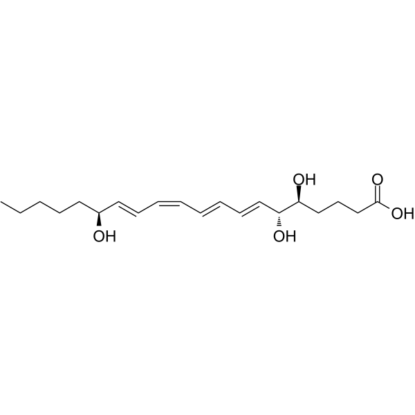
HY-113509
Lipoxin A4Anti-inflammatory Agent -
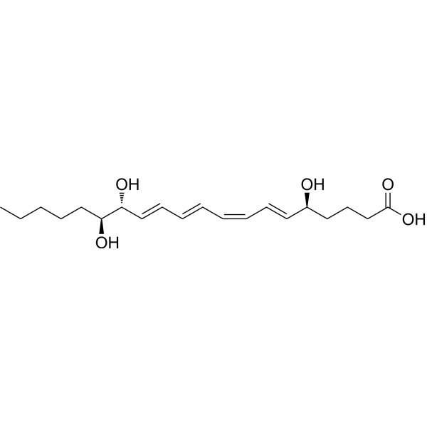
HY-116953
Lipoxin B4Anti-inflammatory Agent -
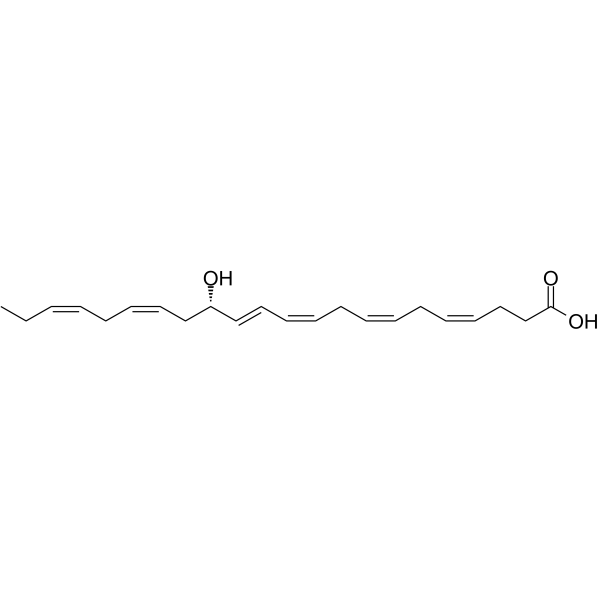
HY-130239
14(S)-HDHADHA Oxygenation Product -
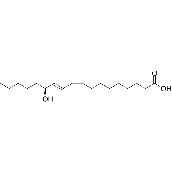
HY-113884B
(S)-Coriolic acidPPARγ Activator/Mitochondrial Dysfunction Inducer -
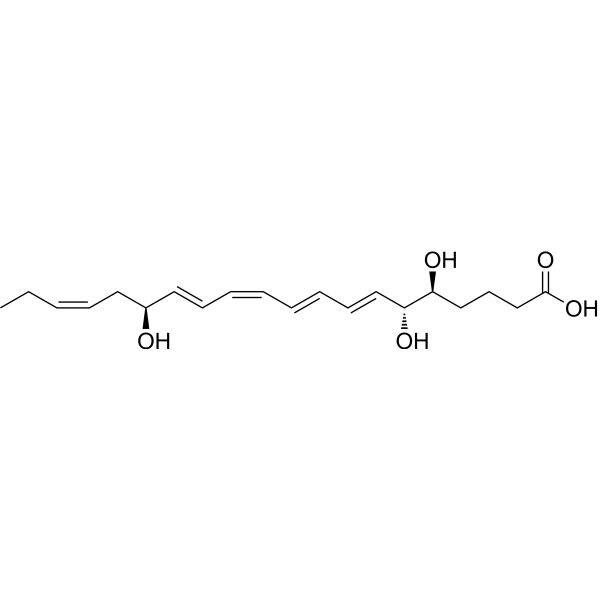
HY-115381
Lipoxin A5Endogenous Metabolite -
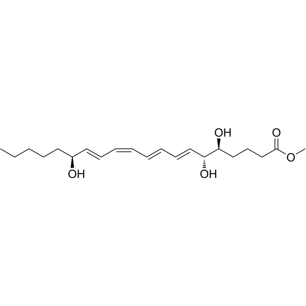
HY-126722
Lipoxin A4 methyl esterLipoxin Analog -

HY-130429
14,15-Leukotriene C4Leukotrienes -
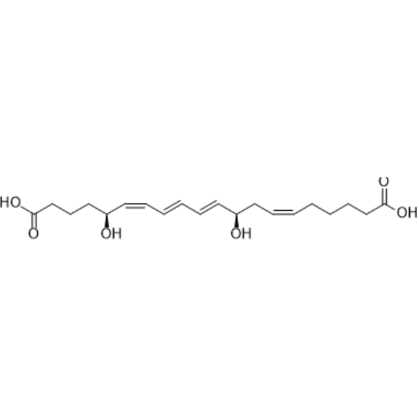
HY-113483
20-Carboxy-Leukotriene B4LTB4 Metabolite -

HY-113490
15-KETE -

HY-118149
9(S)-HpODE -
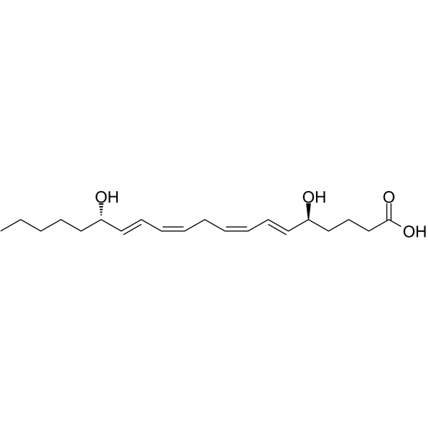
HY-113492
5(S)15(S)-DiHETEPlatelet Aggregation Inhibitor -

HY-113509A
6-epi-Lipoxin A4
All Endogenous Metabolite Isoforms
More

Biological Activity
Description
|
STAT6 |
STAT3 |
JNK1 |
JNK2 |
JNK3 |
ALOX12 |
COX-2 |
ALOX15 |
Human Endogenous Metabolite |
In Vitro
15 (R)-Lipoxin A4 (10 nM; 24 h) reduces the levels of proinflammatory eicosanoids and increases the levels of specific pro-resolving mediators in IL-1β-stimulated tendon matrix cells derived from tendinopathy[1].
15 (R)-Lipoxin A4 (10 nM; 24 h) reduces the levels of proinflammatory eicosanoids and increases the levels of pro-resolving mediators in tendon stromal cells derived from allergic rhinitis following stimulation with IL-1β[1].
15 (R)-Lipoxin A4 (10 nM; 24 h) upregulates the expressions of pro-resolving biosynthetic enzymes ALOX12, ALOX15 and receptor ALX in tendon stromal cells derived from tendinopathy and allergic rhinitis that are stimulated by IL-1β, and the mRNA level of ALOX15 increases significantly in both cell types[1].
MedChemExpress (MCE) has not independently confirmed the accuracy of these methods. They are for reference only. Further protocols information, click here.


Chemical Information
-
CAS No. 171030-11-8
-
Appearance Liquid
-
Molecular Weight 352.47
-
Formula C20H32O5
-
Color Colorless to light yellow
-
SMILES
CCCCC[C@@H](O)/C=C/C=C\C=C\C=C\[C@@H](O)[C@@H](O)CCCC(O)=O
-
Synonyms
15-epi-LXA4
-
Structure Classification
-
Shipping
Room temperature in continental US; may vary elsewhere.
-
Storage
Solution, -20°C, 2 years

Protocols
-
Research Protocol for Inflammation-related Diseases
The NLRP3 inflammasome is a cytosolic innate immune signaling platform that integrates priming signals and danger-signal activation to promote caspase-1 activation, maturation of IL-1β and IL-18, and gasdermin D-mediated pyroptotic cell death. The core experimental logic is to determine whether inflammatory disease phenotypes are driven by increased NLRP3 expression, ASC-containing inflammasome assembly, caspase-1 cleavage, GSDMD cleavage, and extracellular release of IL-1β/IL-18 rather than by nonspecific cell injury alone. The pathway is strongly linked to inflammation-related disease phenotypes because monosodium urate crystals activate NALP3/NLRP3 inflammasome signaling in gout-like crystal inflammation, cholesterol crystals activate NLRP3 inflammasomes in atherogenesis models, and DSS-induced intestinal inflammation has been reported to involve NLRP3 inflammasome activity. However, experimental colitis studies also show context-dependent protective effects of NLRP3 inflammasome co
-
Research Protocol for Neurological Diseases
PINK1/Parkin-mediated mitophagy pathway is a mitochondrial quality-control signaling axis in which mitochondrial depolarization stabilizes PINK1 on damaged mitochondria, activates Parkin recruitment and E3 ubiquitin ligase activity, promotes ubiquitination of outer mitochondrial membrane proteins, recruits selective autophagy adaptors, and drives lysosomal degradation of damaged mitochondria. In neurological disease research, this pathway is experimentally important because neurons, especially dopaminergic neurons, are highly dependent on mitochondrial integrity, and defective mitochondrial turnover can lead to mitochondrial dysfunction, oxidative stress, impaired neuronal survival, α-synuclein accumulation, and neuroinflammatory damage-associated signals. The genetic disease link is strongest in Parkinson’s disease because mutations in PRKN/parkin cause autosomal recessive juvenile parkinsonism, mutations in PINK1 cause hereditary early-onset Parkinson’s disease, and Drosophila studie


Purity & Documentation
-
Data Sheet (267 KB)
-
SDS (393 KB)
- English - EN (393 KB)
- Français - FR (393 KB)
- Deutsch - DE (393 KB)
- Norwegian - NO (393 KB)
- Español - ES (393 KB)
- Swedish - SV (393 KB)
- Italian - IT (393 KB)
- Korean - KR (393 KB)
- Portuguese - PT (393 KB)
-
Handling Instructions (2659 KB)
References
[1]. Dakin SG, et al. 15-Epi-LXA4 and MaR1 counter inflammation in stromal cells from patients with Achilles tendinopathy and rupture. FASEB J. 2019;33(7):8043-8054. [Content Brief]
[2]. Lee JY, et al. Neuronal SphK1 acetylates COX2 and contributes to pathogenesis in a model of Alzheimer's Disease. Nat Commun. 2018;9(1):1479. Published 2018 Apr 16. [Content Brief]

Calculators
Concentration (start) × Volume (start) = Concentration (final) × Volume (final)


15(R)-Lipoxin A4 Related Classifications
HY-113509B Related Classifications
 Powered by Bioz