



Gemifloxacin mesylate

Based on 1 Customer Validation
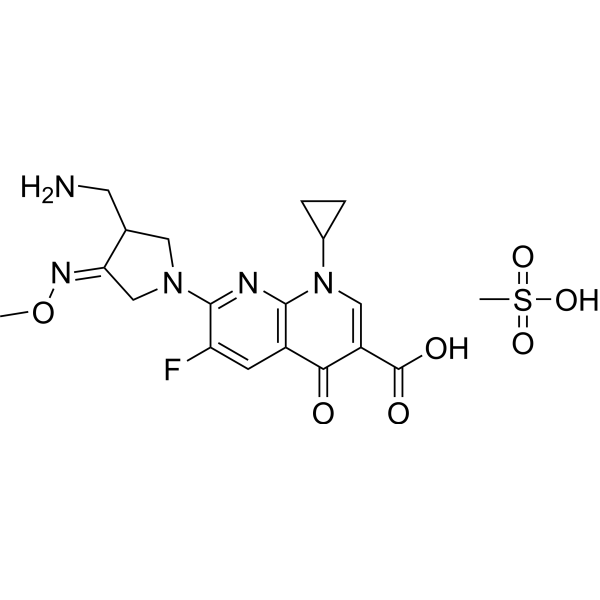
Gemifloxacin mesylate (SB-265805S; LB-20304a) is an orally active broad-spectrum quinolone antibacterial antibiotic. Gemifloxacin mesylate inhibits DNA synthesis by inhibiting DNA gyrase and Topoisomerase IV activities. Gemifloxacin mesylate has potent antibacterial activities against gram-positive bacteria in vitro efficacy study, particularly Streptococci and Staphylococci. Gemifloxacin mesylate has been used in the research of respiratory tract infections.
For research use only. We do not sell to patients.
- Purity : 99.59%
- CAS No.: 210353-53-0
- Formula: C19H24FN5O7S
- Molecular Weight:485.49
-
Storage:
4°C, sealed storage, away from moisture and light
* In solvent : -80°C, 6 months; -20°C, 1 month (sealed storage, away from moisture and light)

To place orders, for customer services and technical support, please contact: MedChemExpress USA
Tel: 609-228-6898 E-mail: [email protected] [email protected]


-
 Biological Activity
-
Chemical Information
-
Solvent & Solubility
- Protocols
- Purity & Documentation
- References
-
 Help & FAQs


-
HY-B0308
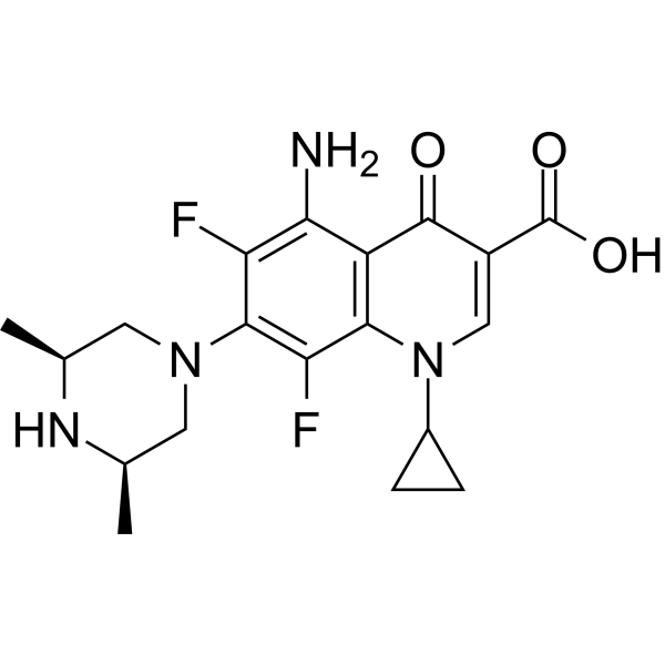
SparfloxacinBacterial Inhibitor -

HY-B1050R
Gemifloxacin mesylate (Standard)Antibacterial Agent -
HY-B1043
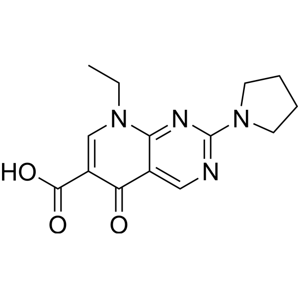
Piromidic acidAntibacterial Agent -
HY-B0159
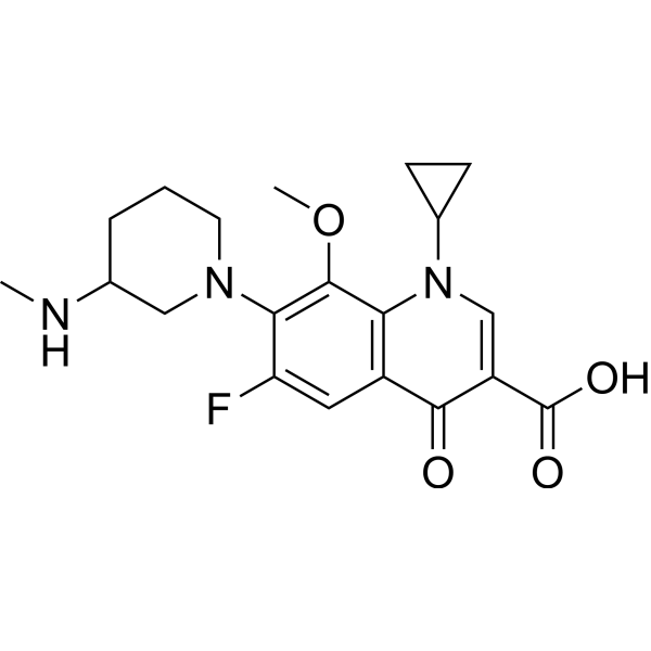
BalofloxacinAntibiotic -
HY-B1387
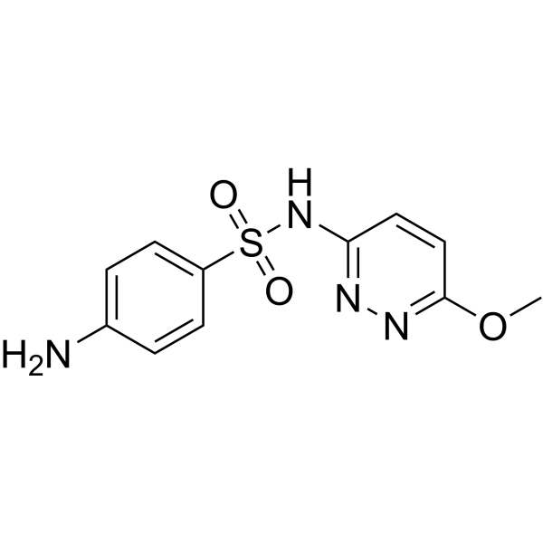
SulfamethoxypyridazineSulfonamide Antibiotic -
HY-13573
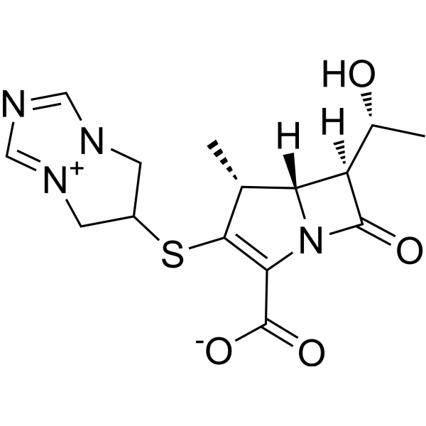
BiapenemAntibiotic -

HY-B1043R
Piromidic acid (Standard)Antibacterial Agent -

HY-B1387R
Sulfamethoxypyridazine (Standard)Reference Standard -

HY-B0159R
Balofloxacin (Standard)Antibiotic -

HY-13573R
Biapenem (Standard)Antibiotic


Biological Activity
Description
IC50 & Target
|
Quinolone |
In Vitro
Gemifloxacin has higher antibacterial activity than Moxifloxacin (HY-66011A) against Streptococcus pneumoniae with a MIC90 of 0.06 μg/mL[2].
Gemifloxacin has highly potent antibacterial activity against Penicillin-resistant strains of S. pneumoniae with a MIC90 of 0.03 μg/mL[2].
MedChemExpress (MCE) has not independently confirmed the accuracy of these methods. They are for reference only. Further protocols information, click here.
In Vivo
Pharmacokinetic Parameters of Gemifloxacin in Sprague-Dawley rats and dogs[1].
| Species | Administration | AUC0-24 (μg/mL·h) | Half-life (h) | Cmax (μg/mL) | Tmax (h) | F (%) |
| Rat | p.o.; 20 mg/kg | 8.50 | 2.33 | 2.44 | 0.33 | 95.3 |
| Dog | p.o.; 4 mg/kg | 7.55 | 5.12 | 1.34 | 1.13 | 71 |
MedChemExpress (MCE) has not independently confirmed the accuracy of these methods. They are for reference only.


Chemical Information
-
CAS No. 210353-53-0
-
Appearance Solid
-
Molecular Weight 485.49
-
Formula C19H24FN5O7S
-
Color White to off-white
-
SMILES
O=C(C1=CN(C2CC2)C3=C(C=C(F)C(N4CC(CN)/C(C4)=N/OC)=N3)C1=O)O.CS(=O)(O)=O
-
Synonyms
SB-265805S; LB-20304a
-
Shipping
Room temperature in continental US; may vary elsewhere.
-
Storage
4°C, sealed storage, away from moisture and light
* In solvent : -80°C, 6 months; -20°C, 1 month (sealed storage, away from moisture and light)


Solvent & Solubility
In Vitro:
DMSO : 100 mg/mL (205.98 mM; Need ultrasonic; Hygroscopic DMSO has a significant impact on the solubility of product, please use newly opened DMSO)
H2O : 50 mg/mL (102.99 mM; Need ultrasonic)
Please refer to the solubility information to select the appropriate solvent. Once prepared, please aliquot and store the solution to prevent product inactivation from repeated freeze-thaw cycles.
Storage method and period of stock solution: -80°C, 6 months; -20°C, 1 month (sealed storage, away from moisture and light). When stored at -80°C, please use it within 6 months. When stored at -20°C, please use it within 1 month.
* Note: If you choose water as the stock solution, please dilute it to the working solution, then filter and sterilize it with a 0.22 μm filter before use.
Please refer to the solubility information to select the appropriate solvent. Once prepared, please aliquot and store the solution to prevent product inactivation from repeated freeze-thaw cycles.
Storage method and period of stock solution: -80°C, 6 months; -20°C, 1 month (sealed storage, away from moisture and light). When stored at -80°C, please use it within 6 months. When stored at -20°C, please use it within 1 month.
* Note: If you choose water as the stock solution, please dilute it to the working solution, then filter and sterilize it with a 0.22 μm filter before use.
Concentration (start) × Volume (start) = Concentration (final) × Volume (final)
In Vivo:
Select the appropriate dissolution method based on your experimental animal and administration route.
- For the following dissolution methods, please ensure to first prepare a clear stock solution using an In Vitro approach and then sequentially add co-solvents:
- To ensure reliable experimental results, the clarified stock solution can be appropriately stored based on storage conditions. As for the working solution for In Vivo experiments, it is recommended to prepare freshly and use it on the same day.
- The percentages shown for the solvents indicate their volumetric ratio in the final prepared solution. If precipitation or phase separation occurs during preparation, heat and/or sonication can be used to aid dissolution.
Add each solvent one by one: 10% DMSO 40% PEG300 5% Tween-80 45% Saline
Solubility: ≥ 2.5 mg/mL (5.15 mM); Clear solution
This protocol yields a clear solution of ≥ 2.5 mg/mL (saturation unknown).
Taking 1 mL working solution as an example, add 100 μL DMSO stock solution (25.0 mg/mL) to 400 μL PEG300, and mix evenly; then add 50 μL Tween-80 and mix evenly; then add 450 μL Saline to adjust the volume to 1 mL.
Preparation of Saline: Dissolve 0.9 g sodium chloride in ddH₂O and dilute to 100 mL to obtain a clear Saline solution.
Add each solvent one by one: 10% DMSO 90% (20% SBE-β-CD in Saline)
Solubility: ≥ 2.5 mg/mL (5.15 mM); Clear solution
This protocol yields a clear solution of ≥ 2.5 mg/mL (saturation unknown).
Taking 1 mL working solution as an example, add 100 μL DMSO stock solution (25.0 mg/mL) to 900 μL 20% SBE-β-CD in Saline, and mix evenly.
Preparation of 20% SBE-β-CD in Saline (4°C, storage for one week): 2 g SBE-β-CD powder is dissolved in 10 mL Saline, completely dissolve until clear.
In Vivo Dissolution Calculator
Please enter the basic information of animal experiments:
-
-
-
-
Recommended: Prepare an additional quantity of animals to account for potential losses during experiments.
Working solution concentration: 0.22 mg/mL
This product has good water solubility, please refer to the measured solubility data in water/PBS/Saline for details.

Protocols
-
Research Protocol for Infectious Diseases
Infectious-disease experiments test how pathogens interact with host barriers, innate immune receptors, inflammatory signaling, pathogen replication, and tissue injury; pattern-recognition receptors such as TLRs, RIG-I-like receptors, NOD-like receptors, and inflammasomes detect microbial molecules and activate NF-κB, interferon, and cytokine responses. The central hypothesis is that infection severity reflects the balance between pathogen burden and host response: protective inflammation restricts pathogen growth, whereas excessive or mislocalized inflammation contributes to tissue damage and disease phenotype. Unresolved questions include which host pathways are protective versus pathogenic, why some infection models fail to translate to human disease, and which combined readouts best predict clinically relevant infection outcomes.
-
EdU Incorporation Assay (Click Chemistry-Based DNA Synthesis Measurement)
The EdU incorporation assay measures DNA synthesis by adding the thymidine analog 5-ethynyl-2′-deoxyuridine to cells or tissues, where it is incorporated into newly synthesized DNA during S phase. Incorporated EdU is detected by copper-catalyzed azide-alkyne cycloaddition, in which a fluorescent azide covalently reacts with the ethynyl group on EdU, allowing S-phase cells to be detected by fluorescence microscopy, flow cytometry, or high-content imaging. EdU detection does not require DNA denaturation or anti-BrdU antibody access, which preserves sample structure and improves compatibility with immunostaining and multiparameter cytometry compared with BrdU-based detection. EdU can be cytotoxic in a cell-type- and exposure-dependent manner, so pulse duration, concentration, and continuous-labeling designs should be validated for each cell type.
-
BrdU Incorporation Assay
Bromodeoxyuridine (BrdU) incorporation assay is based on the principle that BrdU, a thymidine analog, is incorporated into newly synthesized DNA during the S phase of the cell cycle, thereby serving as a marker of DNA replication and cellular proliferation. Incorporated BrdU can be detected using anti-BrdU antibodies following DNA denaturation, enabling visualization or quantification of proliferating cells through immunochemical detection methods such as immunofluorescence or immunohistochemistry.
-
Gram Staining of Tissue Sections
Gram staining of tissue sections is a histochemical technique used to differentiate Gram-positive and Gram-negative bacteria within histological specimens based on differences in bacterial cell wall structure and dye retention, adapted from classical bacteriological Gram staining into tissue-compatible “histological Gram stain” variants. In tissue applications, modifications of the Brown-Hopps and Brown-Brenn methods are commonly used to improve differentiation of microorganisms embedded within host connective tissue and to reduce overstaining or loss of Gram-negative signal, which are known limitations of earlier approaches. The principle relies on crystal violet-iodine complex retention in Gram-positive organisms and subsequent decolorization and counterstaining steps that allow contrast visualization of Gram-negative organisms against tissue background.


Purity & Documentation
-
Data Sheet (274 KB)
-
SDS (419 KB)
- English - EN (419 KB)
- Français - FR (419 KB)
- Deutsch - DE (419 KB)
- Norwegian - NO (419 KB)
- Español - ES (419 KB)
- Swedish - SV (419 KB)
- Italian - IT (419 KB)
- Korean - KR (419 KB)
- Portuguese - PT (419 KB)
-
Handling Instructions (2659 KB)
References
[1]. Hong CY. Discovery of gemifloxacin (Factive, LB20304a): a quinolone of a new generation. Farmaco. 2001 Jan-Feb;56(1-2):41-4. [Content Brief]
[2]. Allen A, Kim I, et al. Multiple-dose pharmacokinetics and tolerability of gemifloxacin administered orally to healthy volunteers. Antimicrob Agents Chemother. 2001 Feb;45(2):540-5. [Content Brief]
[3]. Erdem M, et al. Ingestion of the anti-bacterial agent, gemifloxacin mesylate, leads to increased gst activity and peroxidation products in hemolymph of Galleria mellonella l. (lepidoptera: pyralidae). Arch Insect Biochem Physiol. 2016 Dec;93(4):202-209. [Content Brief]


Complete Stock Solution Preparation Table
Please refer to the solubility information to select the appropriate solvent. Once prepared, please aliquot and store the solution to prevent product inactivation from repeated freeze-thaw cycles.
Storage method and period of stock solution: -80°C, 6 months; -20°C, 1 month (sealed storage, away from moisture and light). When stored at -80°C, please use it within 6 months. When stored at -20°C, please use it within 1 month.
| Optional Solvent | Concentration Solvent Mass | 1 mg | 5 mg | 10 mg | 25 mg |
|---|---|---|---|---|---|
| H2O / DMSO | 1 mM | 2.0598 mL | 10.2989 mL | 20.5977 mL | 51.4944 mL |
| 5 mM | 0.4120 mL | 2.0598 mL | 4.1195 mL | 10.2989 mL | |
| 10 mM | 0.2060 mL | 1.0299 mL | 2.0598 mL | 5.1494 mL | |
| 15 mM | 0.1373 mL | 0.6866 mL | 1.3732 mL | 3.4330 mL | |
| 20 mM | 0.1030 mL | 0.5149 mL | 1.0299 mL | 2.5747 mL | |
| 25 mM | 0.0824 mL | 0.4120 mL | 0.8239 mL | 2.0598 mL | |
| 30 mM | 0.0687 mL | 0.3433 mL | 0.6866 mL | 1.7165 mL | |
| 40 mM | 0.0515 mL | 0.2575 mL | 0.5149 mL | 1.2874 mL | |
| 50 mM | 0.0412 mL | 0.2060 mL | 0.4120 mL | 1.0299 mL | |
| 60 mM | 0.0343 mL | 0.1716 mL | 0.3433 mL | 0.8582 mL | |
| 80 mM | 0.0257 mL | 0.1287 mL | 0.2575 mL | 0.6437 mL | |
| 100 mM | 0.0206 mL | 0.1030 mL | 0.2060 mL | 0.5149 mL |
* Note: If you choose water as the stock solution, please dilute it to the working solution, then filter and sterilize it with a 0.22 μm filter before use.


Gemifloxacin mesylate Related Classifications
HY-B1050 Related Classifications
 Powered by Bioz