



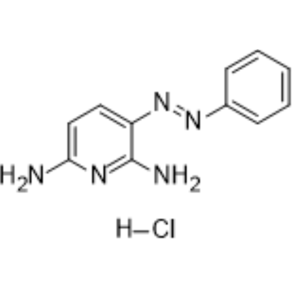
Phenazopyridine hydrochloride
Based on 2 publication(s) in Google Scholar
Phenazopyridine hydrochlorideis a competitive SARM1 inhibitor, with IC50 145 μM. Phenazopyridine hydrochlorideis a TRPM8 antagonist. Phenazopyridine hydrochloride has a local anesthetic/analgesic effect. Phenazopyridine hydrochlorideis used to relieve painful symptoms of conditions such as cystitis and urethritis. Phenazopyridine hydrochloridecan promote neuronal differentiation and can also be used in the study of traumatic brain injury, peripheral neuropathy and neurodegenerative diseases.
For research use only. We do not sell to patients.
- Purity : 99.77%
- CAS No.: 136-40-3
- Formula: C11H12ClN5
- Molecular Weight:249.70
-
Storage:
4°C, sealed storage, away from moisture
* In solvent : -80°C, 6 months; -20°C, 1 month (sealed storage, away from moisture)

To place orders, for customer services and technical support, please contact: MedChemExpress USA
Tel: 609-228-6898 E-mail: [email protected] [email protected]


-
 Biological Activity
-
Chemical Information
-
Solvent & Solubility
- Protocols
- Purity & Documentation
- References
-
 Help & FAQs


-
HY-17421
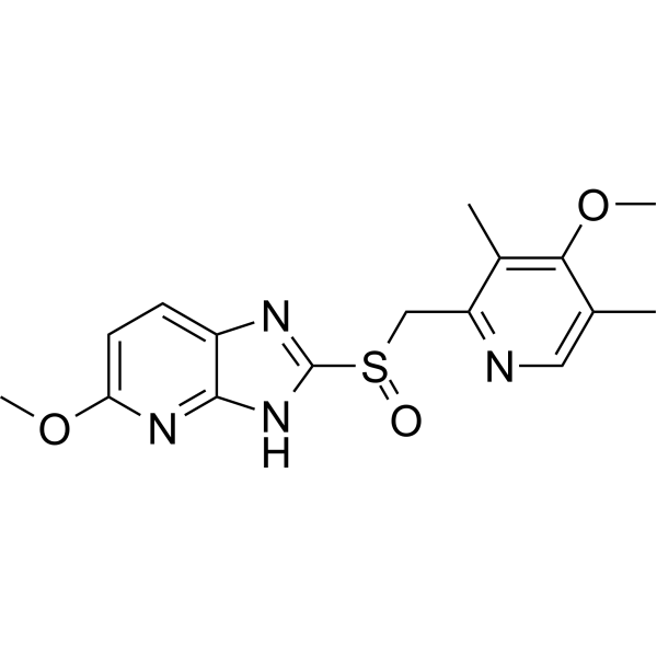
TenatoprazoleProton Pump Inhibitor -
HY-108464A
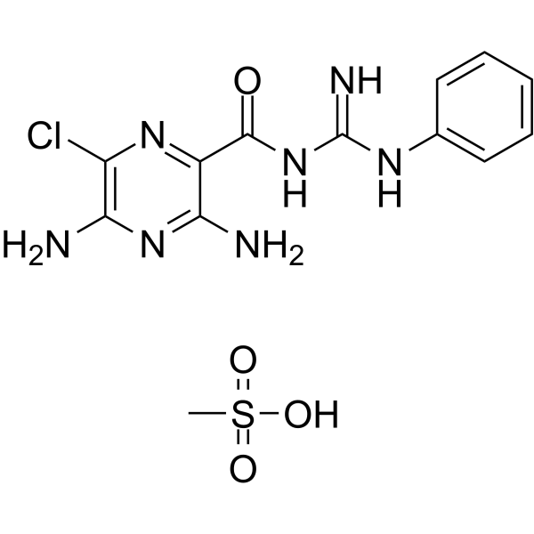
Phenamil methanesulfonateENaC Inhibitor -
HY-12687
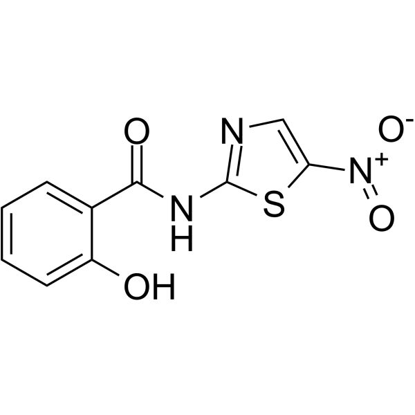
TizoxanideAnti-HIV Activity -
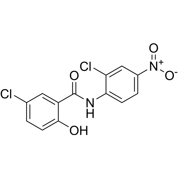
HY-B0497
NiclosamideAntihelminthic Agent -
HY-K0208
Streptavidin Magnetic Beads -
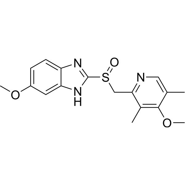
HY-B0113
OmeprazoleH+,K+-ATPase Inhibitor -
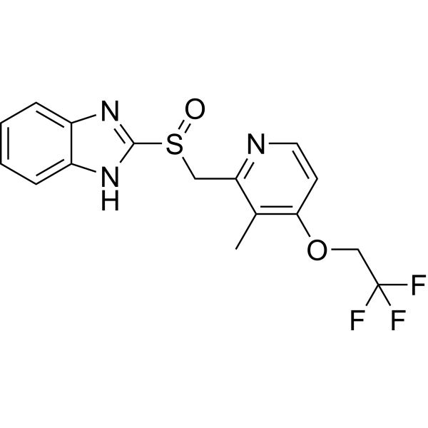
HY-13662
LansoprazoleProton Pump Inhibitor -
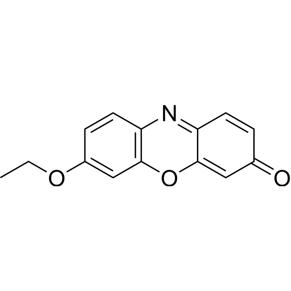
HY-D0145
7-EthoxyresorufinCytochrome P450 Inhibitor -
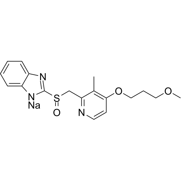
HY-B0656A
Rabeprazole sodiumPump Inhibitor -

HY-B0985R
Phenazopyridine hydrochloride (Standard)Analgesic -
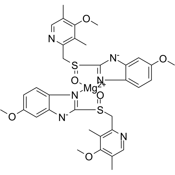
HY-B1446
Esomeprazole magnesiumH+, K+-ATPase Inhibitor -
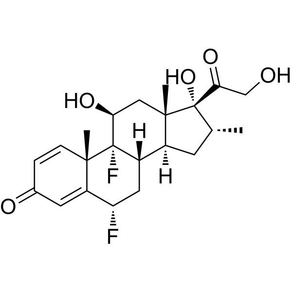
HY-B1051
FlumethasoneGR Agonist
Publications Citing Use of MedChemExpress (MCE) Phenazopyridine hydrochloride
More

Biological Activity
Description
IC50 & Target
EC50: 145 μM (SARM1)[1].
In Vitro
Phenazopyridine (10-30 μM; 12 h) increases mRNA expression of RPS23RG1 in both human SHSY5Y and mouse N2a cells[2].
Phenazopyridine (5-50 μM; 2 min) inhibits menthol induced (50 μM) TRPM8 response in a dose-dependent and reversible manner in HEK293 cells with an IC50 of 9.6 μM[4].
Phenazopyridine hydrochloride(3 μM; 6 weeks) can promote neuronal differentiation in human embryonic stem cells[5].
MedChemExpress (MCE) has not independently confirmed the accuracy of these methods. They are for reference only. Further protocols information, click here.
-
Cell Line:Human ES cells
-
Concentration:3 μM
-
Incubation Time:6 weeks
-
Result:After 10 weeks formed a neural network in the cells.
-
Cell Line:Human ES cells
-
Concentration:3 μM
-
Incubation Time:6 weeks
-
Result:Accelerated emergence of early neuronal markers and decreased markers of undifferentiated and non-neural cells.
In Vivo
Phenazopyridine (0.1-3 mg/kg; i.v.; Single dose) can inhibit mechanosensitive Aδ- fibers, but has no inhibitory effect on C-fibers in Sprague - Dawley rats[3].
MedChemExpress (MCE) has not independently confirmed the accuracy of these methods. They are for reference only.
-
Animal Model:C57BL/6 mice model of Alzheimer's disease[2]
-
Dosage:15 mg/kg
-
Administration:Intraperitoneal injection (i.p.); Once daily for 2 weeks
-
Result:Increased the RPS23RG1 levels in lungs, liver and kidneys of mice.
-
Animal Model:APP/PS1 mice model of Alzheimer's disease[2]
-
Dosage:15 mg/kg
-
Administration:Intracerebroventricularly injection; Once daily for 2 weeks
-
Result:Significantly reduced amyloid plaques.
Increased protein levels of RPS23RG1, PSD-95, and phosphorylated/inactivated GSK-3β, though without affecting levels of tau, phosphorylated tau, and p35.
Clinical Trial
| NCT Number | Sponsor | Condition | Start Date |
Phase

|
|---|---|---|---|---|
| NCT01329991 | Plexxikon| | 2011-05 | PHASE1 |


Chemical Information
-
CAS No. 136-40-3
-
Appearance Solid
-
Molecular Weight 249.70
-
Formula C11H12ClN5
-
Color Pink to red
-
SMILES
NC1=NC(N)=CC=C1/N=N/C2=CC=CC=C2.[H]Cl
-
Shipping
Room temperature in continental US; may vary elsewhere.
-
Storage
4°C, sealed storage, away from moisture
* In solvent : -80°C, 6 months; -20°C, 1 month (sealed storage, away from moisture)


Publications (2)
-
Journal Impact Factor
-
Most Recent
-
Neuropsychopharmacology
Phenazopyridine promotes RPS23RG1/Rps23rg1 transcription and ameliorates Alzheimer-associated phenotypes in mice. [Abstract]2022 Nov;47(12):2042-2050. PMID: 35821069 -
EMBO Rep
2022 Jun 7;23(6):e53932. PMID: 35403787


Solvent & Solubility
In Vitro:
DMSO : 25 mg/mL (100.12 mM; Need ultrasonic; Hygroscopic DMSO has a significant impact on the solubility of product, please use newly opened DMSO)
H2O : 1 mg/mL (4.00 mM; ultrasonic and warming and heat to 60°C)
Please refer to the solubility information to select the appropriate solvent. Once prepared, please aliquot and store the solution to prevent product inactivation from repeated freeze-thaw cycles.
Storage method and period of stock solution: -80°C, 6 months; -20°C, 1 month (sealed storage, away from moisture). When stored at -80°C, please use it within 6 months. When stored at -20°C, please use it within 1 month.
* Note: If you choose water as the stock solution, please dilute it to the working solution, then filter and sterilize it with a 0.22 μm filter before use.
Please refer to the solubility information to select the appropriate solvent. Once prepared, please aliquot and store the solution to prevent product inactivation from repeated freeze-thaw cycles.
Storage method and period of stock solution: -80°C, 6 months; -20°C, 1 month (sealed storage, away from moisture). When stored at -80°C, please use it within 6 months. When stored at -20°C, please use it within 1 month.
* Note: If you choose water as the stock solution, please dilute it to the working solution, then filter and sterilize it with a 0.22 μm filter before use.
Concentration (start) × Volume (start) = Concentration (final) × Volume (final)
In Vivo:
Select the appropriate dissolution method based on your experimental animal and administration route.
- For the following dissolution methods, please ensure to first prepare a clear stock solution using an In Vitro approach and then sequentially add co-solvents:
- To ensure reliable experimental results, the clarified stock solution can be appropriately stored based on storage conditions. As for the working solution for In Vivo experiments, it is recommended to prepare freshly and use it on the same day.
- The percentages shown for the solvents indicate their volumetric ratio in the final prepared solution. If precipitation or phase separation occurs during preparation, heat and/or sonication can be used to aid dissolution.
Add each solvent one by one: 10% DMSO 40% PEG300 5% Tween-80 45% Saline
Solubility: ≥ 2.5 mg/mL (10.01 mM); Clear solution
This protocol yields a clear solution of ≥ 2.5 mg/mL (saturation unknown).
Taking 1 mL working solution as an example, add 100 μL DMSO stock solution (25.0 mg/mL) to 400 μL PEG300, and mix evenly; then add 50 μL Tween-80 and mix evenly; then add 450 μL Saline to adjust the volume to 1 mL.
Preparation of Saline: Dissolve 0.9 g sodium chloride in ddH₂O and dilute to 100 mL to obtain a clear Saline solution.
Add each solvent one by one: 10% DMSO 90% (20% SBE-β-CD in Saline)
Solubility: ≥ 2.5 mg/mL (10.01 mM); Clear solution
This protocol yields a clear solution of ≥ 2.5 mg/mL (saturation unknown).
Taking 1 mL working solution as an example, add 100 μL DMSO stock solution (25.0 mg/mL) to 900 μL 20% SBE-β-CD in Saline, and mix evenly.
Preparation of 20% SBE-β-CD in Saline (4°C, storage for one week): 2 g SBE-β-CD powder is dissolved in 10 mL Saline, completely dissolve until clear.
In Vivo Dissolution Calculator
Please enter the basic information of animal experiments:
-
-
-
-
Recommended: Prepare an additional quantity of animals to account for potential losses during experiments.
Please enter your animal formula composition:
-
%DMSO +
Recommended: Keep the proportion of DMSO in working solution below 2% if your animal is weak.
-
%+
-
+%Tween-80 + +
-
%Saline +
The co-solvents required include: DMSO, . All of co-solvents are available by MedChemExpress (MCE). , Tween 80. All of co-solvents are available by MedChemExpress (MCE).
Working solution concentration: 0.22 mg/mL
Method for preparing stock solution: mg drug dissolved in μL DMSO. Stock solution concentration: mg/mL. * In solvent : -80°C, 6 months; -20°C, 1 month (sealed storage, away from moisture)
1. Take μL DMSO stock solution;
2. Add μL .
μL , mix evenly;
3. Then add μL Tween 80, mix evenly;
4. Then add μL
Please ensure that the stock solution in the first step is dissolved to a clear state, and add co-solvents in sequence. You can use ultrasonic heating (ultrasonic cleaner, recommended frequency 20-40 kHz), vortexing, etc. to assist dissolution.

Protocols
-
PC12 NGF-Induced Neuronal Differentiation Culture
PC12 cells are a rat adrenal pheochromocytoma clonal line that responds to NGF by stopping proliferation and extending branching neurite-like processes; after longer NGF exposure, cells develop long processes and neuronal-like ultrastructural and functional features. NGF-induced differentiation is read out mainly by neurite outgrowth, reduced proliferation, microtubule assembly, and neuronal differentiation-associated proteins such as MAPs, tau, GAP-43, and synapsin-1.
-
iPSC/hPSC-Derived Neuron Differentiation Culture
iPSC/hPSC-derived neuron differentiation culture directs pluripotent cells toward neuroectoderm and then neuronal lineages by suppressing developmental signals that maintain non-neural fates; the classic monolayer dual-SMAD approach blocks BMP and Activin/TGF-β signaling with Noggin or dorsomorphin/LDN193189 plus SB431542, producing PAX6-positive neural progenitors that can be further matured into neurons. The readout is generated by morphology, neural progenitor markers, neuronal markers, subtype markers, and functional assays: PAX6/SOX1/NESTIN indicate neural progenitor induction, βIII-tubulin/TUJ1 and MAP2 indicate neuronal differentiation, cortical programs can be assessed by FOXG1, TBR1, CTIP2, SATB2, and synaptic maturation can be assessed by synaptic proteins, calcium activity, multielectrode arrays, or patch-clamp electrophysiology.
-
Cell Cytotoxicity Assay
Cytotoxicity assays are usually based on the assessment of cell membrane damage, which can also be indirectly detected by measuring cell viability. Detection methods include MTT assay, CKK-8 assay, LDH assay and ATP assay, etc.
-
SH-SY5Y Neuronal Differentiation Culture
SH-SY5Y neuronal differentiation culture uses sequential exposure to retinoic acid and neurotrophic factors to reduce proliferative neuroblastoma-like behavior and induce neuron-like morphology, including neurite extension, neuronal marker expression, and, in RA/BDNF protocols, greater synaptic-marker expression than undifferentiated culture. Retinoic acid is commonly used as the initiating differentiation cue, while BDNF in serum-reduced or serum-free medium supports later maturation and neurotrophic-factor-dependent neuron-like survival.
-
Human pluripotent stem cell neural induction and neuron differentiation
Human pluripotent stem cell neural induction can be achieved by blocking BMP and TGFβ/Activin/Nodal SMAD signaling, which suppresses non-neural differentiation and promotes early neuroectodermal identity; the expected readout is loss of pluripotency markers such as OCT4 and induction of neural markers such as PAX6, followed by neural progenitor and neuron marker acquisition during differentiation. This protocol uses dual-SMAD neural induction as the core induction method, followed by cortical neuron differentiation as a representative neuron differentiation model; published cortical protocols describe generation of cortical progenitors, temporally ordered cortical projection neurons, action-potential firing, synaptogenesis, and neural network formation over an approximately 80-day process.
-
SH-SY5Y neuronal-like differentiation
SH-SY5Y neuronal-like differentiation uses defined culture conditions to shift proliferative human neuroblastoma cells toward a neuron-like state, mainly assessed by reduced proliferation, neurite extension, neuronal-marker expression, and, in some protocols, increased dependence on neurotrophic support. Retinoic acid (RA) is commonly used for the first differentiation phase, and sequential RA followed by brain-derived neurotrophic factor (BDNF) in serum-free medium is a well-characterized approach for generating neuron-like SH-SY5Y cultures with extensive neurite outgrowth. The primary readouts are morphology-based neurite outgrowth and marker-based confirmation using proteins such as βIII-tubulin, MAP2, GAP43, synaptophysin, NeuN, NSE, TH, or related neuronal/synaptic markers, depending on the study endpoint.
-
Cell differentiation
Cell differentiation refers to the process in which cells of the same origin gradually produce cell groups with different morphological structure and functional characteristics.
-
PC12 NGF-induced neuronal-like differentiation
PC12 cells are a rat adrenal pheochromocytoma-derived clonal cell line that responds to nerve growth factor by stopping proliferation and extending neurites, producing a sympathetic neuron-like phenotype used to study neuronal differentiation and neurite outgrowth. NGF acts through TrkA-dependent signaling, and neurite outgrowth is associated with ERK/Akt signaling, microtubule organization, neuronal-marker expression, and increased electrophysiological neuronal features such as sodium-channel density. The main assay readout is morphological differentiation, usually measured as the percentage of neurite-bearing cells, neurite length, neurite number, or total neurite length per cell. Additional readouts include GAP-43, tyrosine hydroxylase, βIII-tubulin, neurofilament, synapsin I, synaptophysin, ERK phosphorylation, Akt phosphorylation, and sodium-channel current density.
-
Directly Induced Neuron Culture
Directly induced neuron culture converts somatic cells, most commonly fibroblasts, into induced neurons without passing through a pluripotent or neural progenitor stage; classic evidence shows that mouse fibroblasts can be converted by Ascl1, Brn2/Pou3f2, and Myt1l, human fibroblasts can be converted by defined neuronal transcription factors, and human fibroblasts can also be converted by miR-9/9-124 with neurogenic or subtype-specifying transcription factors. The readout is acquisition of neuronal identity and function, assessed by neuronal morphology, neuronal markers such as Tuj1/βIII-tubulin, MAP2, synapsin, and subtype markers when relevant, together with functional assays such as action-potential firing, synaptic activity, and electrophysiology.


Purity & Documentation
-
Data Sheet (280 KB)
-
SDS (420 KB)
- English - EN (420 KB)
- Français - FR (420 KB)
- Deutsch - DE (420 KB)
- Norwegian - NO (420 KB)
- Español - ES (420 KB)
- Swedish - SV (420 KB)
- Italian - IT (420 KB)
- Korean - KR (420 KB)
- Portuguese - PT (420 KB)
-
Handling Instructions (2659 KB)
References
[1]. Loring H S, et al. Identification of the first noncompetitive SARM1 inhibitors[J]. Bioorganic & Medicinal Chemistry, 2020, 28(18): 115644. [Content Brief]
[2]. Wang C, et al. Phenazopyridine promotes RPS23RG1/Rps23rg1 transcription and ameliorates Alzheimer-associated phenotypes in mice[J]. Neuropsychopharmacology, 2022, 47(12): 2042-2050. [Content Brief]
[3]. Aizawa N, et al. Effects of phenazopyridine on rat bladder primary afferent activity, and comparison with lidocaine and acetaminophen[J]. Neurourology and Urodynamics, 2010, 29(8): 1445-1450. [Content Brief]
[4]. Luyts N, et al. Inhibition of TRPM8 by the urinary tract analgesic drug phenazopyridine[J]. European Journal of Pharmacology, 2023, 942: 175512. [Content Brief]
[5]. Suter, David M et al. Phenazopyridine hydrochlorideinduces and synchronizes neuronal differentiation of embryonic stem cells. Journal of cellular and molecular medicine vol. 13,9B (2009): 3517-27. [Content Brief]


Complete Stock Solution Preparation Table
Please refer to the solubility information to select the appropriate solvent. Once prepared, please aliquot and store the solution to prevent product inactivation from repeated freeze-thaw cycles.
Storage method and period of stock solution: -80°C, 6 months; -20°C, 1 month (sealed storage, away from moisture). When stored at -80°C, please use it within 6 months. When stored at -20°C, please use it within 1 month.
| Optional Solvent | Concentration Solvent Mass | 1 mg | 5 mg | 10 mg | 25 mg |
|---|---|---|---|---|---|
| H2O / DMSO | 1 mM | 4.0048 mL | 20.0240 mL | 40.0481 mL | 100.1201 mL |
| DMSO | 5 mM | 0.8010 mL | 4.0048 mL | 8.0096 mL | 20.0240 mL |
| 10 mM | 0.4005 mL | 2.0024 mL | 4.0048 mL | 10.0120 mL | |
| 15 mM | 0.2670 mL | 1.3349 mL | 2.6699 mL | 6.6747 mL | |
| 20 mM | 0.2002 mL | 1.0012 mL | 2.0024 mL | 5.0060 mL | |
| 25 mM | 0.1602 mL | 0.8010 mL | 1.6019 mL | 4.0048 mL | |
| 30 mM | 0.1335 mL | 0.6675 mL | 1.3349 mL | 3.3373 mL | |
| 40 mM | 0.1001 mL | 0.5006 mL | 1.0012 mL | 2.5030 mL | |
| 50 mM | 0.0801 mL | 0.4005 mL | 0.8010 mL | 2.0024 mL | |
| 60 mM | 0.0667 mL | 0.3337 mL | 0.6675 mL | 1.6687 mL | |
| 80 mM | 0.0501 mL | 0.2503 mL | 0.5006 mL | 1.2515 mL | |
| 100 mM | 0.0400 mL | 0.2002 mL | 0.4005 mL | 1.0012 mL |
* Note: If you choose water as the stock solution, please dilute it to the working solution, then filter and sterilize it with a 0.22 μm filter before use.


Phenazopyridine hydrochloride Related Classifications
HY-B0985 Related Classifications
 Powered by Bioz