



Andecaliximab

Based on 1 Customer Validation
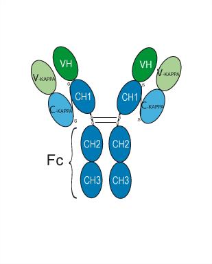
Andecaliximab is a recombinant chimeric IgG4 monoclonal antibody (mAb) targets matrix metalloproteinase 9 (MMP9). Andecaliximab shows the antifibrotic efficacy in idiopathic pulmonary fibrosis mouse models. Andecaliximab can be used for the research of gastric adenocarcinoma and idiopathic pulmonary fibrosis (IPF).
For research use only. We do not sell to patients.
- Purity : 98.57%
- CAS No.: 1518996-49-0
- Molecular Weight:150 kDa
-
Storage:
Please store the product under the recommended conditions in the Certificate of Analysis.


To place orders, for customer services and technical support, please contact: MedChemExpress USA
Tel: 609-228-6898 E-mail: [email protected] [email protected]


-
 Biological Activity
-
Chemical Information
- Protocols
- Purity & Documentation
- References
-
 Help & FAQs





Biological Activity
Description
Isotype
Human IgG4 kappa
Recommend Isotype Controls
Species Reactivity
Human
IC50 & Target
MMP9
In Vitro
In Vivo
MedChemExpress (MCE) has not independently confirmed the accuracy of these methods. They are for reference only.
-
Animal Model:Humanized nonobese diabetic, SCID, IL-2 receptor g (NSG) mouse model of IPF[2]
-
Dosage:20 mg/kg
-
Administration:Intraperitoneal injection; 20 mg/kg, once a week for once
-
Result:Decreased SMAD2 phosphorylation and increased surfactant protein C. Reduced MMP9+ cells, including MMP9+, CCR10+ and MMP9+CD45- and EpCAM+CCR10+ subpopulations.
Clinical Trial
| NCT Number | Sponsor | Condition | Start Date |
Phase

|
|---|---|---|---|---|
| NCT01329991 | 2011-05 | PHASE1 |
Gene ID
Accession
Conjugated
Unconjugated
Reconsititution
The product can be reconstituted/diluted with sterile PBS or saline.
Format
-
Human IgG4 kappa
Application
ELISA, FACS, Functional assay
Verified Bioactivity
-
 Immobilized MMP-9 Protein, Human (HEK293, HY-P73300) can bind Andecaliximab. The EC50 for this effect is 5.72 ng/mL.


Chemical Information
-
CAS No. 1518996-49-0
-
Appearance Liquid
-
Molecular Weight 150 kDa
-
Color Colorless to light yellow
-
SMILES
[Andecaliximab]
-
Synonyms
Anti-MMP9 Reference Antibody (andecaliximab)
-
Shipping
Shipping with dry ice.
-
Formulation
Please refer to the lot-specific COA for specific buffer information.
-
Storage
Please store the product under the recommended conditions in the Certificate of Analysis.


Purity & Documentation
-
Data Sheet (260 KB)
-
SDS (251 KB)
- English - EN (251 KB)
- Français - FR (251 KB)
- Deutsch - DE (251 KB)
- Norwegian - NO (251 KB)
- Español - ES (251 KB)
- Swedish - SV (251 KB)
- Italian - IT (251 KB)
- Korean - KR (251 KB)
- Portuguese - PT (251 KB)
-
Inhibitory Antibodies User Guide (603 KB)
References

Calculators
Concentration (start) × Volume (start) = Concentration (final) × Volume (final)


Andecaliximab Related Classifications
HY-P99351 Related Classifications
 Powered by Bioz