



Arvanil

Based on 1 Customer Validation
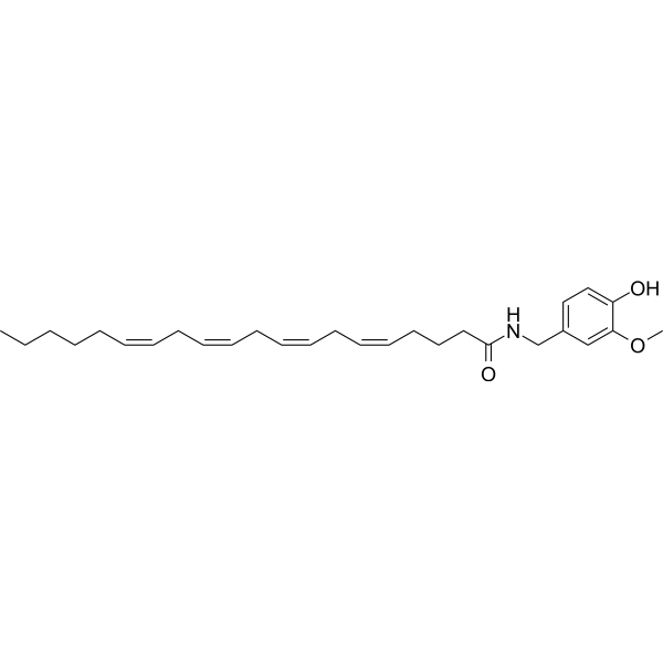
Arvanil (N-Vanillylarachidonamide) is a mixed agonist of CB1 and TRPV1 receptors. Arvanil downregulates CD25, HLA-DR, CD134/OX40, blocks G1/S phase transition, and induces phosphorylation of Akt. Arvanil does not induce apoptosis in cells. Arvanil inhibits lymphocyte activation and ameliorates autoimmune encephalomyelitis. Arvanil can be used in research related to Huntington's disease, vomiting, and multiple sclerosis.
For research use only. We do not sell to patients.
- Purity : 99.1%
- CAS No.: 128007-31-8
- Formula: C28H41NO3
- Molecular Weight:439.63
-
Storage:
Solution, -20°C, 2 years

To place orders, for customer services and technical support, please contact: MedChemExpress USA
Tel: 609-228-6898 E-mail: [email protected] [email protected]


-
 Biological Activity
-
Chemical Information
- Protocols
- Purity & Documentation
- References
-
 Help & FAQs


-
HY-14167
GW842166XCannabinoid Receptor Agonist -
HY-15443
AM251CB1 Receptor Antagonist -
HY-103332
N-ArachidonylglycineGPR18 Agonist -
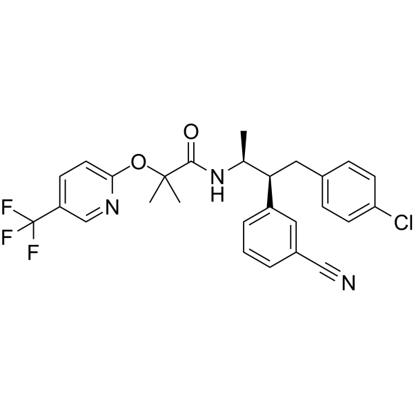
HY-10013
TaranabantCB1 Receptor Invert Agonist -

HY-121476
Arachidonoyl SerinolMAGL Inhibitor -
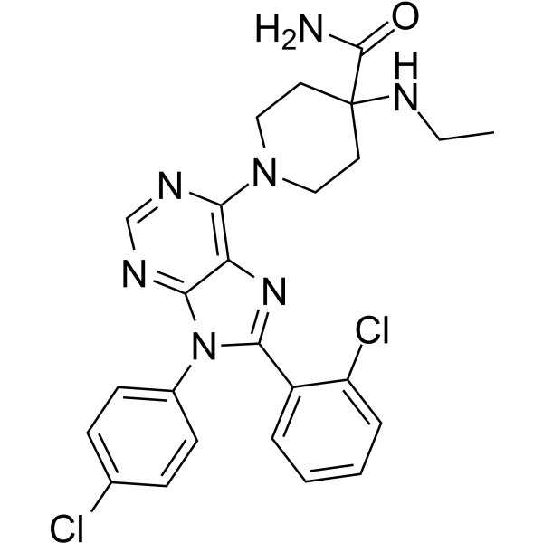
HY-10871
OtenabantCannabinoid Receptor Antagonist -
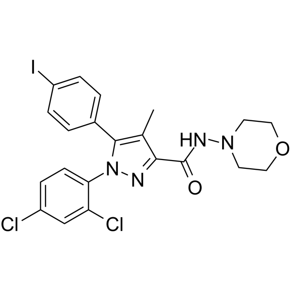
HY-13505
AM281CB1 Antagonist -
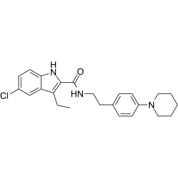
HY-13288
Org 27569Cannabinoid Receptor Modulator -
HY-W011040
LY320135CB1 Antagonist -

HY-103335
O1918 -
HY-103337
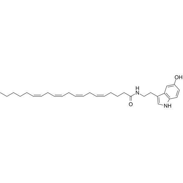
N-ArachidonoylserotoninFAAH Inhibitor/TRPV1 Antagonist -
HY-110004
ACEABiochemical Assay Reagent
All Orexin Receptor (OX Receptor) Isoforms
More

Biological Activity
Description
IC50 & Target
[1]|
TRPV1 |
CB1 |
Cellular Effect
|
Cell Line

|
Type | Value | Description | References |
|---|---|---|---|---|
| DMS-114 | IC50 |
15 μM
Compound: 10
|
Antiproliferative activity in human DMS-114 cells assessed as cell growth inhibition by MTT assay
Antiproliferative activity in human DMS-114 cells assessed as cell growth inhibition by MTT assay
|
[PMID: 33508189] |
| EFM-19 | IC50 |
0.55 μM
Compound: 10
|
Antiproliferative activity in human EFM-19 cells assessed as cell growth inhibition by MTT assay
Antiproliferative activity in human EFM-19 cells assessed as cell growth inhibition by MTT assay
|
[PMID: 33508189] |
| MCF7 | IC50 |
0.4 μM
Compound: 10
|
Antiproliferative activity in human MCF7 cells assessed as cell growth inhibition by MTT assay
Antiproliferative activity in human MCF7 cells assessed as cell growth inhibition by MTT assay
|
[PMID: 33508189] |
| PPC-1 | IC50 |
1 μM
Compound: 10
|
Antiproliferative activity in human PPC-1 cells assessed as cell growth inhibition
Antiproliferative activity in human PPC-1 cells assessed as cell growth inhibition
|
[PMID: 33508189] |
| T47D | IC50 |
0.35 μM
Compound: 10
|
Antiproliferative activity in human T47D cells assessed as cell growth inhibition by MTT assay
Antiproliferative activity in human T47D cells assessed as cell growth inhibition by MTT assay
|
[PMID: 33508189] |
| TSU | IC50 |
100 nM
Compound: 10
|
Antiproliferative activity in human TSU cells assessed as cell growth inhibition
Antiproliferative activity in human TSU cells assessed as cell growth inhibition
|
[PMID: 33508189] |
In Vitro
Arvanil (10 μM; 48 h) potently inhibits the proliferation of OKT3-stimulated peripheral blood mononuclear cells (PBMCs), with an inhibition rate of 75.3% at 10 μM, and this effect is superior to that of anandamide and capsaicin at the same concentration[3].
Arvanil (0.3-30 μM; 48 h) inhibits the proliferation of CD3CD28-stimulated human peripheral blood mononuclear cells (hPBMCs) in a dose-dependent manner, with an inhibition rate of 64.0% at 10 μM. It also reduces IFN-γ production, but does not affect IL-5 levels at this concentration[3].
Arvanil (3-10 μM; 48 h) inhibits the proliferation of mouse CTLL-2 cells stimulated by IL-2, and significant inhibitory effect is observed at the concentration of 10 μM[3].
Arvanil (10 μM) inhibits the upregulation of CD25, HLA-DR and CD134/OX40 activation markers on CD4+ T cells in OKT3-stimulated human peripheral blood mononuclear cells (hPBMCs)[3].
Arvanil (10 μM; 48 h) does not induce apoptosis in CD4+ T cells derived from OKT3-stimulated human peripheral blood mononuclear cells (hPBMCs)[3].
Arvanil (10 μM; 48 h) blocks G1/S phase transition in OKT3-stimulated human peripheral blood mononuclear cells (hPBMCs)[3].
MedChemExpress (MCE) has not independently confirmed the accuracy of these methods. They are for reference only. Further protocols information, click here.
-
Cell Line:murine CTLL-2 cells stimulated with interleukin-2 (IL-2)
-
Concentration:3-10 μM
-
Incubation Time:48 h
-
Result:Inhibited IL-2-stimulated CTLL-2 proliferation, with statistically significant inhibition observed at 10 μM (p<0.05); the inhibitory effect at 3 μM was less pronounced.
-
Cell Line:OKT3-stimulated hPBMCs
-
Concentration:10 μM
-
Incubation Time:48 h
-
Result:Increased G1-phase cells to 86% and reduced S-phase cells to 13%.
In Vivo
Arvanil (2 mg/kg; i.p.; single acute dose) reduces locomotor and exploratory activity in normal rats, an effect associated with increased GABA content in the globus pallidus[1].
Arvanil (1-2 mg/kg; i.p.; single dose 15 min pre-M6G) dose-dependently inhibits morphine 6 glucuronide-induced emesis and vomiting in ferrets via activation of both CB1 and TRPV1 receptors, with the 2 mg/kg dose producing a significant reduction in emetic and vomiting episodes that is reversed by selective antagonists of these receptors[2].
Arvanil (0.5 mg/kg; given on 2 separate days: day -3 and day +3 relative to EAE induction) treatment significantly ameliorates experimental autoimmune encephalomyelitis in female SJL/J mice, reducing the cumulative disease index by 48.2%, peak clinical score by 56.7%, maximal body weight loss by 66.8%, and central nervous system inflammatory foci by 51.3%[3].
MedChemExpress (MCE) has not independently confirmed the accuracy of these methods. They are for reference only.
-
Animal Model:Sprague-Dawley (male, 3-4 months old, 300-400 g, bilateral intrastriatal injection of 375 nmol 3-nitropropionic acid per striatum, used 14 days post-lesion)[1]
-
Dosage:2 mg/kg
-
Administration:i.p.; single acute dose
-
Result:Significantly reduced increased ambulatory activity (F(1,21)=18.74, P < 0.0005).
Increased time spent in inactivity (F(1,21)=40.10, P < 0.0001).
Enhanced lesion-induced reductions in stereotypic activity (F(1,22)=21.91, P < 0.0001) and hole entries (F(1,22)=43.18, P < 0.0001).
Increased glutamate contents in the globus pallidus (F(1,21)=5.89, P < 0.05).
Did not reverse GABA, glutamate, or dopamine deficits in the caudate-putamen.
-
Animal Model:Sprague-Dawley (male, 3-4 months old, 300-400 g, bilateral intrastriatal injection of saline vehicle, allowed 14 days to recover)[1]
-
Dosage:2 mg/kg
-
Administration:i.p.; single acute dose
-
Result:Significantly reduced ambulatory activity, stereotypic activity, and number of hole entries.
Increased time spent in inactivity.
Increased GABA contents in the globus pallidus (F(1,19)=3.89, P < 0.05).
Tended to increase globus pallidus GAD activity.
Showed no effects on dopamine, DOPAC, TH activity, GABA, GAD activity, or glutamate contents in the caudate-putamen.
-
Animal Model:Mustela putorius furo (adult male, 900-1500 g)[2]
-
Dosage:1 mg/kg; 2 mg/kg
-
Administration:i.p.; single dose 15 min pre-M6G
-
Result:Reduced M6G-induced emetic episodes compared to M6G alone.
Dose-dependently reduced M6G-induced emetic episodes to a greater extent than the 1 mg/kg dose; this anti-emetic effect was reversed by pre-treatment with 5 mg/kg AM251 or 0.1 mg/kg IRTX, with emetic episode counts returning to levels not significantly different from M6G alone.
Reduced M6G-induced vomiting episodes from 1.8 to 0.8 (P < 0.05); this effect was reversed by 5 mg/kg AM251 (to 1.4 episodes, P < 0.05 vs arvanil alone) and completely abolished by 0.1 mg/kg IRTX (to 1.8 episodes, P < 0.05 vs arvanil alone).
Reduced M6G-induced tongue licking movements from 6.6 licks/h to 2.3 licks/h (P < 0.01 vs M6G); this effect was reversed by CB1 and TRPV1 antagonists.
Dose-dependently reduced voluntary activity minutes in a 1-hour observation period; this sedative effect was not reversed by AM251 or IRTX.
Pre-treatment with 5 mg/kg AM630 did not reduce the anti-emetic effects of arvanil (3.5 emetic episodes vs 1.8 emetic episodes with arvanil alone, P > 0.05).


Chemical Information
-
CAS No. 128007-31-8
-
Appearance Liquid
-
Molecular Weight 439.63
-
Formula C28H41NO3
-
Color Colorless to light yellow
-
SMILES
CCCCC/C=C\C/C=C\C/C=C\C/C=C\CCCC(NCC1=CC=C(O)C(OC)=C1)=O
-
Synonyms
N-Vanillylarachidonamide
-
Shipping
Room temperature in continental US; may vary elsewhere.
-
Storage
Solution, -20°C, 2 years

Protocols
-
Kinase activity and phosphorylation assays
Kinase activity assays measure the ability of kinases to transfer phosphate groups from ATP to specific substrates, while phosphorylation assays detect the presence and levels of phosphorylated proteins. Common methods include radiolabeled ATP incorporation (e. g. ,), ADP release detection via bioluminescence (e. g. ,[3]), enzyme-linked immunosorbent assays (ELISA) for phospho-specific epitopes (e. g. ,[6]), and microtiter-based formats for high-throughput screening (e. g. ,[8]). The ADP-Glo assay quantifies kinase activity by measuring ADP produced during phosphorylation using a luciferase-based system. Radiometric assays involve autoradiography or scintillation counting after incorporation of 32P-labeled ATP into substrate proteins. ELISA-based approaches rely on phospho-specific antibodies to detect activated kinases in cell lysates or purified samples.
-
EdU Incorporation Assay (Click Chemistry-Based DNA Synthesis Measurement)
The EdU incorporation assay measures DNA synthesis by adding the thymidine analog 5-ethynyl-2′-deoxyuridine to cells or tissues, where it is incorporated into newly synthesized DNA during S phase. Incorporated EdU is detected by copper-catalyzed azide-alkyne cycloaddition, in which a fluorescent azide covalently reacts with the ethynyl group on EdU, allowing S-phase cells to be detected by fluorescence microscopy, flow cytometry, or high-content imaging. EdU detection does not require DNA denaturation or anti-BrdU antibody access, which preserves sample structure and improves compatibility with immunostaining and multiparameter cytometry compared with BrdU-based detection. EdU can be cytotoxic in a cell-type- and exposure-dependent manner, so pulse duration, concentration, and continuous-labeling designs should be validated for each cell type.
-
Western Blot
Western blotting (WB) is a commonly used experimental method in molecular biology, biochemistry, and immunogenetics for identifying and quantifying target proteins. It combines gel electrophoresis with immunoassay, enabling researchers to analyze protein expression, post-translational modifications, and molecular weight.
-
Apoptosis
Apoptosis, also called programmed cell death, is generally characterized by distinct morphological characteristics.
-
TUNEL staining for apoptotic DNA fragmentation
TUNEL staining detects DNA strand breaks by using terminal deoxynucleotidyl transferase to add labeled nucleotides to exposed 3′-OH DNA termini, generating either microscopic staining in fixed cells or tissue sections, or fluorescence/cytometric signal in cell suspensions. TUNEL positivity reflects DNA fragmentation but should not be interpreted alone as definitive apoptosis, because TUNEL can also label necrotic, autolytic, mechanically damaged, or DNA-repair-associated DNA breaks.
-
Flow cytometric DNA-content cell-cycle staining
Flow cytometric DNA-content cell-cycle staining measures the fluorescence intensity of DNA-bound fluorochromes in single cells or nuclei to estimate DNA content distributions, allowing assignment of populations to G0/G1, S, and G2/M phases by DNA histogram deconvolution. Propidium iodide (PI) intercalates into DNA, and PI fluorescence is proportional to cellular DNA content when staining is performed under conditions that make DNA accessible and minimize non-DNA signal. Cells with G2/M DNA content are expected to show approximately twice the fluorescence intensity of G0/G1 cells, while S-phase cells occupy intermediate fluorescence values. PI-based DNA-content analysis can also detect cells with fractional DNA content, often reported as sub-G1, when DNA fragmentation and extraction during staining reduce retained DNA signal in apoptotic cells. DAPI is an alternative DNA fluorochrome for univariate DNA-content analysis, while bivariate approaches combining DNA content with proliferation
-
Annexin V plus membrane-impermeant dye apoptosis staining
Annexin V-based apoptosis assays rely on the detection of phosphatidylserine (PS) externalization from the inner leaflet of the plasma membrane to the outer leaflet, an early biochemical hallmark of apoptosis. Fluorescently labeled Annexin V binds PS in a calcium-dependent manner, enabling identification of early apoptotic cells by flow cytometry or fluorescence microscopy. When combined with a membrane-impermeant DNA-binding dye (e. g. , propidium iodide), this approach allows discrimination between viable (Annexin V−/dye−), early apoptotic (Annexin V+/dye−), and late apoptotic or necrotic (Annexin V+/dye+) cell populations by assessing membrane integrity and PS exposure.
-
BrdU Incorporation Assay
Bromodeoxyuridine (BrdU) incorporation assay is based on the principle that BrdU, a thymidine analog, is incorporated into newly synthesized DNA during the S phase of the cell cycle, thereby serving as a marker of DNA replication and cellular proliferation. Incorporated BrdU can be detected using anti-BrdU antibodies following DNA denaturation, enabling visualization or quantification of proliferating cells through immunochemical detection methods such as immunofluorescence or immunohistochemistry.
-
Apoptosis Solutions
Apoptosis is a regulated, generally non-lytic cell-death pathway that removes unwanted, damaged, infected, or abnormal cells through coordinated morphological changes, caspase activation, DNA fragmentation, and membrane remodeling. The intrinsic apoptosis pathway is controlled mainly by mitochondrial outer membrane permeabilization, BCL-2 family proteins, cytochrome c release, apoptosome formation, caspase-9 activation, and downstream executioner caspase-3/7 activation. The extrinsic apoptosis pathway is initiated by death receptors such as Fas, TNFR, and TRAIL receptors, which recruit adaptor proteins and activate caspase-8 before engaging executioner caspases or mitochondrial amplification through BID cleavage. Apoptosis is linked to many phenotypes, including cancer cell killing, tissue homeostasis, immune regulation, neurodegeneration, infection response, and treatment-induced cytotoxicity; unresolved questions include how apoptosis interacts with necroptosis, pyroptosis, ferroptos
-
Protocol for Cell Cycle
Cell-cycle analysis by flow cytometry measures DNA content in single cells to estimate the fraction of cells in G0/G1, S, and G2/M phases. Propidium iodide intercalates into DNA, and after RNA removal with RNase, fluorescence intensity reflects cellular DNA content: 2N cells are assigned to G0/G1, cells between 2N and 4N to S phase, and 4N cells to G2/M. DNA-content analysis alone cannot reliably separate G0 from G1 or G2 from M. Ki-67 can distinguish quiescent G0 cells from cycling cells, EdU or BrdU incorporation marks active DNA synthesis in S phase, and phospho-histone H3 staining identifies mitotic cells within the 4N population.
-
Protocol for Kinase activity and phosphorylation assays
Kinase activity assays measure transfer of phosphate from ATP to a protein or peptide substrate, generating phosphorylated substrate, ADP, or incorporated radiolabeled phosphate as the readout; phosphorylation assays measure site-specific phosphorylation in cells or tissues as a proxy for kinase-pathway activation, inhibition, or substrate regulation. Phosphorylation can be detected by phospho-specific Western blot, immunoprecipitation kinase assay, phospho-immunofluorescence, phospho-flow cytometry, luminescent ADP detection, radiolabeled ATP incorporation, or reporter-based pathway assays, and these readouts can be applied to cancer cells, primary neurons, mouse tumors, organoids, inflammatory macrophages, ferroptosis studies, and mitophagy studies when the kinase target is biologically relevant.


Purity & Documentation
-
Data Sheet (275 KB)
-
SDS (545 KB)
- English - EN (545 KB)
- Français - FR (545 KB)
- Deutsch - DE (545 KB)
- Norwegian - NO (545 KB)
- Español - ES (545 KB)
- Swedish - SV (545 KB)
- Italian - IT (545 KB)
- Korean - KR (545 KB)
- Portuguese - PT (545 KB)
-
Handling Instructions (2659 KB)
References
[1]. de Lago E, et al. Arvanil, a hybrid endocannabinoid and vanilloid compound, behaves as an antihyperkinetic agent in a rat model of Huntington's disease. Brain Res. 2005;1050(1-2):210-216. [Content Brief]
[2]. Sharkey KA, et al. Arvanil, anandamide and N-arachidonoyl-dopamine (NADA) inhibit emesis through cannabinoid CB1 and vanilloid TRPV1 receptors in the ferret. Eur J Neurosci. 2007;25(9):2773-2782. [Content Brief]
[3]. Malfitano AM, et al. Arvanil inhibits T lymphocyte activation and ameliorates autoimmune encephalomyelitis. J Neuroimmunol. 2006;171(1-2):110-119. [Content Brief]

Calculators
Concentration (start) × Volume (start) = Concentration (final) × Volume (final)


Arvanil Related Classifications
HY-103333 Related Classifications
 Powered by Bioz
Keywords