



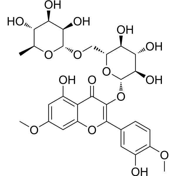
Ombuoside

Based on 1 Customer Validation
Ombuoside has antioxidant properties, inhibiting ROS production and apoptosis. Ombuoside exerts neuroprotective effects through the ERK-JNK-caspase-3 system. Ombuoside promotes Dopamine biosynthesis through TH and CREB activation. Ombuoside exhibits antimicrobial activity against several Gram-positive and Gram-negative bacteria, as well as Candida albicans
Nur für Forschungszwecke. Wir verkaufen nicht an Patienten.
- Reinheit : 99.68%
- CAS. Nr.: 20188-85-6
- Formel: C29H34O16
- Molecular Weight:638.57
-
Speicherung:
4°C, protect from light
* In solvent : -80°C, 6 months; -20°C, 1 month (protect from light)

To place orders, for customer services and technical support, please contact: MedChemExpress USA
Tel: 609-228-6898 E-mail: [email protected] [email protected]


-
 Biologische Aktivität
-
Chemical Information
-
Lösungsmittel & Löslichkeit
- Protokoll
- Reinheit & Dokumentation
- Verweise
-
 Help & FAQs


Alle Caspase Isoform-spezifische Produkte anzeigen
More

Biologische Aktivität
Beschreibung
IC50 & Target
[2]|
JNK1 |
ERK1 |
ERK2 |
Caspase 3 |
In Vitro
Ombuoside (0-100 μM, 24-26 h) inhibits HEI-OC1 cells growth only treatment with 80 or 100 μM, increases the survival of Cisplatin (HY-17394)-treated HEI-OC1 cells, with the 30 μM dose demonstrating the most protective effect[1].
Ombuoside (30 μM, 24 h) reduces Cisplatin-induced apoptosis, oxidative stress via the mitochondria-related apoptotic pathway in HEI-OC1 cells[1].
Ombuoside (30 μM, 24 h) protects cochlear explants from Cisplatin-induced hair cell damage by inhibiting apoptosis[1].
Ombuoside (1-100 μM, 1-72 h) reduces PC12 cells viability to 80.4% of the control level at 100 µM, increases the cell viability in L-DOPA (HY-N0304)- induced PC12 cells[2].
Ombuoside (1-10 μM, 0-6 h) decreases phosphorylation of ERK1/2 and JNK1/2, the expression of cleaved-caspase-3, increases SOD activity in L-DOPA- induced PC12 cells[2].
Ombuoside (1-10 μM, 0-24 h) increases Dopamine (HY-B0451) levels in a time- and dose-dependent manner, increases the phosphorylation of TH (Ser40) and CREB (Ser133) level in PC12 cells[3].
MedChemExpress (MCE) has not independently confirmed the accuracy of these methods. They are for reference only.
-
Cell Line:HEI-OC1 cells
-
Concentration:30 μM + Cisplatin (30 μM)
-
Incubation Time:24 h
-
Result:Reduced Cisplatin-induced death and apoptosis.
Significantly decreased the expression of Bax, Bak, cytochrome c, caspase-3, increased the expression of Bcl2.
-
Cell Line:HEI-OC1 cells
-
Concentration:30 μM + Cisplatin (30 μMl)
-
Incubation Time:24 h
-
Result:Reduced CellROX green-positive cells and MitoSOX red-positive cells, reduced ROS levels.
Increased MitoTracker Red fluorescent signal, alleviates mitochondrial damage.
-
Cell Line:HEI-OC1 cells
-
Concentration:1, 5, and 10 µM
-
Incubation Time:0, 0.5, 1, 3, 6 h
-
Result:Increased phosphorylated ERK1/2 at 0.5 and 1 h and then returned to the control level at 3 h.
Decreased phosphorylation of ERK1/2 by cotreatments with 1, 5, and 10 µM to 1.28-, 1.17-, and 1.08-folds of the control level.
Decreased phosphorylation of JNK1/2 by cotreatments with 1, 5, and 10 µMto 1.28-, 1.15-, and 1.06-folds of the control level.
Reduced the expression of cleaved-caspase-3 at 1, 5, and 10 µM to 1.20-, 1.09-, and 1.02-folds of the control level.
Increased SOD activity at 1, 5, and 10 µM to 80.0%, 88.0%, and 90.8% of the control level.
Significantly increased the phosphorylation of TH (Ser40) and CREB (Ser133) level.
In Vivo
MedChemExpress (MCE) has not independently confirmed the accuracy of these methods. They are for reference only.
-
Animal Model:Zebrafish model[1]
-
Dosage:30 μM + Cisplatin (30 μM)
-
Administration:24 h
-
Result:Showed a protective effect on the hair cells of zebrafish neuromasts, resulting in a higher number of hair cells and more regularly shaped cells, compared with those in the Cisplatin group.
Decreased the expression of noxal, baxb, and tp53, upregulated bcl2a.


Chemical Information
-
CAS. Nr. 20188-85-6
-
Appearance Solid
-
Molecular Weight 638.57
-
Formel C29H34O16
-
Color White to light yellow
-
SMILES
O=C1C(O[C@@H]([C@@H]([C@H]2O)O)O[C@@H]([C@H]2O)CO[C@H](O[C@H]3C)[C@@H]([C@@H]([C@H]3O)O)O)=C(C(C=C4)=CC(O)=C4OC)OC5=CC(OC)=CC(O)=C15
-
Structure Classification
-
Versand
Room temperature in continental US; may vary elsewhere.
-
Speicherung
4°C, protect from light
* In solvent : -80°C, 6 months; -20°C, 1 month (protect from light)


Lösungsmittel & Löslichkeit
In Vitro:
DMSO : 50 mg/mL (78.30 mM; Need ultrasonic; Hygroscopic DMSO has a significant impact on the solubility of product, please use newly opened DMSO)
Please refer to the solubility information to select the appropriate solvent. Once prepared, please aliquot and store the solution to prevent product inactivation from repeated freeze-thaw cycles.
Storage method and period of stock solution: -80°C, 6 months; -20°C, 1 month (protect from light). When stored at -80°C, please use it within 6 months. When stored at -20°C, please use it within 1 month.
Please refer to the solubility information to select the appropriate solvent. Once prepared, please aliquot and store the solution to prevent product inactivation from repeated freeze-thaw cycles.
Storage method and period of stock solution: -80°C, 6 months; -20°C, 1 month (protect from light). When stored at -80°C, please use it within 6 months. When stored at -20°C, please use it within 1 month.
Konzentration (Stammlösung) × Volumen (Stammlösung) = Konzentration (Ziellösung) × Volumen (Ziellösung)
In Vivo:
Select the appropriate dissolution method based on your experimental animal and administration route.
- For the following dissolution methods, please ensure to first prepare a clear stock solution using an In Vitro approach and then sequentially add co-solvents:
- To ensure reliable experimental results, the clarified stock solution can be appropriately stored based on storage conditions. As for the working solution for In Vivo experiments, it is recommended to prepare freshly and use it on the same day.
- The percentages shown for the solvents indicate their volumetric ratio in the final prepared solution. If precipitation or phase separation occurs during preparation, heat and/or sonication can be used to aid dissolution.
Add each solvent one by one: 10% DMSO 90% (20% SBE-β-CD in Saline)
Solubility: ≥ 1.25 mg/mL (1.96 mM); Clear solution
This protocol yields a clear solution of ≥ 1.25 mg/mL (saturation unknown).
Taking 1 mL working solution as an example, add 100 μL DMSO stock solution (12.5 mg/mL) to 900 μL 20% SBE-β-CD in Saline, and mix evenly.
Preparation of 20% SBE-β-CD in Saline (4°C, storage for one week): 2 g SBE-β-CD powder is dissolved in 10 mL Saline, completely dissolve until clear.
In Vivo Dissolution Calculator
Please enter the basic information of animal experiments:
-
-
-
-
Recommended: Prepare an additional quantity of animals to account for potential losses during experiments.
Please enter your animal formula composition:
-
%DMSO +
Recommended: Keep the proportion of DMSO in working solution below 2% if your animal is weak.
-
%+
-
+%Tween-80 + +
-
%Saline +
The co-solvents required include: DMSO, . All of co-solvents are available by MedChemExpress (MCE). , Tween 80. All of co-solvents are available by MedChemExpress (MCE).
Working solution concentration: 0.22 mg/mL
Method for preparing stock solution: mg drug dissolved in μL DMSO. Stock solution concentration: mg/mL. * In solvent : -80°C, 6 months; -20°C, 1 month (protect from light)
1. Take μL DMSO stock solution;
2. Add μL .
μL , mix evenly;
3. Then add μL Tween 80, mix evenly;
4. Then add μL
Please ensure that the stock solution in the first step is dissolved to a clear state, and add co-solvents in sequence. You can use ultrasonic heating (ultrasonic cleaner, recommended frequency 20-40 kHz), vortexing, etc. to assist dissolution.


Reinheit & Dokumentation
-
Data Sheet (288 KB)
-
SDS (393 KB)
- English - EN (393 KB)
- Français - FR (393 KB)
- Deutsch - DE (393 KB)
- Norwegian - NO (393 KB)
- Español - ES (393 KB)
- Swedish - SV (393 KB)
- Italian - IT (393 KB)
- Korean - KR (393 KB)
- Portuguese - PT (393 KB)
-
Handling Instructions (2659 KB)
Verweise
[1]. Wu X, et al. New application of ombuoside in protecting auditory cells from cisplatin-induced ototoxicity via the apoptosis pathway. Heliyon. 2024 Oct 9;10(20):e39166. [Content Brief]
[2]. Davaasambuu U, et al.Ombuoside from Gynostemma pentaphyllum Protects PC12 Cells from L-DOPA-Induced Neurotoxicity. Planta Med. 2018 Sep;84(14):1007-1012. [Content Brief]
[4]. Amaro-Luis JM, et al. Isolation, identification and antimicrobial activity of ombuoside from Stevia triflora. Ann Pharm Fr. 1997;55(6):262-8. [Content Brief]
[5]. Abreu PM, et al. Antioxidant compounds from Ebenus pinnata. Fitoterapia. 2007 Jan;78(1):32-4. [Content Brief]


Complete Stock Solution Preparation Table
Please refer to the solubility information to select the appropriate solvent. Once prepared, please aliquot and store the solution to prevent product inactivation from repeated freeze-thaw cycles.
Storage method and period of stock solution: -80°C, 6 months; -20°C, 1 month (protect from light). When stored at -80°C, please use it within 6 months. When stored at -20°C, please use it within 1 month.
| Optional Solvent | Concentration Solvent Mass | 1 mg | 5 mg | 10 mg | 25 mg |
|---|---|---|---|---|---|
| DMSO | 1 mM | 1.5660 mL | 7.8300 mL | 15.6600 mL | 39.1500 mL |
| 5 mM | 0.3132 mL | 1.5660 mL | 3.1320 mL | 7.8300 mL | |
| 10 mM | 0.1566 mL | 0.7830 mL | 1.5660 mL | 3.9150 mL | |
| 15 mM | 0.1044 mL | 0.5220 mL | 1.0440 mL | 2.6100 mL | |
| 20 mM | 0.0783 mL | 0.3915 mL | 0.7830 mL | 1.9575 mL | |
| 25 mM | 0.0626 mL | 0.3132 mL | 0.6264 mL | 1.5660 mL | |
| 30 mM | 0.0522 mL | 0.2610 mL | 0.5220 mL | 1.3050 mL | |
| 40 mM | 0.0391 mL | 0.1957 mL | 0.3915 mL | 0.9787 mL | |
| 50 mM | 0.0313 mL | 0.1566 mL | 0.3132 mL | 0.7830 mL | |
| 60 mM | 0.0261 mL | 0.1305 mL | 0.2610 mL | 0.6525 mL |


Ombuoside Related Classifications
HY-N3138 Related Classifications
 Powered by Bioz