



BML-278

Based on 1 Customer Validation
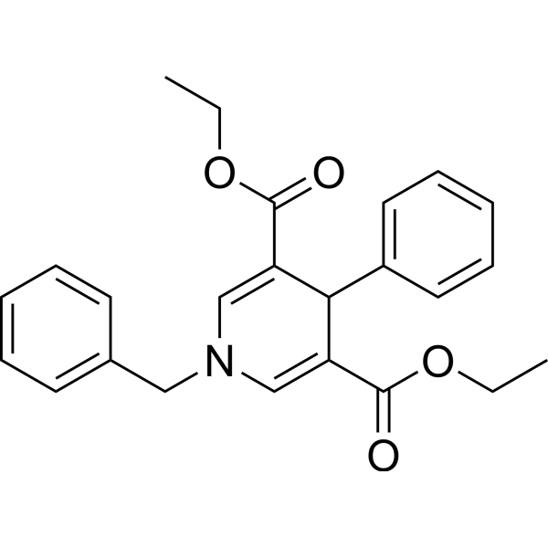
BML-278 is a SIRT1 activator (EC150: 1 μM). BML-278 increases H3K9 methylation and inhibits H3K9 acetylation in both the paternal and maternal pronucleus. BML-278 improves early embryonic development. BML-278 arrests the cell cycle at the G1/S phase, and reduces senescence in primary human mesenchymal cells. BML-278 reduces tubulin acetylation in U937 cells. BML-278 also increases mitochondrial density in murine C2C12 myoblasts.
For research use only. We do not sell to patients.
- Purity : 98.55%
- CAS No.: 120533-76-8
- Formula: C24H25NO4
- Molecular Weight:391.46
-
Storage:Powder -20°C, 3 years , 4°C, 2 years ; In solvent -80°C, 6 months , -20°C, 1 month

To place orders, for customer services and technical support, please contact: MedChemExpress USA
Tel: 609-228-6898 E-mail: [email protected] [email protected]


-
 Biological Activity
-
Chemical Information
-
Solvent & Solubility
- Protocols
- Purity & Documentation
- References
-
 Help & FAQs


-
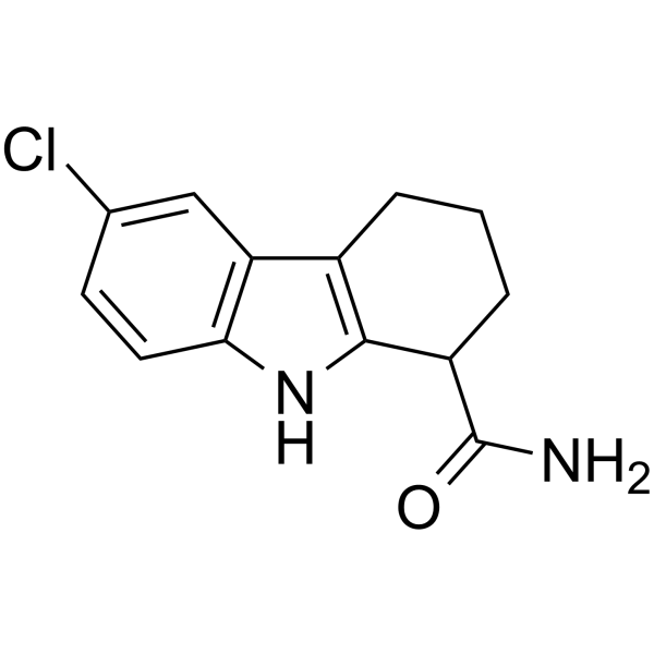
HY-15452
SelisistatSirT1/Sir2 Inhibitor -
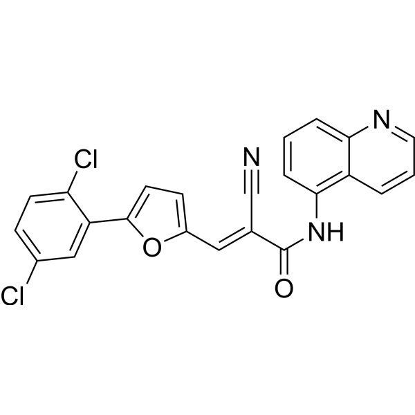
HY-100578
AGK2SIRT2 Inhibitor -
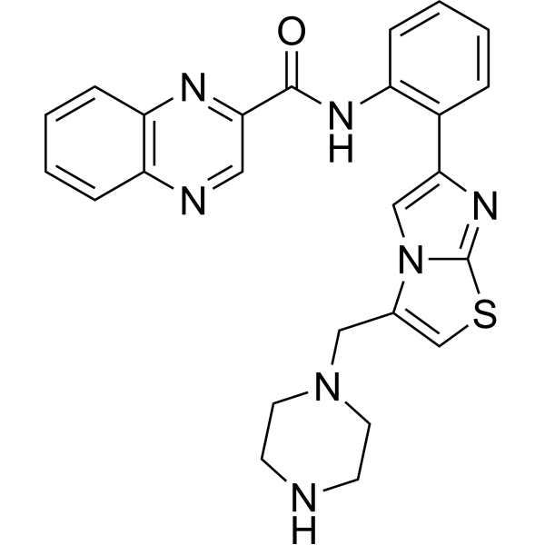
HY-10532
SRT 1720SIRT1 Activator -
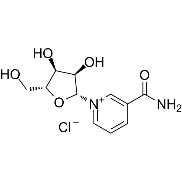
HY-123033A
Nicotinamide riboside chlorideEndogenous Metabolite -
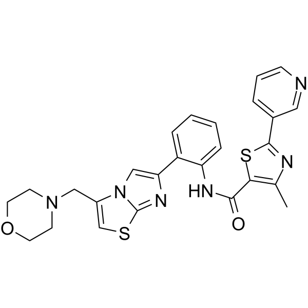
HY-15262
SRT 2104SIRT1 Activator -
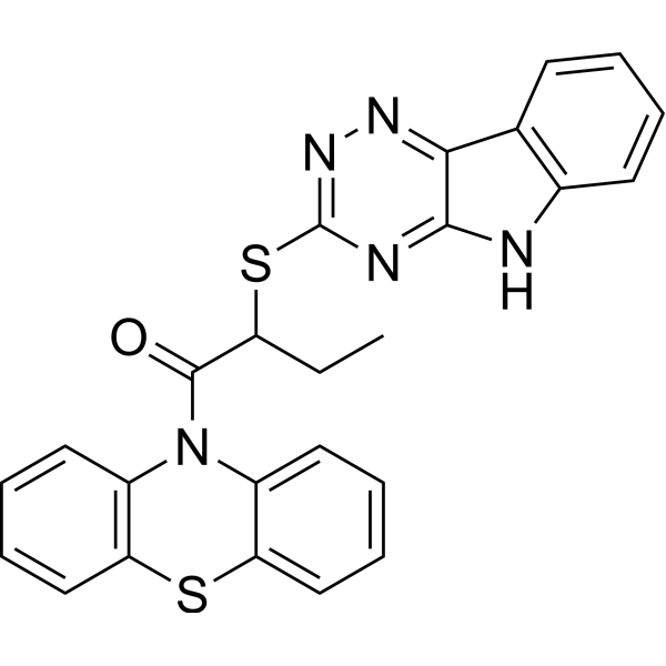
HY-15869
InauhzinSirtuin Inhibitor -
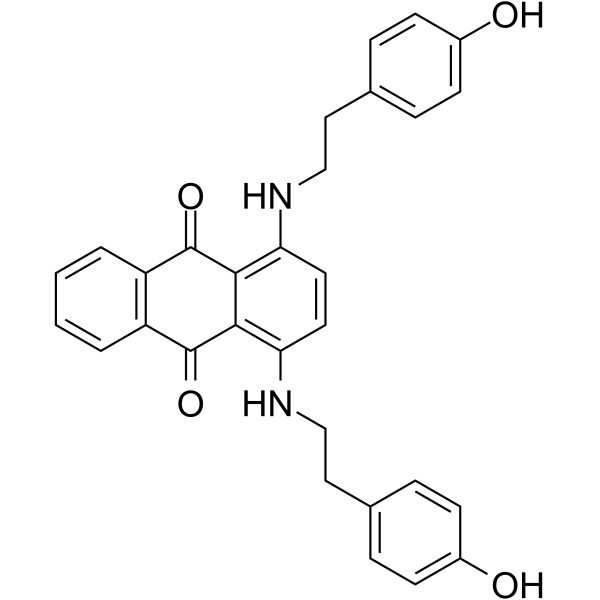
HY-108986
JFD00244SIRT2 Inhibitor -
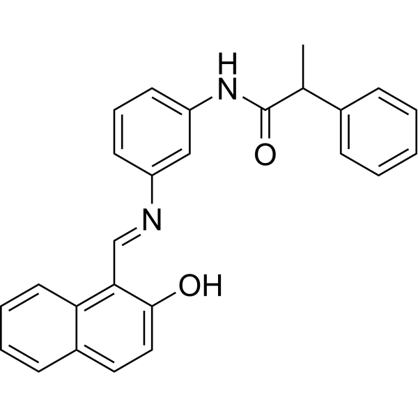
HY-101073
SalermideSirt1/Sirt2 Inhibitor -
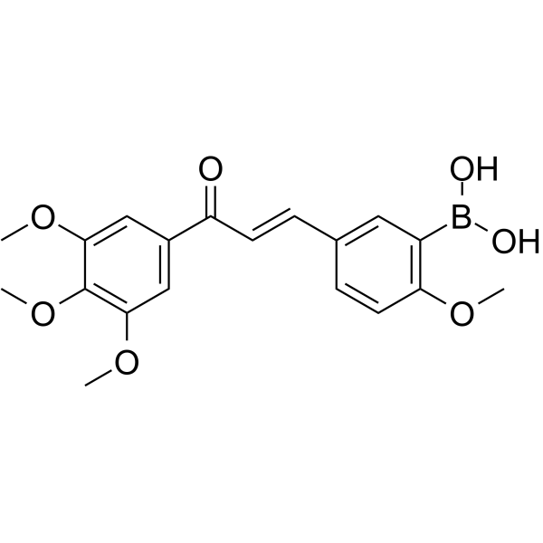
HY-19634
YK-3-237SIRT1 Activator -
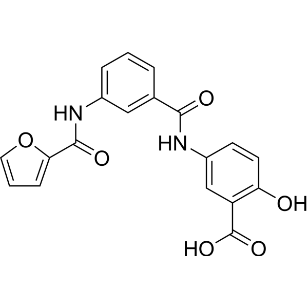
HY-107454
OSS_128167SIRT6 Inhibitor -
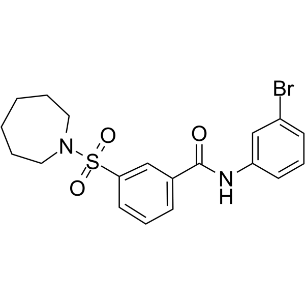
HY-16691
AK-7Sirtuin Inhibitor -
HY-15452A
(S)-SelisistatSIRT1 Inhibitor
All Histone Methyltransferase Isoforms
More

Biological Activity
Description
IC50 & Target
SIRT1[1].


Chemical Information
-
CAS No. 120533-76-8
-
Appearance Solid
-
Molecular Weight 391.46
-
Formula C24H25NO4
-
Color Light yellow to yellow
-
SMILES
CCOC(C1=CN(C=C(C(OCC)=O)C1C2=CC=CC=C2)CC3=CC=CC=C3)=O
-
Shipping
Room temperature in continental US; may vary elsewhere.
-
Storage
Powder -20°C 3 years 4°C 2 years In solvent -80°C 6 months -20°C 1 month


Solvent & Solubility
In Vitro:
DMSO : 10 mg/mL (25.55 mM; ultrasonic and warming and heat to 60°C; Hygroscopic DMSO has a significant impact on the solubility of product, please use newly opened DMSO)
Please refer to the solubility information to select the appropriate solvent. Once prepared, please aliquot and store the solution to prevent product inactivation from repeated freeze-thaw cycles.
Storage method and period of stock solution: -80°C, 6 months; -20°C, 1 month. When stored at -80°C, please use it within 6 months. When stored at -20°C, please use it within 1 month.
Please refer to the solubility information to select the appropriate solvent. Once prepared, please aliquot and store the solution to prevent product inactivation from repeated freeze-thaw cycles.
Storage method and period of stock solution: -80°C, 6 months; -20°C, 1 month. When stored at -80°C, please use it within 6 months. When stored at -20°C, please use it within 1 month.
Concentration (start) × Volume (start) = Concentration (final) × Volume (final)
In Vivo:
Select the appropriate dissolution method based on your experimental animal and administration route.
- For the following dissolution methods, please ensure to first prepare a clear stock solution using an In Vitro approach and then sequentially add co-solvents:
- To ensure reliable experimental results, the clarified stock solution can be appropriately stored based on storage conditions. As for the working solution for In Vivo experiments, it is recommended to prepare freshly and use it on the same day.
- The percentages shown for the solvents indicate their volumetric ratio in the final prepared solution. If precipitation or phase separation occurs during preparation, heat and/or sonication can be used to aid dissolution.
In Vivo Dissolution Calculator
Please enter the basic information of animal experiments:
-
-
-
-
Recommended: Prepare an additional quantity of animals to account for potential losses during experiments.
Please enter your animal formula composition:
-
%DMSO +
Recommended: Keep the proportion of DMSO in working solution below 2% if your animal is weak.
-
%+
-
+%Tween-80 + +
-
%Saline +
The co-solvents required include: DMSO, . All of co-solvents are available by MedChemExpress (MCE). , Tween 80. All of co-solvents are available by MedChemExpress (MCE).
Working solution concentration: 0.22 mg/mL
Method for preparing stock solution: mg drug dissolved in μL DMSO. Stock solution concentration: mg/mL.
1. Take μL DMSO stock solution;
2. Add μL .
μL , mix evenly;
3. Then add μL Tween 80, mix evenly;
4. Then add μL
Please ensure that the stock solution in the first step is dissolved to a clear state, and add co-solvents in sequence. You can use ultrasonic heating (ultrasonic cleaner, recommended frequency 20-40 kHz), vortexing, etc. to assist dissolution.

Protocols
-
EdU Incorporation Assay (Click Chemistry-Based DNA Synthesis Measurement)
The EdU incorporation assay measures DNA synthesis by adding the thymidine analog 5-ethynyl-2′-deoxyuridine to cells or tissues, where it is incorporated into newly synthesized DNA during S phase. Incorporated EdU is detected by copper-catalyzed azide-alkyne cycloaddition, in which a fluorescent azide covalently reacts with the ethynyl group on EdU, allowing S-phase cells to be detected by fluorescence microscopy, flow cytometry, or high-content imaging. EdU detection does not require DNA denaturation or anti-BrdU antibody access, which preserves sample structure and improves compatibility with immunostaining and multiparameter cytometry compared with BrdU-based detection. EdU can be cytotoxic in a cell-type- and exposure-dependent manner, so pulse duration, concentration, and continuous-labeling designs should be validated for each cell type.
-
Flow cytometric DNA-content cell-cycle staining
Flow cytometric DNA-content cell-cycle staining measures the fluorescence intensity of DNA-bound fluorochromes in single cells or nuclei to estimate DNA content distributions, allowing assignment of populations to G0/G1, S, and G2/M phases by DNA histogram deconvolution. Propidium iodide (PI) intercalates into DNA, and PI fluorescence is proportional to cellular DNA content when staining is performed under conditions that make DNA accessible and minimize non-DNA signal. Cells with G2/M DNA content are expected to show approximately twice the fluorescence intensity of G0/G1 cells, while S-phase cells occupy intermediate fluorescence values. PI-based DNA-content analysis can also detect cells with fractional DNA content, often reported as sub-G1, when DNA fragmentation and extraction during staining reduce retained DNA signal in apoptotic cells. DAPI is an alternative DNA fluorochrome for univariate DNA-content analysis, while bivariate approaches combining DNA content with proliferation
-
BrdU Incorporation Assay
Bromodeoxyuridine (BrdU) incorporation assay is based on the principle that BrdU, a thymidine analog, is incorporated into newly synthesized DNA during the S phase of the cell cycle, thereby serving as a marker of DNA replication and cellular proliferation. Incorporated BrdU can be detected using anti-BrdU antibodies following DNA denaturation, enabling visualization or quantification of proliferating cells through immunochemical detection methods such as immunofluorescence or immunohistochemistry.
-
Protocol for Cell Cycle
Cell-cycle analysis by flow cytometry measures DNA content in single cells to estimate the fraction of cells in G0/G1, S, and G2/M phases. Propidium iodide intercalates into DNA, and after RNA removal with RNase, fluorescence intensity reflects cellular DNA content: 2N cells are assigned to G0/G1, cells between 2N and 4N to S phase, and 4N cells to G2/M. DNA-content analysis alone cannot reliably separate G0 from G1 or G2 from M. Ki-67 can distinguish quiescent G0 cells from cycling cells, EdU or BrdU incorporation marks active DNA synthesis in S phase, and phospho-histone H3 staining identifies mitotic cells within the 4N population.
-
Senescence-associated β-galactosidase staining
Senescence-associated β-galactosidase staining detects β-galactosidase activity that is histochemically visible at pH 6. 0 in senescent cells, where X-gal cleavage produces an insoluble blue precipitate observable by bright-field microscopy. This activity reflects increased lysosomal β-galactosidase/lysosomal mass rather than a senescence-essential enzyme, because GLB1 depletion or genetic lysosomal β-galactosidase deficiency can abolish SA-β-gal staining while cells still undergo senescence. SA-β-gal was originally reported in senescent but not presenescent fibroblasts and keratinocytes, absent from quiescent fibroblasts and terminally differentiated keratinocytes, and increased with donor age in human skin samples. Because SA-β-gal can also appear in some non-senescent or tissue-specific contexts, interpretation should be paired with experimental controls and, when possible, independent senescence markers.


Purity & Documentation
-
Data Sheet (275 KB)
-
SDS (396 KB)
- English - EN (396 KB)
- Français - FR (396 KB)
- Deutsch - DE (396 KB)
- Norwegian - NO (396 KB)
- Español - ES (396 KB)
- Swedish - SV (396 KB)
- Italian - IT (396 KB)
- Korean - KR (396 KB)
- Portuguese - PT (396 KB)
-
Handling Instructions (2659 KB)
References
[1]. Adamkova K, et al. SIRT1-dependent modulation of methylation and acetylation of histone H3 on lysine 9 (H3K9) in the zygotic pronuclei improves porcine embryo development. J Anim Sci Biotechnol. 2017 Nov 1;8:83. [Content Brief]
[2]. Mai A, et al. Study of 1,4-dihydropyridine structural scaffold: discovery of novel sirtuin activators and inhibitors. J Med Chem. 2009 Sep 10;52(17):5496-504. [Content Brief]


Complete Stock Solution Preparation Table
Please refer to the solubility information to select the appropriate solvent. Once prepared, please aliquot and store the solution to prevent product inactivation from repeated freeze-thaw cycles.
Storage method and period of stock solution: -80°C, 6 months; -20°C, 1 month. When stored at -80°C, please use it within 6 months. When stored at -20°C, please use it within 1 month.
| Optional Solvent | Concentration Solvent Mass | 1 mg | 5 mg | 10 mg | 25 mg |
|---|---|---|---|---|---|
| DMSO | 1 mM | 2.5545 mL | 12.7727 mL | 25.5454 mL | 63.8635 mL |
| 5 mM | 0.5109 mL | 2.5545 mL | 5.1091 mL | 12.7727 mL | |
| 10 mM | 0.2555 mL | 1.2773 mL | 2.5545 mL | 6.3863 mL | |
| 15 mM | 0.1703 mL | 0.8515 mL | 1.7030 mL | 4.2576 mL | |
| 20 mM | 0.1277 mL | 0.6386 mL | 1.2773 mL | 3.1932 mL | |
| 25 mM | 0.1022 mL | 0.5109 mL | 1.0218 mL | 2.5545 mL |


BML-278 Related Classifications
HY-W025074 Related Classifications
 Powered by Bioz