



Procyanidin C2
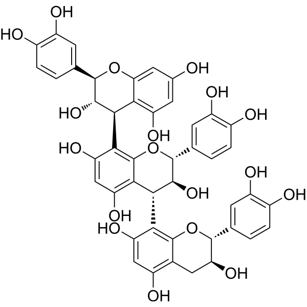
Procyanidin C2 (Procyanidine C2) is a lipid metabolism regulator and antioxidant with free radical scavenging activity. Procyanidin C2 down-regulates ACC, SREBP-1c, FAS, SCD-1 and PPARγ. Procyanidin C2 increases the level of phosphorylated AMPKα and inhibits the level of phosphorylated mTOR. Procyanidin C2 reduces lipid accumulation, alleviates oxidative stress, enhances fatty acid oxidation and improves mitochondrial function. Procyanidin C2 can be used in the research of non-alcoholic fatty liver disease.
For research use only. We do not sell to patients.
- CAS No.: 37064-31-6
- Formula: C45H38O18
- Molecular Weight:866.77
-
Storage:
Please store the product under the recommended conditions in the Certificate of Analysis.

To place orders, for customer services and technical support, please contact: MedChemExpress USA
Tel: 609-228-6898 E-mail: [email protected] [email protected]


-
 Biological Activity
-
Chemical Information
- Protocols
- Purity & Documentation
- References
-
 Help & FAQs


All AMPK Isoforms
More

Biological Activity
Description
In Vitro
Procyanidin C2 (10 μM) reduces lipid accumulation, TG levels, and TC levels in Oleic acid (HY-N1446)-induced HepG2 cells[1].
Procyanidin C2 (10 μM) improves redox balance in oleic acid-induced HepG2 cells by increasing SOD activity, decreasing MDA levels, and reducing intracellular and mitochondrial-derived ROS[1].
Procyanidin C2 (10 μM) improves mitochondrial function in oleic acid-induced HepG2 cells by increasing mitochondrial membrane potential and mass, and restoring mitochondrial ultrastructure[1].
Procyanidin C2 (10 μM) regulates lipid metabolism in oleic acid-induced HepG2 cells by reducing lipogenic protein expression, increasing fatty acid oxidation-related protein expression, enhancing oxidative stress response proteins, and modulating the AMPK/mTOR signaling pathway via increased p-AMPKα, increased p-JNK, and decreased p-mTOR[1].
Procyanidin C2 (fractions derived from 200 μg/mL GSPE stock; 5 min) displays obvious antioxidant activity in a cell-free ABTS radical cation scavenging assay[2].
MedChemExpress (MCE) has not independently confirmed the accuracy of these methods. They are for reference only. Further protocols information, click here.
In Vivo
MedChemExpress (MCE) has not independently confirmed the accuracy of these methods. They are for reference only.


Chemical Information
-
CAS No. 37064-31-6
-
Molecular Weight 866.77
-
Formula C45H38O18
-
SMILES
OC1=C([C@@H](C2=C(O[C@H](C3=CC(O)=C(O)C=C3)[C@@H](O)C4)C4=C(O)C=C2O)[C@H](O)[C@@H](C5=CC(O)=C(O)C=C5)O6)C6=C([C@@H]([C@@H]([C@@H](C7=CC(O)=C(O)C=C7)O8)O)C(C8=CC(O)=C9)=C9O)C(O)=C1
-
Synonyms
Procyanidine C2
-
Structure Classification
-
Initial Source
-
Shipping
Room temperature in continental US; may vary elsewhere.
-
Storage
Please store the product under the recommended conditions in the Certificate of Analysis.

Protocols
-
3T3-L1 preadipocyte-to-adipocyte differentiation
3T3-L1 preadipocytes are induced to differentiate after growth arrest using adipogenic media containing insulin, dexamethasone, and IBMX; differentiation is assessed by lipid-droplet accumulation, triglyceride increase, Oil Red O staining, and adipocyte-marker induction such as PPARγ and C/EBPα.
-
Mitochondrial membrane-potential fluorescent assay
Mitochondrial membrane potential fluorescent assays estimate ΔΨm in living cells using lipophilic cationic dyes such as TMRM, TMRE, rhodamine 123, and JC-1, which accumulate in mitochondria according to membrane polarization; loss of signal after FCCP or CCCP treatment is interpreted as mitochondrial depolarization. TMRM/TMRE and rhodamine 123 are commonly used for semi-quantitative live-cell microscopy or flow cytometry, while JC-1 can report a shift from red aggregate fluorescence to green monomer fluorescence during depolarization; interpretation requires controls because dye concentration, quenching mode, cell type, dye efflux, and mitochondrial mass can affect fluorescence independently of ΔΨm.
-
ROS/oxidative-stress fluorescent staining
ROS/oxidative-stress fluorescent staining uses cell-permeant fluorogenic probes that become fluorescent after oxidation inside cells or tissues; commonly used examples include DCFH-DA/DCFDA for broad cellular oxidant detection, DHE for superoxide-related signal detection, MitoSOX for mitochondrial superoxide-related signal detection, and CellROX probes for oxidative-stress-associated fluorescence readouts. The assay detects probe oxidation rather than a single ROS species unless the probe and analysis method have been chemically validated for that species. DCFH-DA enters cells, is deacetylated by intracellular esterases to DCFH, and produces fluorescent DCF after oxidation, so the readout is used as an operational measure of total cellular oxidative stress rather than a species-specific ROS measurement. DHE and MitoSOX can report superoxide-related oxidation, but red fluorescence alone can include non-specific ethidium-like oxidation products; HPLC or optimized spectral approaches are
-
Mitochondrial membrane-potential and mitochondrial mass staining
Mitochondrial membrane potential staining measures the electrochemical polarization across the mitochondrial inner membrane in live cells using lipophilic cationic fluorescent probes; early rhodamine-based work showed that selective mitochondrial dye accumulation is lost when the mitochondrial transmembrane potential is dissipated. JC-1 reports mitochondrial polarization by shifting from green monomer fluorescence to red J-aggregate fluorescence as dye concentration increases within energized mitochondria; therefore, the red/green fluorescence ratio is used as a relative readout of mitochondrial membrane potential. TMRE or TMRM staining provides a single-channel relative readout because these cationic rhodamine esters accumulate in polarized mitochondria, and lower fluorescence indicates reduced mitochondrial polarization when acquisition and dye-loading conditions are controlled. Mitochondrial mass staining is commonly performed with MitoTracker Green FM or related MitoTracker dyes as
-
Research Protocol for Metabolic Diseases
AMP-activated protein kinase, AMPK, is a conserved cellular energy sensor that responds to reduced cellular energy status and coordinates metabolism by increasing ATP-generating catabolic pathways while suppressing ATP-consuming anabolic processes. In metabolic disease research, the AMPK pathway is experimentally relevant because it regulates hepatic lipid synthesis, fatty acid oxidation, glucose production, skeletal-muscle glucose disposal, mTORC1-linked biosynthesis, autophagy, mitochondrial homeostasis, and whole-body energy balance. The central pathway logic is that energy stress, metformin, exercise-like stimulation, or direct AMPK activators increase AMPKα Thr172 phosphorylation and downstream substrate phosphorylation, including ACC and RAPTOR. Phosphorylation of ACC suppresses lipogenesis and supports fatty acid oxidation, whereas phosphorylation of RAPTOR suppresses mTORC1 signaling and links cellular energy status to growth and protein synthesis control. The pathway is linked
-
Lipid Droplets: Oil Red O/Sudan Dye Lipid Staining
Lipid droplets are intracellular organelles with a neutral-lipid core that stores triacylglycerols and sterol esters, and Oil Red O or Sudan dyes detect these hydrophobic lipid deposits by partitioning into retained lipids in fresh or frozen specimens. Oil Red O stains neutral triglycerides and lipids in frozen tissue sections or air-dried cytologic preparations, while Sudan Black B has also been used as a histochemical fat stain for lipid-rich tissue structures.


Purity & Documentation
References
[1]. Tie F, et al. B-Type Trimeric Procyanidins Attenuate Nonalcoholic Hepatic Steatosis Through AMPK/mTOR Signaling Pathway in Oleic Acid-Induced HepG2 Cells and High-Fat Diet- Fed Zebrafish. Plant Foods Hum Nutr. 2025;80(1):44. Published 2025 Jan 23. [Content Brief]
[2]. Gao J, et al. Bioactivity-based antioxidative components screening and evaluation in grape seed proanthocyanidin extract. J Food Sci Technol. 2017;54(9):2645-2652. [Content Brief]

Calculators
Concentration (start) × Volume (start) = Concentration (final) × Volume (final)


Procyanidin C2 Related Classifications
HY-N7521 Related Classifications
 Powered by Bioz
Keywords