



Funalenone
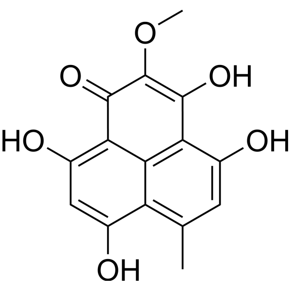
Funalenone (BMS-304245) is a MraY + MurG inhibitor with an IC50 of 25.5 μM in a MraY + MurG membrane plate assay. Funalenone inhibits Staphylococcus aureus (A15090) with an MIC of 64 μg/mL. Funalenone also inhibits MMP-1 with an IC50 of 170 μM.
Nos produits utilisent uniquement pour la recherche. Nous ne vendons pas aux patients.
- CAS No.: 259728-61-5
- Formule: C15H12O6
- Masse moléculaire:288.25
-
Stockage:
Please store the product under the recommended conditions in the Certificate of Analysis.

To place orders, for customer services and technical support, please contact: MedChemExpress USA
Tel: 609-228-6898 E-mail: [email protected] [email protected]


-
 Activité biologique
-
Chemical Information
- Protocole
- Pureté et documentation
- Références
-
 Help & FAQs


-
HY-17605
BictegravirHIV-1 Integrase Inhibitor -
HY-10570S
Nevirapine-d4Deuterium Labeled Nevirapine -
HY-N2532
DiphyllinV-ATPase/HIV-1 Inhibitor -
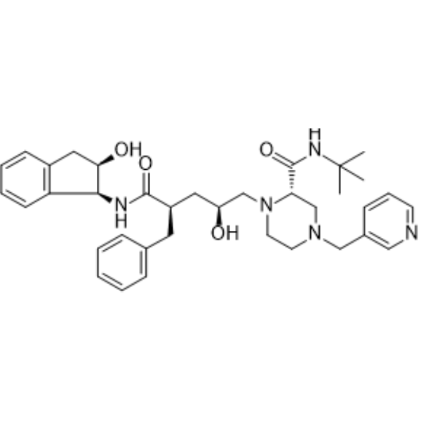
HY-B0689
IndinavirHIV Protease Inhibitor -
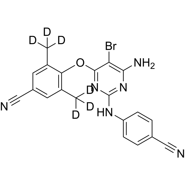
HY-132508S
Etravirine-d8Stable Isotope -
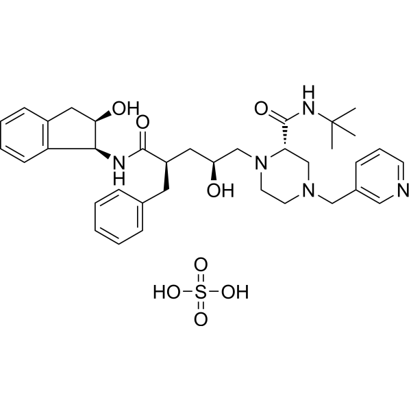
HY-B0689A
Indinavir sulfateHIV Protease Inhibitor -
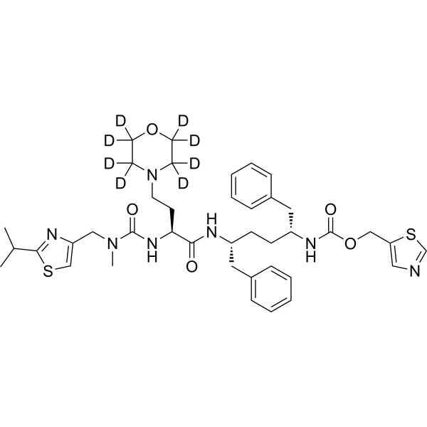
HY-10493S
Cobicistat-d8CYP3A Inhibitor -

HY-17605R
Bictegravir (Standard)HIV-1 Integrase Inhibitor -
HY-W714002
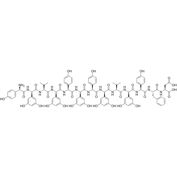
FeglymycinAntibiotic/Antiviral peptide -
HY-N10268
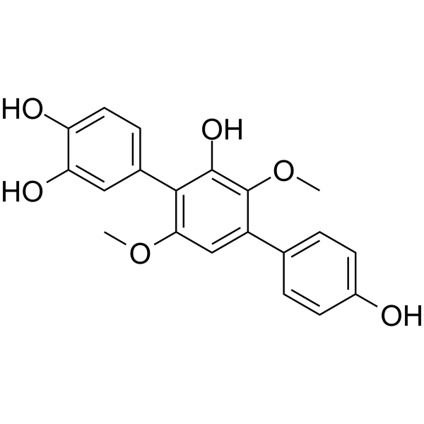
3-HydroxyterphenyllinAnti-cancer Agent


Activité biologique
Description
|
collagenases 1 |
HIV Integrase 10 μM (IC50) |


Chemical Information
-
CAS No. 259728-61-5
-
Masse moléculaire 288.25
-
Formule C15H12O6
-
SMILES
O=C1C(OC)=C(O)C2=C3C1=C(O)C=C(O)C3=C(C)C=C2O
-
Synonyms
BMS-304245
-
Livraison
Room temperature in continental US; may vary elsewhere.
-
Stockage
Please store the product under the recommended conditions in the Certificate of Analysis.


Pureté et documentation
-
Fiche technique (273 KB)
-
SDS (252 KB)
- Français - FR (252 KB)
- Deutsch - DE (252 KB)
- Norwegian - NO (252 KB)
- Español - ES (252 KB)
- Swedish - SV (252 KB)
- Italian - IT (252 KB)
- Korean - KR (252 KB)
- Portuguese - PT (252 KB)
-
Instruction de manipulation (2659 KB)
Références
[1]. J Inokoshi, et al. Funalenone, a novel collagenase inhibitor produced by Aspergillus niger. J Antibiot (Tokyo). 1999 Dec;52(12):1095-100. [Content Brief]
[2]. Zawadzke LE, et al. Targeting the MraY and MurG bacterial enzymes for antimicrobial therapeutic intervention. Anal Biochem. 2003 Mar 15;314(2):243-52. [Content Brief]
[3]. Kazuro Shiomi, et al. Fungal phenalenones inhibit HIV-1 integrase. J Antibiot (Tokyo). 2005 Jan;58(1):65-8. [Content Brief]
[4]. Inokoshi J, et al. Funalenone, a novel collagenase inhibitor produced by Aspergillus niger. J Antibiot (Tokyo). 1999 Dec;52(12):1095-100. [Content Brief]

Calculators
Concentration (start) × Volume (start) = Concentration (final) × Volume (final)


Funalenone Related Classifications
HY-121096 Related Classifications
 Powered by Bioz