



Callistephin chloride
Based on 2 publication(s) in Google Scholar
Callistephin (Pelargonidin 3-O-glucoside) chloride is an anthocyanin. Callistephin chloride regulates the expression of inflammatory (reducing iNOS/TNF-α/COX-2) and apoptosis-related proteins by inhibiting p38 phosphorylation, and enhances the protective effect of Isoflurane (HY-A0134) on microglial cell damage. Callistephin chloride significantly reduces ROS levels, eliminates DPPH free radicals, protects retinal pigment epithelial cells, and inhibits lipid peroxidation. Callistephin chloride can alleviate glutamate excitotoxicity, reduce neuronal apoptosis, and protect cerebellar granule neurons. Callistephin chloride can inhibit the proliferation and metastasis of breast cancer cells by inducing apoptosis.
For research use only. We do not sell to patients.
- Purity : 98.31%
- CAS No.: 18466-51-8
- Formula: C21H21ClO10
- Molecular Weight:468.84
-
Storage:
4°C, sealed storage, away from moisture and light
* In solvent : -80°C, 6 months; -20°C, 1 month (sealed storage, away from moisture and light)

To place orders, for customer services and technical support, please contact: MedChemExpress USA
Tel: 609-228-6898 E-mail: [email protected] [email protected]


-
 Biological Activity
-
Chemical Information
-
Solvent & Solubility
- Protocols
- Purity & Documentation
- References
-
 Help & FAQs


-
HY-W040127
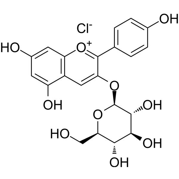
Peonidin 3-O-glucoside chlorideInsulin Secretagogue -
HY-N0640
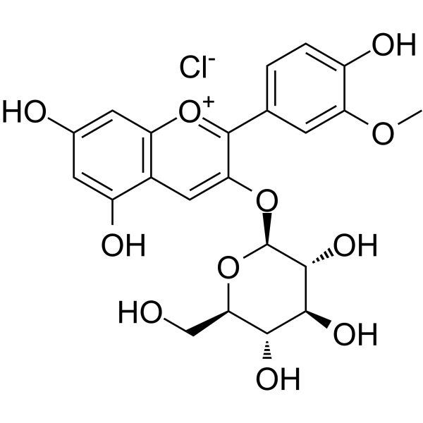
Kuromanin chlorideBlood glucose regulator -
HY-125740
Malvidin-3-glucoside chlorideNF-κB Inhibitor -
HY-N2409
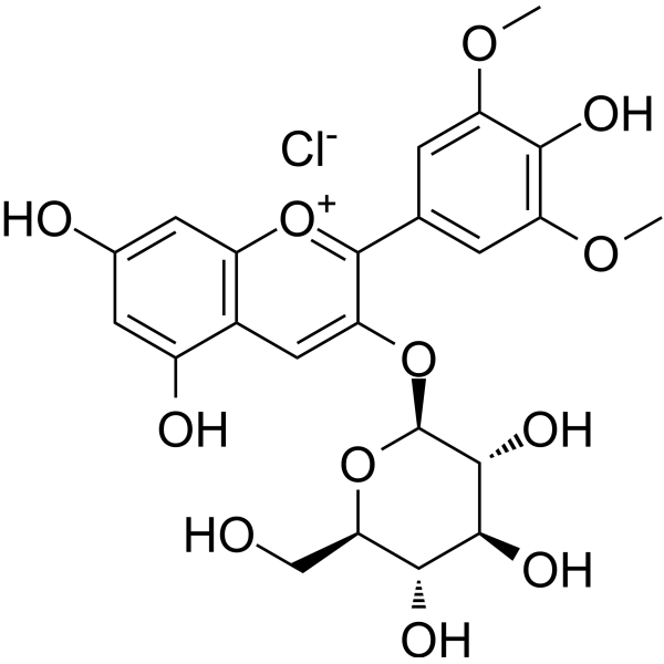
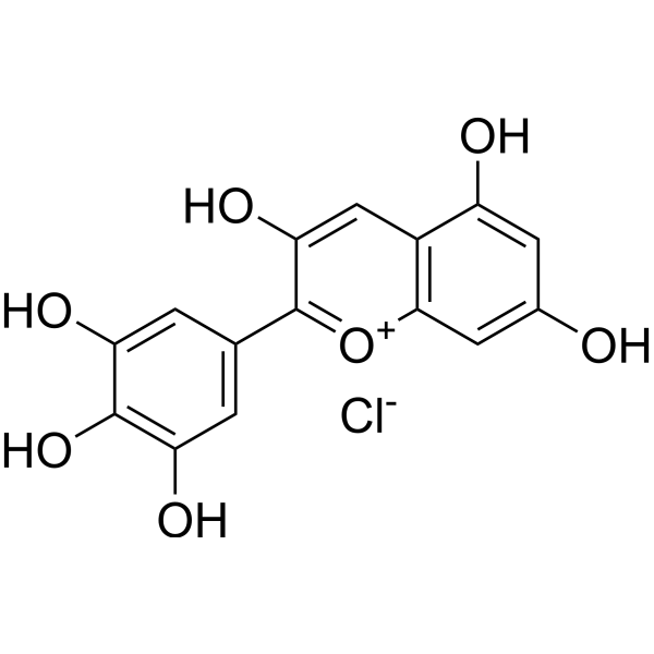
Delphinidin chlorideAnthocyanidin -
HY-129138
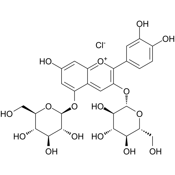
Cyanidin 3,5-diglucoside chlorideAnthocyanin -
HY-N1445
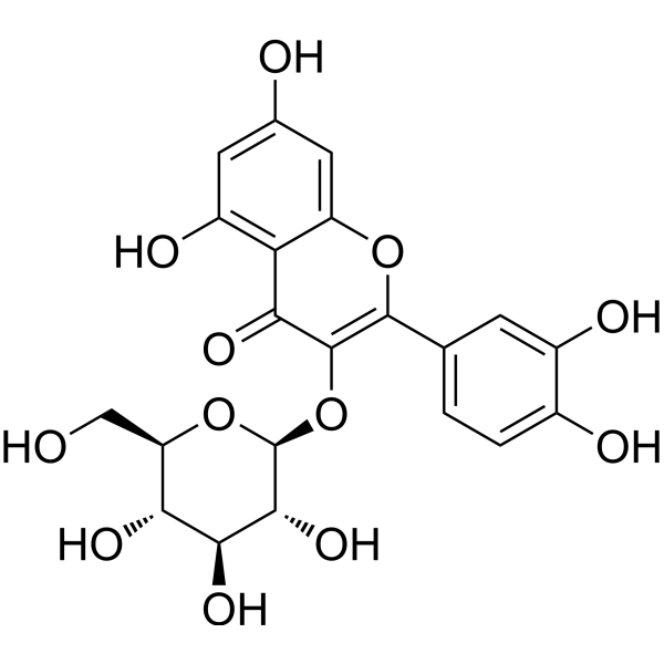
IsoquercitrinNO Synthase Inhibitor -
HY-N8408
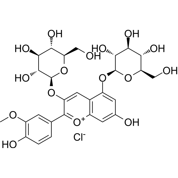
Peonidin 3,5-diglucosideAnthocyanin -
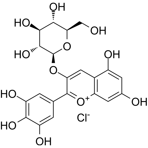
HY-108052
Delphinidin 3-glucoside chlorideEGFR Inhibitor -
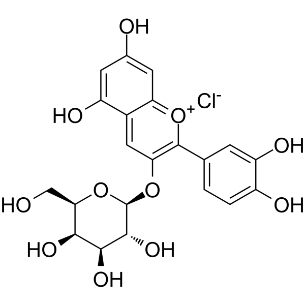
HY-N4142
Cyanidin-3-O-galactoside chlorideAntioxidant Agent -
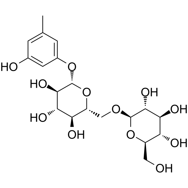
HY-N4123
Orcinol gentiobioside -
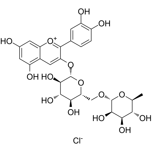
HY-105935
Keracyanin chlorideAntioxidant Agent -
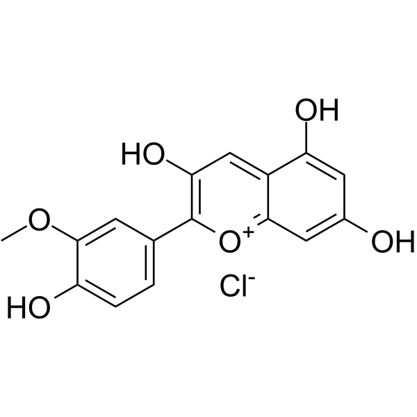
HY-N2459
Peonidin chlorideAnthocyanin Compound
Publications Citing Use of MedChemExpress (MCE) Callistephin chloride
More

Biological Activity
Description
IC50 & Target
[2]|
COX-2 |
p38 |
iNOS |
In Vitro
Callistephin chloride (5-100 μg/mL) significantly alters the morphology of red blood cells, increases the density of the membrane's hydrophilic regions, and has no obvious effect on the hydrophobic regions of the membrane[1].
Callistephin chloride (5-50 μg/mL, 30 min) inhibits erythrocyte membrane lipid oxidation induced with UVC and AAPH radical with IC50s of 17.7 and 5.8 μg/mL[1].
Callistephin (0-400 μM μM, 24 h) chloride enhances the protective effects of Isoflurane by modulating engulfment and apoptosis in C8-B4 cells involved in the modulation of inducible nitric oxide synthase, cytochrome c oxidase subunit 2, tumor necrosis factor α and NF-κB[2].
Callistephin (0.1-1 μg/mL, 30 min) chloride can effectively protect retinal pigment epithelium (RPE) cells by decreasing intracellular ROS and increasing mitochondrial redox activity in blue-light irradiated ARPE-19 cells[3].
Callistephin (100 μM μM, 24 h) chloride significantly protects cerebellar granule neurons (CGNs) from the toxicity of glutamate but has no capacity to defend neurons from nitric oxide (NO)-induced apoptosis[4].
Callistephin-rich extracts (0-200 μg/mL, 24-72 h) chloride inhibits the proliferation of MCF-7 cells, induces cell apoptosis, and exhibits DPPH radical scavenging activity and iron ion reduction ability[5].
MedChemExpress (MCE) has not independently confirmed the accuracy of these methods. They are for reference only. Further protocols information, click here.
-
Cell Line:C8-B4 cells treated with LPS (HY-D1056) and IFN-γ
-
Concentration:100 μM
-
Incubation Time:24 h
-
Result:Reduced the expressions of iNOS, TNF-α and COX-2, but slightly increase NF-κB.
-
Cell Line:C8-B4 cells treated with LPS (HY-D1056) and IFN-γ
-
Concentration:100 μM
-
Incubation Time:24 h
-
Result:Significantly increased the expression of iNOS and COX-2.
Effectively inhibited p38 phosphorylation.
Maintained the level of Bcl-xL and reduced PARP cleavage.
-
Cell Line:cerebellar granule neurons (CGNs)
-
Concentration:100 μM
-
Incubation Time:24 h
-
Result:Reduced CGN apoptosis to 20%.
In Vivo
MedChemExpress (MCE) has not independently confirmed the accuracy of these methods. They are for reference only.
-
Animal Model:Hepatocyte damage assay established in adult male SD rats[4]
-
Dosage:3 mg/kg
-
Administration:Intraperitoneal injection (i.p.), single dose, 2 weeks after Aβ injection
-
Result:Significantly increased the number of NeuroD1-positive cells in the Aβ group.


Chemical Information
-
CAS No. 18466-51-8
-
Appearance Solid
-
Molecular Weight 468.84
-
Formula C21H21ClO10
-
Color Brown to black
-
SMILES
OC1=CC(O)=CC2=C1C=C(O[C@@H]3O[C@H](CO)[C@@H](O)[C@H](O)[C@H]3O)C(C4=CC=C(O)C=C4)=[O+]2.[Cl-]
-
Synonyms
Pelargonidin-3-O-glucoside chloride
-
Structure Classification
-
Initial Source
-
Shipping
Room temperature in continental US; may vary elsewhere.
-
Storage
4°C, sealed storage, away from moisture and light
* In solvent : -80°C, 6 months; -20°C, 1 month (sealed storage, away from moisture and light)


Publications (2)
-
Journal Impact Factor
-
Most Recent
-
Food Chem
Flavonoid-mediated metabolic underpinning quality variation in red bud-sport pear mutants. [Abstract]2025 Oct 15:489:144992. PMID: 40466530 -


Solvent & Solubility
In Vitro:
DMSO : ≥ 100 mg/mL (213.29 mM; Hygroscopic DMSO has a significant impact on the solubility of product, please use newly opened DMSO)
H2O : 50 mg/mL (106.65 mM; Need ultrasonic)
* "≥" means soluble, but saturation unknown.
Please refer to the solubility information to select the appropriate solvent. Once prepared, please aliquot and store the solution to prevent product inactivation from repeated freeze-thaw cycles.
Storage method and period of stock solution: -80°C, 6 months; -20°C, 1 month (sealed storage, away from moisture and light). When stored at -80°C, please use it within 6 months. When stored at -20°C, please use it within 1 month.
* Note: If you choose water as the stock solution, please dilute it to the working solution, then filter and sterilize it with a 0.22 μm filter before use.
Please refer to the solubility information to select the appropriate solvent. Once prepared, please aliquot and store the solution to prevent product inactivation from repeated freeze-thaw cycles.
Storage method and period of stock solution: -80°C, 6 months; -20°C, 1 month (sealed storage, away from moisture and light). When stored at -80°C, please use it within 6 months. When stored at -20°C, please use it within 1 month.
* Note: If you choose water as the stock solution, please dilute it to the working solution, then filter and sterilize it with a 0.22 μm filter before use.
Concentration (start) × Volume (start) = Concentration (final) × Volume (final)
In Vivo:
Select the appropriate dissolution method based on your experimental animal and administration route.
- For the following dissolution methods, please ensure to first prepare a clear stock solution using an In Vitro approach and then sequentially add co-solvents:
- To ensure reliable experimental results, the clarified stock solution can be appropriately stored based on storage conditions. As for the working solution for In Vivo experiments, it is recommended to prepare freshly and use it on the same day.
- The percentages shown for the solvents indicate their volumetric ratio in the final prepared solution. If precipitation or phase separation occurs during preparation, heat and/or sonication can be used to aid dissolution.
Add each solvent one by one: 10% DMSO 40% PEG300 5% Tween-80 45% Saline
Solubility: ≥ 2.5 mg/mL (5.33 mM); Clear solution
This protocol yields a clear solution of ≥ 2.5 mg/mL (saturation unknown).
Taking 1 mL working solution as an example, add 100 μL DMSO stock solution (25.0 mg/mL) to 400 μL PEG300, and mix evenly; then add 50 μL Tween-80 and mix evenly; then add 450 μL Saline to adjust the volume to 1 mL.
Preparation of Saline: Dissolve 0.9 g sodium chloride in ddH₂O and dilute to 100 mL to obtain a clear Saline solution.
Add each solvent one by one: 10% DMSO 90% (20% SBE-β-CD in Saline)
Solubility: ≥ 2.5 mg/mL (5.33 mM); Clear solution
This protocol yields a clear solution of ≥ 2.5 mg/mL (saturation unknown).
Taking 1 mL working solution as an example, add 100 μL DMSO stock solution (25.0 mg/mL) to 900 μL 20% SBE-β-CD in Saline, and mix evenly.
Preparation of 20% SBE-β-CD in Saline (4°C, storage for one week): 2 g SBE-β-CD powder is dissolved in 10 mL Saline, completely dissolve until clear.
In Vivo Dissolution Calculator
Please enter the basic information of animal experiments:
-
-
-
-
Recommended: Prepare an additional quantity of animals to account for potential losses during experiments.
Working solution concentration: 0.22 mg/mL
This product has good water solubility, please refer to the measured solubility data in water/PBS/Saline for details.

Protocols
-
RNA extraction experimental
By lysing cells, releasing RNA, and removing impurities such as proteins and DNA, high-purity RNA products are finally obtained. The commonly used traditional method is the guanidine isothiocyanate/phenol/chloroform method (Trizol), which is suitable for a variety of animal materials including animal tissues, microorganisms, cultured cells, etc., and most plant materials.
-
Western Blot
Western blotting (WB) is a commonly used experimental method in molecular biology, biochemistry, and immunogenetics for identifying and quantifying target proteins. It combines gel electrophoresis with immunoassay, enabling researchers to analyze protein expression, post-translational modifications, and molecular weight.
-
Annexin V plus membrane-impermeant dye apoptosis staining
Annexin V-based apoptosis assays rely on the detection of phosphatidylserine (PS) externalization from the inner leaflet of the plasma membrane to the outer leaflet, an early biochemical hallmark of apoptosis. Fluorescently labeled Annexin V binds PS in a calcium-dependent manner, enabling identification of early apoptotic cells by flow cytometry or fluorescence microscopy. When combined with a membrane-impermeant DNA-binding dye (e. g. , propidium iodide), this approach allows discrimination between viable (Annexin V−/dye−), early apoptotic (Annexin V+/dye−), and late apoptotic or necrotic (Annexin V+/dye+) cell populations by assessing membrane integrity and PS exposure.
-
ROS/oxidative-stress fluorescent staining
ROS/oxidative-stress fluorescent staining uses cell-permeant fluorogenic probes that become fluorescent after oxidation inside cells or tissues; commonly used examples include DCFH-DA/DCFDA for broad cellular oxidant detection, DHE for superoxide-related signal detection, MitoSOX for mitochondrial superoxide-related signal detection, and CellROX probes for oxidative-stress-associated fluorescence readouts. The assay detects probe oxidation rather than a single ROS species unless the probe and analysis method have been chemically validated for that species. DCFH-DA enters cells, is deacetylated by intracellular esterases to DCFH, and produces fluorescent DCF after oxidation, so the readout is used as an operational measure of total cellular oxidative stress rather than a species-specific ROS measurement. DHE and MitoSOX can report superoxide-related oxidation, but red fluorescence alone can include non-specific ethidium-like oxidation products; HPLC or optimized spectral approaches are
-
Breast Cancer Modeling
Breast cancer is a heterogeneous cancer, and it has been distinguished into four subtypes: luminal A, luminal B, HER2-positive and basal-like. Molecular mutations, epigenetic alterations, hormone exposure and immune microenvironment are related to the progression of breast cancer.
-
Cerebellar Granule Neuron Culture
Cerebellar granule neuron culture is a primary neuronal culture method in which postnatal rodent cerebella are dissected, meninges are removed, tissue is enzymatically and mechanically dissociated, and neurons are plated on poly-cation-coated surfaces in medium that supports neuronal attachment, maturation, neurite-network formation, and biochemical or imaging analysis. Cultured CGNs are used to study neuronal development, survival, apoptosis, differentiation, neurotransmitter release, and toxin-sensitive synaptic proteins; mature cultures develop dense neuritic networks, neuronal activity, glutamate release, and neuronal marker expression. A common survival paradigm uses depolarizing extracellular potassium: CGNs maintained in high potassium medium survive and differentiate, whereas switching mature cultures from 25 mM KCl to 5 mM KCl induces apoptotic death that can be used as a readout of activity-dependent neuronal survival.
-
Cell Viability Determination by MTT Colorimetric Assay
The following protocol uses the MTT colorimetric assay as a classic literature-established method for assessing cell viability/metabolic activity in cultured mammalian cells. MTT[3-(4,5-dimethylthiazol-2-yl)-2,5-diphenyltetrazolium bromide] is reduced by metabolically active cells to a colored formazan product; the amount of formazan is quantified spectrophotometrically and provides an indirect measure of metabolically active viable cells. Importantly, MTT reduction reflects cellular oxidoreductase/metabolic activity rather than an absolute direct count of living cells, so changes in cellular metabolism can alter the signal independently of cell number.
-
Kinase activity and phosphorylation assays
Kinase activity assays measure the ability of kinases to transfer phosphate groups from ATP to specific substrates, while phosphorylation assays detect the presence and levels of phosphorylated proteins. Common methods include radiolabeled ATP incorporation (e. g. ,), ADP release detection via bioluminescence (e. g. ,[3]), enzyme-linked immunosorbent assays (ELISA) for phospho-specific epitopes (e. g. ,[6]), and microtiter-based formats for high-throughput screening (e. g. ,[8]). The ADP-Glo assay quantifies kinase activity by measuring ADP produced during phosphorylation using a luciferase-based system. Radiometric assays involve autoradiography or scintillation counting after incorporation of 32P-labeled ATP into substrate proteins. ELISA-based approaches rely on phospho-specific antibodies to detect activated kinases in cell lysates or purified samples.
-
Apoptosis
Apoptosis, also called programmed cell death, is generally characterized by distinct morphological characteristics.
-
TUNEL staining for apoptotic DNA fragmentation
TUNEL staining detects DNA strand breaks by using terminal deoxynucleotidyl transferase to add labeled nucleotides to exposed 3′-OH DNA termini, generating either microscopic staining in fixed cells or tissue sections, or fluorescence/cytometric signal in cell suspensions. TUNEL positivity reflects DNA fragmentation but should not be interpreted alone as definitive apoptosis, because TUNEL can also label necrotic, autolytic, mechanically damaged, or DNA-repair-associated DNA breaks.
-
Apoptosis Solutions
Apoptosis is a regulated, generally non-lytic cell-death pathway that removes unwanted, damaged, infected, or abnormal cells through coordinated morphological changes, caspase activation, DNA fragmentation, and membrane remodeling. The intrinsic apoptosis pathway is controlled mainly by mitochondrial outer membrane permeabilization, BCL-2 family proteins, cytochrome c release, apoptosome formation, caspase-9 activation, and downstream executioner caspase-3/7 activation. The extrinsic apoptosis pathway is initiated by death receptors such as Fas, TNFR, and TRAIL receptors, which recruit adaptor proteins and activate caspase-8 before engaging executioner caspases or mitochondrial amplification through BID cleavage. Apoptosis is linked to many phenotypes, including cancer cell killing, tissue homeostasis, immune regulation, neurodegeneration, infection response, and treatment-induced cytotoxicity; unresolved questions include how apoptosis interacts with necroptosis, pyroptosis, ferroptos
-
Protocol for Kinase activity and phosphorylation assays
Kinase activity assays measure transfer of phosphate from ATP to a protein or peptide substrate, generating phosphorylated substrate, ADP, or incorporated radiolabeled phosphate as the readout; phosphorylation assays measure site-specific phosphorylation in cells or tissues as a proxy for kinase-pathway activation, inhibition, or substrate regulation. Phosphorylation can be detected by phospho-specific Western blot, immunoprecipitation kinase assay, phospho-immunofluorescence, phospho-flow cytometry, luminescent ADP detection, radiolabeled ATP incorporation, or reporter-based pathway assays, and these readouts can be applied to cancer cells, primary neurons, mouse tumors, organoids, inflammatory macrophages, ferroptosis studies, and mitophagy studies when the kinase target is biologically relevant.
-
Ferroptosis Solutions
Ferroptosis is an iron-dependent, non-apoptotic form of regulated cell death characterized by lethal lipid peroxidation and sensitivity to suppression by iron chelators or lipophilic radical-trapping antioxidants. The core pathway links cystine uptake through system Xc−, glutathione availability, GPX4-dependent detoxification of phospholipid hydroperoxides, iron-dependent oxidative reactions, and polyunsaturated-phospholipid metabolism into a cell-death program that is biochemically and morphologically distinct from apoptosis, necrosis, and autophagy. The ferroptosis pathway is experimentally linked to phenotype through chemical and genetic perturbation. Erastin induces ferroptosis by inhibiting cystine uptake through system Xc− and weakening antioxidant defenses, while GPX4 inhibition or depletion causes lipid peroxide accumulation and ferroptotic cancer-cell death. ACSL4 and oxidizable arachidonoyl- or adrenoyl-containing phosphatidylethanolamines shape ferroptosis sensitivity by con
-
Orthotopic Cell-Line Xenograft
Orthotopic cell-line xenograft models involve implantation of human cancer cell lines into the anatomically corresponding organ of immunodeficient mice to reproduce tumor growth within a native microenvironment, enabling more clinically relevant tumor behavior compared with subcutaneous models. These models are widely used because orthotopic placement better recapitulates tumor progression, including invasion and metastatic spread, which are often underrepresented in heterotopic implantation systems. Compared with conventional xenografts, orthotopic implantation is described as more technically complex but provides improved simulation of tumor-microenvironment interactions and metastatic behavior, making it particularly valuable for translational oncology research. Surgical orthotopic implantation approaches have been emphasized as enabling faithful reproduction of clinical cancer features, including metastasis and disease progression patterns that align with the tumor’s organ of origi


Purity & Documentation
-
Data Sheet (288 KB)
-
SDS (396 KB)
- English - EN (396 KB)
- Français - FR (396 KB)
- Deutsch - DE (396 KB)
- Norwegian - NO (396 KB)
- Español - ES (396 KB)
- Swedish - SV (396 KB)
- Italian - IT (396 KB)
- Korean - KR (396 KB)
- Portuguese - PT (396 KB)
-
Handling Instructions (2659 KB)
References
[1]. Bonarska-Kujawa D, et, al. Interaction of selected anthocyanins with erythrocytes and liposome membranes. Cell Mol Biol Lett. 2012 Jun;17(2):289-308. [Content Brief]
[2]. Zhao L, et al. Callistephin enhances the protective effects of isoflurane on microglial injury through downregulation of inflammation and apoptosis. Mol Med Rep. 2019 Jul;20(1):802-812. [Content Brief]
[3]. Yacout SM, Gaillard ER. The Anthocyanins, Oenin and Callistephin, Protect RPE Cells Against Oxidative Stress. Photochem Photobiol. 2017 Mar;93(2):590-599. [Content Brief]
[4]. Winter AN, et al. Chemical basis for the disparate neuroprotective effects of the anthocyanins, callistephin and kuromanin, against nitrosative stress. Free Radic Biol Med. 2017 Feb;103:23-34. [Content Brief]
[5]. Eroglu Ozkan E, et al. Antiproliferative effects of Turkish pomegranate (Punica granatum L.) extracts on MCF-7 human breast cancer cell lines with focus on antioxidant potential and bioactive compounds analyzed by LC-MS/MS. J Food Biochem. 2021 Sep;45(9):e13904. [Content Brief]


Complete Stock Solution Preparation Table
Please refer to the solubility information to select the appropriate solvent. Once prepared, please aliquot and store the solution to prevent product inactivation from repeated freeze-thaw cycles.
Storage method and period of stock solution: -80°C, 6 months; -20°C, 1 month (sealed storage, away from moisture and light). When stored at -80°C, please use it within 6 months. When stored at -20°C, please use it within 1 month.
| Optional Solvent | Concentration Solvent Mass | 1 mg | 5 mg | 10 mg | 25 mg |
|---|---|---|---|---|---|
| H2O / DMSO | 1 mM | 2.1329 mL | 10.6646 mL | 21.3292 mL | 53.3231 mL |
| 5 mM | 0.4266 mL | 2.1329 mL | 4.2658 mL | 10.6646 mL | |
| 10 mM | 0.2133 mL | 1.0665 mL | 2.1329 mL | 5.3323 mL | |
| 15 mM | 0.1422 mL | 0.7110 mL | 1.4219 mL | 3.5549 mL | |
| 20 mM | 0.1066 mL | 0.5332 mL | 1.0665 mL | 2.6662 mL | |
| 25 mM | 0.0853 mL | 0.4266 mL | 0.8532 mL | 2.1329 mL | |
| 30 mM | 0.0711 mL | 0.3555 mL | 0.7110 mL | 1.7774 mL | |
| 40 mM | 0.0533 mL | 0.2666 mL | 0.5332 mL | 1.3331 mL | |
| 50 mM | 0.0427 mL | 0.2133 mL | 0.4266 mL | 1.0665 mL | |
| 60 mM | 0.0355 mL | 0.1777 mL | 0.3555 mL | 0.8887 mL | |
| 80 mM | 0.0267 mL | 0.1333 mL | 0.2666 mL | 0.6665 mL | |
| 100 mM | 0.0213 mL | 0.1066 mL | 0.2133 mL | 0.5332 mL |
* Note: If you choose water as the stock solution, please dilute it to the working solution, then filter and sterilize it with a 0.22 μm filter before use.


Callistephin chloride Related Classifications
HY-W040045 Related Classifications
 Powered by Bioz