



Napabucasin
Based on 29 publication(s) in Google Scholar
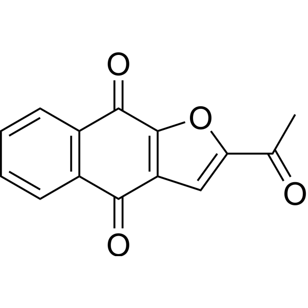
Napabucasin (BBI608; BB608) is an orally active STAT3 inhibitor. Napabucasin inhibits STAT3/RelA signaling and induces cancer cell apoptosis, cell cycle arrest, and ROS/DNA damage, inhibiting cancer cell proliferation, migration, and invasion. Napabucasin, upon activation by NQO1/POR, enhances Docetaxel (HY-B0011) sensitivity and inhibits BCR-ABL1-positive leukemia self-renewal. Napabucasin recruits cytotoxic T/B lymphocytes, enhances immune checkpoint blockade, prevents liver metastasis, and radiosensitizes diffuse midline glioma. Napabucasin can be used in research on cancers such as glioblastoma, pancreatic ductal adenocarcinoma, and prostate cancer.
Nur für Forschungszwecke. Wir verkaufen nicht an Patienten.
- Reinheit : 99.94%
- CAS. Nr.: 83280-65-3
- Formel: C14H8O4
- Molecular Weight:240.21
-
Speicherung:Powder -20°C, 3 years , 4°C, 2 years ; In solvent -80°C, 2 years , -20°C, 1 year

To place orders, for customer services and technical support, please contact: MedChemExpress USA
Tel: 609-228-6898 E-mail: [email protected] [email protected]


-
 Biologische Aktivität
-
Chemical Information
-
Lösungsmittel & Löslichkeit
- Protokoll
- Reinheit & Dokumentation
- Verweise
-
 Help & FAQs


-
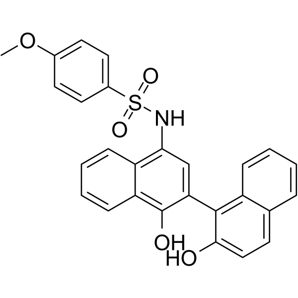
HY-112288
C188-9STAT3 Inhibitor -
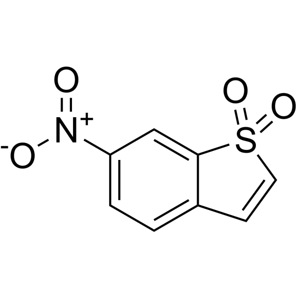
HY-13818
StatticSTAT3 Inhibitor -
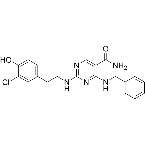
HY-100614
AS1517499STAT6 Inhibitor -
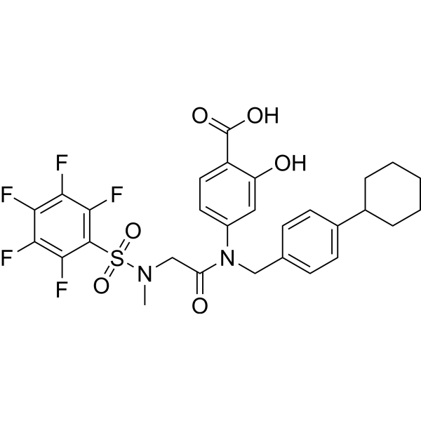
HY-100493
BP-1-102STAT3 Inhibitor -
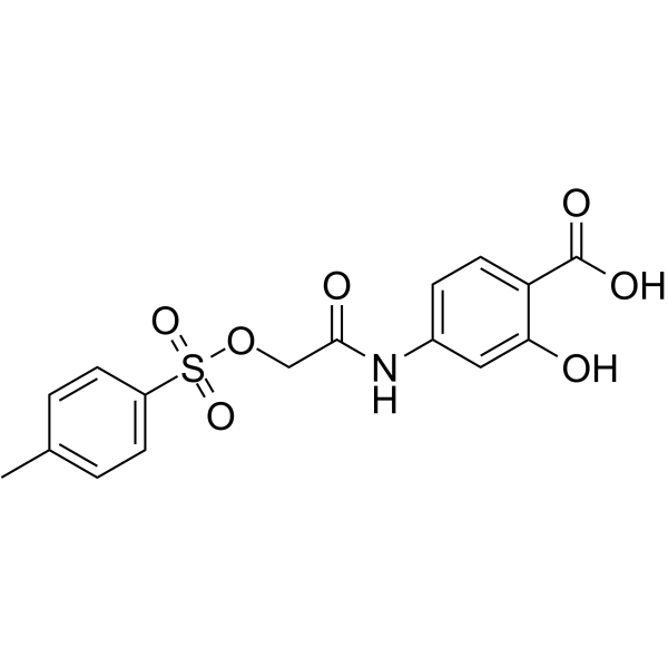
HY-15146
NSC 74859STAT3 Inhibitor -
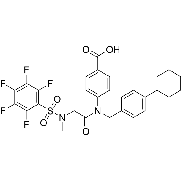
HY-16975
SH-4-54STAT3/5 Inhibitor -
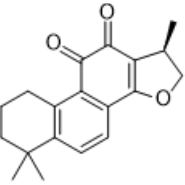
HY-N0174
CryptotanshinoneAutophagy Inducer -
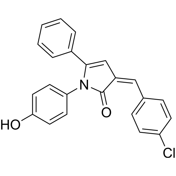
HY-103128
inS3-54A18STAT3 Inhibitor -
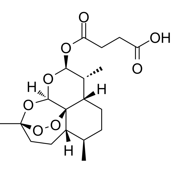
HY-N0193
ArtesunateVirus Protease Inhibitor -
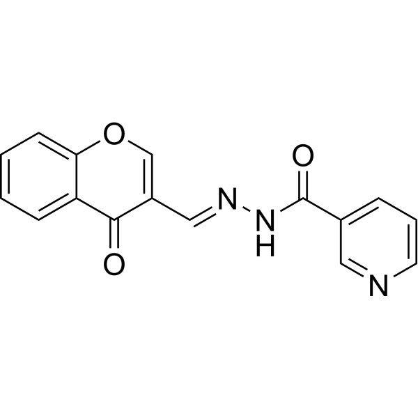
HY-101853
STAT5-IN-1STAT5 Inhibitor -
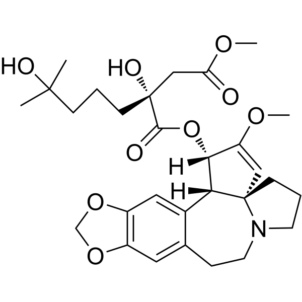
HY-14944
HomoharringtonineSTAT Inhibitor -
HY-50856
RuxolitinibJAK1/2 Inhibitor
Publications Citing Use of MedChemExpress (MCE) Napabucasin
More- Cell. 2024 Apr 25;187(9):2288-2304.e27. [Abstract]
- J Clin Invest. 2022 Oct 17;132(20):e157917. [Abstract]
- J Exp Clin Cancer Res. 2019 Jul 5;38(1):289. [Abstract]
- Cell Death Dis. 2026 Jun 27;17(1):603. [Abstract]
- Cell Death Dis. 2025 Mar 12;16(1):170. [Abstract]
- Phytomedicine. 2023 Jun:114:154769. [Abstract]
- Oncogene. 2023 May;42(20):1649-1660. [Abstract]
- Clin Transl Med. 2022 Oct;12(10):e1036. [Abstract]
- Cell Rep. 2022 Dec 13;41(11):111821. [Abstract]
- J Med Chem. 2020 Sep 10;63(17):9838-9855. [Abstract]
- Sci Signal. 2025 Jun 3;18(889):eadv4579. [Abstract]
- Cells. 2020 Mar 15;9(3):722. [Abstract]
- Cancer Biol Ther. 2026 Dec 31;27(1):2665867. [Abstract]
- Int Immunopharmacol. 2026 Jan 1;168(Pt 1):115811. [Abstract]
- Oncol Rep. 2025 Aug;54(2):97. [Abstract]
- J Biol Chem. 2024 Jun;300(6):107351. [Abstract]
- J Nat Prod. 2022 Apr 22;85(4):765-775. [Abstract]
- Cancer Med. 2016 Jun;5(6):1251-8. [Abstract]
- J Cancer. 2023 Oct 2;14(17):3309-3320. [Abstract]
- J Clin Med. 2019 Nov 2;8(11):1847. [Abstract]
- Med Sci Monit. 2019 Nov 24:25:8905-8912. [Abstract]
- Onco Targets Ther. 2020 Oct 16:13:10567-10580. [Abstract]
- Arch Oral Biol. 2026 Feb:182:106467. [Abstract]
- Acta Cir Bras. 2024 Sep 30:39:e396624. [Abstract]
- Res Sq. 2026 Jul 3.
- bioRxiv. 2026 Mar 26.
- Universidad de Las Palmas de Gran Canaria. 2023 May 24.
- University of Michigan. 2020 Oct.
- Methods Mol Biol. 2019:1866:285-310. [Abstract]
 Customer Validation & Images

-
Cell Proliferation/Viability Assay

-
WB

-
IF

-
Cell Proliferation/Viability Assay

-
Flow Cytometry


Biologische Aktivität
Beschreibung
IC50 & Target
|
STAT3 |
Cellular Effect
|
Cell Line

|
Type | Value | Description | References |
|---|---|---|---|---|
| U-87MG ATCC | IC50 |
6.4 μM
|
Antiproliferative activity against human U87MG GBM cells assessed as viability reduction incubated 24, 48, and 72 hrs by CCK-8 assay.
Antiproliferative activity against human U87MG GBM cells assessed as viability reduction incubated 24, 48, and 72 hrs by CCK-8 assay.
|
31277685 |
| LN-229 | IC50 |
5.6 μM
|
Antiproliferative activity against human LN229 GBM cells assessed as viability reduction incubated 24, 48, and 72 hrs by CCK-8 assay.
Antiproliferative activity against human LN229 GBM cells assessed as viability reduction incubated 24, 48, and 72 hrs by CCK-8 assay.
|
31277685 |
| PC-3 | IC50 |
3.67 μM
|
Inhibition of viability in human PC-3 prostate cancer bulk cells by napabucasin with IC50 determined by cell viability assay.
Inhibition of viability in human PC-3 prostate cancer bulk cells by napabucasin with IC50 determined by cell viability assay.
|
26899963 |
| PC-3 | IC50 |
1.35 μM
|
Inhibition of viability in human PC-3 prostate cancer stem cells by napabucasin with IC50 determined by cell viability assay.
Inhibition of viability in human PC-3 prostate cancer stem cells by napabucasin with IC50 determined by cell viability assay.
|
26899963 |
| A549/CDDP | IC50 |
0.19 μM
|
Antiproliferative activity against human chemoresistant A549/DDP NSCLC cells assessed as viability reduction after 48 hrs by CCK-8 assay.
Antiproliferative activity against human chemoresistant A549/DDP NSCLC cells assessed as viability reduction after 48 hrs by CCK-8 assay.
|
37748583 |
In Vitro
Napabucasin (5 μM; 48 h) reduces EdU incorporation in U87MG and LN229 cells[1].
Napabucasin (5 μM; 10 d) inhibits colony formation in U87MG and LN229 cells[1].
Napabucasin (1 μM; 1 d) attenuates wound healing in U87MG and LN229 cells[1].
Napabucasin (1 μM; 24 h) reduces the invasive ability of U87MG and LN229 cells through Matrigel and decreases MMP2/MMP9 protein levels[1].
Napabucasin (1 μM; 48 h) inhibits the invasive ability of U87MG and LN229 cells in a 3D spheroid environment[1].
Napabucasin (1-10 μM; 48 h) reduces STAT3 protein levels in U87MG and LN229 cells in a dose-dependent manner[1].
Napabucasin (5 μM; 10 d) inhibits the self-renewal and sphere formation of U87MG-GSCs and LN229-GSCs[1].
Napabucasin (5 μM; 10 d) downregulates the mRNA of STAT3 and stemness-related genes in U87MG-GSCs and LN229-GSCs[1].
Napabucasin (5 μM; 10 d) reduces STAT3, β-catenin, and c-Myc protein levels in U87MG-GSCs and LN229-GSCs[1].
Napabucasin (0.5 µM; 6 h) induces NQO1-dependent γH2AX DNA damage in MiaPaCa2 Rosa26 cells, but not in NQO1-71 or NQO1-163 clones[2].
Napabucasin (1 μM; 48-120 h) inhibits the proliferation of PC-3 and 22RV1 prostate cancer cells[3].
Napabucasin (1 μM; 24 h) inhibits colony formation in PC-3 and 22RV1 prostate cancer cells[3].
Napabucasin (18 h) reduces the migration of PC-3 and 22RV1 prostate cancer cells[3].
Napabucasin (1 μM; 48 h) increases the sensitivity of PC-3 to docetaxel, reducing the docetaxel IC50 from 22.269 nM to 9.516 nM[3].
Napabucasin (1 μM; 24 h) reduces stemness-high cancer cells by 10-fold and 7-fold in PC-3 and 22RV1 sphere cultures, respectively[3].
Napabucasin inhibits side population cells of 22RV1 prostate cancer cells[3].
Napabucasin inhibits CD133+/CD44+ prostate cancer stem-like cells in PC-3[3].
Napabucasin inhibits PrCSC sphere formation enriched from PC-3 and 22RV1[3].
Napabucasin shows lower IC50 values in 22RV1 and PC-3 cancer stem cells (1.043 and 1.35 μM) than in bulk cancer cells (1.57 and 3.67 μM)[3].
Napabucasin (0-2 μM; 24 h) dose-dependently downregulates β-catenin, KLF-4, NANOG, Survivin, and C-myc protein expression in PC-3-derived PrCSCs[3].
Napabucasin downregulates the mRNA expression of Nanog, Klf4, survivin, and β-catenin in PC-3 high-stemness cancer cells[3].
Napabucasin (0.17-1.01 μM; 48 h) inhibits the proliferation of NSCLC cell lines, including the chemotherapy-resistant A549/DDP and A549/GEM cells, with IC50 values of 0.19 μM and 0.17 μM, respectively[4].
Napabucasin (0.2-0.4 μM; 2 weeks) inhibits colony formation of NSCLC cells[4].
Napabucasin (0.2 μM; 24 h) partially inhibits the migration of A549 and H1975 cells[4].
Napabucasin (0.2 μM; 48 h) reduces CD44+ cells to 2% in A549/DDP and A549/GEM chemoresistant NSCLC cells and decreases SP cells[4].
Napabucasin (0.2 μM; 48 h) inhibits the expression of p-STAT3, β-catenin, and SOX-2 in A549/DDP cells[4].
Napabucasin (2 µM; 72 h) inhibits secondary colony formation after serial replating of CD34+CD38-CD19+ cells derived from T315I BCR-ABL1 and WT-BCR-ABL1[6].
Napabucasin (72 h) is a potent in vitro cytotoxic agent in DMG cultures, with IC50 values of 0.80, 1.05, 0.84, and 1.44 µM in DIPG36, DIPG6, DIPG17, and SF8628 cells, respectively[7].
Napabucasin (0.2 µM; 14 days) radiosensitizes DIPG36 and DIPG-KAPP cells, with Bliss synergy scores of 16.7 and 17.36, respectively[7].
Napabucasin (5 μM; 48 h) increases cleaved caspase-3 immunofluorescence in U87MG and LN229 cells[1].
Napabucasin (5 μM; 48 h) reduces STAT3 expression in U87MG and LN229 cells[1].
Napabucasin (5 μM; 48 h) decreases total RelA and phosphorylated RelA immunofluorescence in U87MG and LN229 cells[1].
Napabucasin (48 h) increases apoptosis in PC-3 and 22RV1 prostate cancer cells[3].
Napabucasin inhibits STAT3 phosphorylation and Bcl-2 expression in A549 and H1975 cells[4].
Napabucasin (5 μM; 48 h) reduces total RelA and phosphorylated RelA protein levels in U87MG and LN229 cells[1].
Napabucasin (0.2 μM; 48 h) induces apoptosis in A549 and H1975 cells and increases G2/M phase arrest, especially in H1975 cells[4].
Napabucasin induces G1 phase accumulation and reduces S phase in PC-3 and 22RV1 prostate cancer cells[3].
Napabucasin (5 µM; 6 h) induces ROS and cytotoxicity in NQO1-deficient Panc1 cells; POR knockdown attenuates this effect, whereas TXNRD1 knockdown sensitizes the cells[2].
Napabucasin (1 µM; 24 h) combined with 4 Gy RT enhances ROS production in DIPG36 and DIPG-KAPP cells[7].
NQO1 depletion abolishes Napabucasin (1 µM; 24 h)-mediated ROS production in DIPG36 cells[7].
Napabucasin (24-48 h) activates ROS-related transcriptional programs in DIPG6 cells[7].
Napabucasin (1 µM; 6 h) induces SRXN1, HMOX1, and GCLM expression in DIPG36 cells in an NQO1-dependent manner[7].
Napabucasin is a POR substrate with a KM of 2.6 µM[2].
Napabucasin (4 μM) upregulates CCL21 mRNA, protein, and secretion in primary mouse hepatocytes and AML12 mouse hepatocytes, and upregulates CCL21 expression in MIHA human hepatocytes[5].
Napabucasin (4 μM) induces c-Fos in primary hepatocytes and AML12 mouse hepatocytes; c-Fos knockdown inhibits CCL21 mRNA and protein secretion in AML12 cells[5].
MedChemExpress (MCE) has not independently confirmed the accuracy of these methods. They are for reference only.
-
Cell Line:human GBM cell lines U87MG and LN229
-
Concentration:1, 5, 10 μM
-
Incubation Time:24, 48, 72 h
-
Result:Impaired U87MG and LN229 proliferation in a time- and dose-dependent manner.
IC50 values were 6.4 μM in U87MG cells and 5.6 μM in LN229 cells.
-
Cell Line:human GBM cell lines U87MG and LN229
-
Concentration:5 μM
-
Incubation Time:48 h
-
Result:Decreased the fraction of EdU-positive U87MG cells to 12.3% and LN229 cells to 11.7%.
-
Cell Line:human GBM cell lines U87MG and LN229
-
Concentration:5 μM
-
Incubation Time:10 d
-
Result:After Napabucasin exposure, colony-forming U87MG cells were 19.0% and LN229 cells were 14.7%.
-
Cell Line:human GBM cell lines U87MG and LN229
-
Concentration:1 μM
-
Incubation Time:24 h
-
Result:Reduced the percentage of invaded U87MG cells to 26.5% and LN229 cells to 21.5%.
Western blotting showed that MMP2 and MMP9 expression was decreased after Napabucasin exposure.
-
Cell Line:human GBM cell lines U87MG and LN229
-
Concentration:1 μM
-
Incubation Time:48 h
-
Result:Napabucasin-exposed spheroids showed impaired invasive behavior consistent with previous invasion results.
-
Cell Line:human GBM cell lines U87MG and LN229
-
Concentration:5 μM
-
Incubation Time:48 h
-
Result:Increased G0/G1 phase in U87MG cells to 76.7% and reduced S phase to 11.9%; in LN229 cells, G0/G1 increased to 72.3% and S phase decreased to 15.0%.
-
Cell Line:human GBM cell lines U87MG and LN229
-
Concentration:5 μM
-
Incubation Time:48 h
-
Result:Increased the apoptotic ratio in U87MG cells to 35.9% and in LN229 cells to 34.2%.
-
Cell Line:human GBM cell lines U87MG and LN229
-
Concentration:5 μM
-
Incubation Time:48 h
-
Result:Reduced CDK4 and cyclin D1 protein levels and enhanced cleaved caspase-3 and cleaved PARP levels.\nRelative total RelA levels were 0.47 in U87MG cells and 0.43 in LN229 cells; relative phospho-RelA levels were 0.15 in U87MG cells and 0.20 in LN229 cells.
-
Cell Line:human GBM cell lines U87MG and LN229
-
Concentration:5 μM
-
Incubation Time:48 h
-
Result:Cleaved caspase-3 was widely distributed in Napabucasin-exposed U87MG and LN229 cells.\nReduced STAT3 immunofluorescence in both U87MG and LN229 cells.\nDecreased total and phospho-RelA fluorescence intensities in U87MG and LN229 cells; RelA colocalized with STAT3 in both phosphorylated and non-phosphorylated states.
-
Cell Line:human GBM cell lines U87MG and LN229
-
Concentration:1, 5, 10 μM
-
Incubation Time:48 h
-
Result:Relative STAT3 protein levels in U87MG cells were 0.60, 0.20, and 0.13 at 1, 5, and 10 μM, respectively.
In LN229 cells, relative STAT3 levels were 1.08, 0.14, and 0.01 at 1, 5, and 10 μM, respectively.
-
Cell Line:U87MG-GSCs and LN229-GSCs
-
Concentration:5 μM
-
Incubation Time:10 d
-
Result:Decreased mRNA levels in U87MG-GSCs and LN229-GSCs, respectively: STAT3 to 11.7% and 17.4%; β-catenin to 21.4% and 21.5%; c-Myc to 19.4% and 18.6%; SOX2 to 41.2% and 40.5%; OCT4 to 19.8% and 25.0%; Nestin to 35.0% and 33.9%.
-
Cell Line:U87MG-GSCs and LN229-GSCs
-
Concentration:5 μM
-
Incubation Time:10 d
-
Result:Decreased protein expression of STAT3, β-catenin, and c-Myc in both U87MG-GSCs and LN229-GSCs.
-
Cell Line:PC-3 and 22RV1 cells
-
Concentration:1 μM
-
Incubation Time:48, 72, 96, and 120 h
-
Result:Inhibited proliferation of PC-3 and 22RV1 cells at 48, 72, 96, and 120 h compared with control.
-
Cell Line:PC-3 cells
-
Concentration:1 μM
-
Incubation Time:48 h
-
Result:Docetaxel IC50 was 22.269 nM in DMSO control and 9.516 nM in PC-3 cells exposed to napabucasin.
-
Cell Line:PrCSCs isolated from PC-3 cells
-
Concentration:0, 1, 2 μM
-
Incubation Time:24 h
-
Result:Decreased protein expression of β-catenin, KLF-4, NANOG, Survivin, and C-myc in PrCSCs isolated from PC-3 cells in a dose-dependent manner.
-
Cell Line:NSCLC cells
-
Concentration:0.2 μM or 0.4 μM
-
Incubation Time:2 weeks
-
Result:BBI608 significantly inhibited colony formation of NSCLC cells compared with the control group.
-
Cell Line:A549 and H1975 cells
-
Concentration:0.2 μM
-
Incubation Time:24 h
-
Result:BBI608 reduced the migration rate of A549 cells and H1975 cells by 8% and 13%, respectively, compared to the control group.
-
Cell Line:A549 and H1975 cells
-
Concentration:0.2 μM
-
Incubation Time:48 h
-
Result:Significantly induced apoptosis in both A549 and H1975 cells.
Resulted in an increase in the number of cells in the G2/M phase, markedly in H1975 cells, indicating cell cycle arrest.
-
Cell Line:A549/DDP cells
-
Concentration:0.2 μM
-
Incubation Time:48 h
-
Result:The results demonstrated that BBI608 significantly inhibited the expression of p-STAT3, β-catenin and SOX-2 in A549/DDP cells.
-
Cell Line:DIPG36 WT and NQO1-depleted cells
-
Concentration:1 µM
-
Incubation Time:6 h
-
Result:Increased SRXN1, HMOX1, and GCLM expression by 3.4-, 23.7-, and 4.0-fold, respectively.
NQO1 depletion abrogated this transcriptional effect.
In Vivo
Napabucasin (40 mg/kg; intraperitoneal injection; once every 3 days) inhibits the growth of PC-3 prostate cancer xenograft tumors in nude mice and tumor-derived sphere formation[3].
Napabucasin (40 mg/kg; intraperitoneal injection; once every 3 days) inhibits the growth of 22RV1 prostate cancer xenograft tumors in nude mice and tumor-derived sphere formation[3].
Napabucasin (20 mg/kg; oral; daily; administered 7 days before tumor inoculation) inhibits liver metastatic colonization and prolongs survival in C57BL/6J mice in the MC38 splenic liver metastasis model[5].
Napabucasin (20 mg/kg; oral; daily; administered 7 days before tumor inoculation) does not inhibit MC38 lung metastasis in C57BL/6J mice and does not affect subcutaneous MC38 tumor growth or lymphocyte infiltration in C57BL/6J mice[5].
Napabucasin (20 mg/kg; oral; daily; administered 7 days before tumor inoculation or continuously) reduces liver metastasis in the poorly immunogenic CT26/BALB/c model under both the pre-exposure discontinuation and continuous administration regimens[5].
Napabucasin (20 mg/kg; oral; for 28 consecutive days) reduces the CD34+CD38- leukemia cell population and prevents long-term leukemia engraftment in secondary recipients[6].
Napabucasin (10 mg/kg; intraperitoneal injection; daily; for 5 consecutive days) in combination with radiotherapy improves local tumor control in DIPG36 and DIPG-KAPP subcutaneous xenograft models[7].
MedChemExpress (MCE) has not independently confirmed the accuracy of these methods. They are for reference only.
-
Animal Model:nude (female, 6 weeks old)[1]
-
Dosage:40 mg/kg in 200 μL PBS
-
Administration:i.p.; every other day; starting 7 days after tumor cell implantation
-
Result:Nude mice bearing intracranial U87MG xenografts exhibited significantly slower glioma proliferation by bioluminescence imaging.
Had a prolonged survival period (42.5 days) compared with the vehicle group (30.5 days).
Immunohistochemical analyses demonstrated downregulated expression levels of STAT3, MMP2, Ki-67, cyclin D1, β-catenin, and c-Myc.
-
Animal Model:Nude (Charles River Laboratory, stock number 24102242)[2]
-
Dosage:200 mg/kg
-
Administration:p.o. (oral gavage); once daily; for 24 days
-
Result:Rintratumoral GSH:GSSG ratio in MiaPaCa2 Rosa26 xenografts, indicating ROS generation; this reduction was not observed in tumors derived from NQO1-knockout cells.
After 24 days, qPCR analysis of snap-frozen tumor samples from MiaPaCa2 Rosa26 xenografts (n=6) and MiaPaCa2 NQO1-71 xenografts (n=6) showed increased expression of HMOX1 and other NRF2 target genes in-exposed MiaPaCa2 Rosa26 tumors but not in NQO1-71 tumors.
The ROS-induced stress response was observed in MiaPaCa2 Rosa26 xenografts but not in MiaPaCa2 NQO1-knockout xenografts.
-
Animal Model:nude (immunodeficient)[3]
-
Dosage:40 mg/kg
-
Administration:intraperitoneal (i.p.); once every 3 days
-
Result:Significantly reduced xenograft tumor growth and tumor volume compared with PBS.
No differences were observed between the this compound and docetaxel groups in PC-3 mouse xenograft tumor volume.
Significantly reduced tumor weight compared with PBS.
In tumor-derived cells, this compound inhibited spherogenesis, with numbers of spheres significantly decreased compared with PBS; docetaxel had no obvious effect on spherogenesis.
-
Animal Model:nude (immunodeficient)[3]
-
Dosage:40 mg/kg
-
Administration:intraperitoneal (i.p.); once every 3 days
-
Result:Significantly reduced xenograft tumor growth and tumor volume compared with PBS.
In 22RV1 mouse xenografts, tumor volume in the this compound group was lower than in the docetaxel group.
Significantly reduced tumor weight compared with PBS.
In tumor-derived cells, this compound inhibited spherogenesis, with numbers of spheres significantly decreased compared with PBS; docetaxel had no obvious effect on spherogenesis.
-
Animal Model:C57BL/6J (male, 6-week-old)[5]
-
Dosage:20 mg/kg
-
Administration:p.o.; daily; 7 days before tumor inoculation; discontinued thereafter
-
Result:Showed reduced liver metastasis, with lower liver gross weight, fewer metastatic nodules, and lower liver tumor net weight compared with controls.
No effect was observed on the primary tumor at the spleen.
H&E staining showed smaller metastatic areas in this compound-pretreated livers.
CD8+ T-cell infiltration was increased in liver metastases.
this compound-pretreated mice showed extended survival compared with controls.
In ex vivo restimulation, T cells from the MC38 liver micro-metastasis model showed increased polyfunctional CD8+ T cells; triple-positive IFN-γ+TNF-α+IL-2+ CD8+ T cells were 4.6% in control and 10.9% in this compound groups.
Did not show reduced lung metastases.
No differences in lung/body weight or metastatic area were observed between this compound-pretreated and control groups.
Had no effect on subcutaneous tumor growth.
No differences in tumor volume or tumor weight were observed between groups.
Lymphocyte infiltration (CD3+ and CD8+ cells) also showed no difference between this compound-pretreated and control groups.
-
Animal Model:CT26/BALB/c (male, 6-week-old)[5]
-
Dosage:20 mg/kg
-
Administration:p.o.; daily; 7 days before tumor inoculation; continuous dosing
-
Result:Showed reduced liver metastasis compared with controls.
Continuous dosing also reduced metastatic burden.
The pretreated-withdrawal group still showed inhibition of liver metastasis compared with controls.
-
Animal Model:C57BL/6J (male, 6-week-old)[5]
-
Dosage:20 mg/kg
-
Administration:p.o.; daily; 7 days
-
Result:RNA sequencing of liver tissue showed 554 upregulated genes and 176 downregulated genes after this compound exposure.
STAT3-related genes showed minimal changes; immune-related pathways were enriched.
Flow cytometry showed increased CD3+ cells (13.0% control vs 23.1% this compound), CD8+CD3+ cells (4.43% vs 16.9%), and B220+CD3− cells (24.5% vs 42.2%).
GZMB+CD8+ T cells increased from 43.3% to 70.3%, and IFN-γ+CD8+ T cells increased from 11.4% to 33.1%.
Hepatic Ccl21a and Ccr7 expression increased after this compound exposure, while Ccl21a showed no change in spleen, heart, lung, small intestine, or colon.
Immunohistochemistry localized increased CCL21 expression in hepatocytes.
-
Animal Model:NOD/SCID[6]
-
Dosage:20 mg/kg
-
Administration:p.o.; for 28 days
-
Result:BBI608 reduced the population of CD34+CD38- cells.
Secondary recipient mice receiving human CD45+ spleen cells from this compound-exposed donors showed no detectable leukemia engraftment, whereas recipients of vehicle-exposed donor cells engrafted with leukemia.
-
Animal Model:NOD/SCID[6]
-
Dosage:20 mg/kg
-
Administration:p.o.; for 14 days
-
Result:Induced expression of p21Cip1 and cleaved PARP.
Reduced expression of BMI-1, phospho-Stat3, c-Myc, Sox-2, and Bcl-XL.
-
Animal Model:athymic nude (6-week-old)[7]
-
Dosage:10 mg/kg
-
Administration:i.p.; daily; 5 consecutive days
-
Result:Combination RT plus this compound improved local tumor control starting at 8 weeks post-initiation for DIPG36.
-
Animal Model:B6[Cg]-Tyrc-2J/J (6-week-old)[7]
-
Dosage:10 mg/kg
-
Administration:i.p.; daily; 5 consecutive days
-
Result:Combination RT plus this compound improved local tumor control starting at 4 weeks post-initiation for DIPG-KAPP.
- Cell migration
- Somatic Cell Culture
- Detection of Apoptosis
- TUNEL staining for apoptotic DNA fragmentation
- Flow cytometric DNA-content cell-cycle staining
- Apoptosis Solutions
- Cell invasion
- MTT Cell Proliferation Assay
- hPSC maintenance and expansion
- Genotoxicity/Mutagenicity Study
- Annexin V plus membrane-impermeant dye apoptosis staining
- BrdU Incorporation Assay
- ROS/oxidative-stress fluorescent staining
- CFSE Dye Dilution Proliferation Assay
- Dye-dilution cell tracking and proliferation staining
- Protocol for Cell Cycle
- CCK-8/WST-8 Cell Proliferation Assay
- Cell Counting-Based Growth Curve Assay
- Colony Formation (Clonogenic) Assay
- Research Protocol for Cancer Immunology
- Cell Viability Determination by MTT Colorimetric Assay
- EdU Incorporation Assay (Click Chemistry-Based DNA Synthesis Measurement)
- Ki-67 Immunostaining Proliferation Assay
- PCNA Immunodetection Proliferation Assay
- Protocol for Cell Counting and Cell Density Analysis
- Splenic/Portal-Vein Liver Metastasis Xenograft
- Orthotopic Cell-Line Xenograft
Clinical Trial
| NCT Number | Sponsor | Condition | Start Date |
Phase
|
|---|---|---|---|---|
| NCT01329991 | 2011-05 | PHASE1 |


Chemical Information
-
CAS. Nr. 83280-65-3
-
Appearance Solid
-
Molecular Weight 240.21
-
Formel C14H8O4
-
Color Light yellow to green yellow
-
SMILES
O=C(C1=C2OC(C(C)=O)=C1)C3=C(C2=O)C=CC=C3
-
Synonyms
BBI608; BB608
-
Versand
Room temperature in continental US; may vary elsewhere.
-
Speicherung
Powder -20°C 3 years 4°C 2 years In solvent -80°C 2 years -20°C 1 year


Publications (29)
-
Journal Impact Factor
-
Most Recent
-
Cell
Cancer SLC6A6-mediated taurine uptake transactivates immune checkpoint genes and induces exhaustion in CD8+ T cells. [Abstract]2024 Apr 25;187(9):2288-2304.e27. PMID: 38565142 -
J Clin Invest
IL20RB mediates tumoral response to osteoclastic niches and promotes bone metastasis of lung cancer. [Abstract]2022 Oct 17;132(20):e157917. PMID: 36006737 -
J Exp Clin Cancer Res
Napabucasin, a novel STAT3 inhibitor suppresses proliferation, invasion and stemness of glioblastoma cells. [Abstract]2019 Jul 5;38(1):289. PMID: 31277685
Napabucasin purchased from MedChemExpress. Usage Cited in: J Exp Clin Cancer Res. 2019 Jul 5;38(1):289. [Abstract]
Proliferation of glioma cells treated with Napabucasin (0-10 μM, 24-48 h) and detected by CCK-8 assays. Half maximal inhibitory concentrations (IC50) of Napabucasin in U87MG and LN229 cells were 6.4 and 5.6 μM respectively.
Napabucasin purchased from MedChemExpress. Usage Cited in: J Exp Clin Cancer Res. 2019 Jul 5;38(1):289. [Abstract]
After treatment with the indicated doses of Napabucasin (0, 1, 5, and 10 μM) for 48 h, protein levels of STAT3 in U87MG and LN229 cells were evaluated by western blotting.
Napabucasin purchased from MedChemExpress. Usage Cited in: J Exp Clin Cancer Res. 2019 Jul 5;38(1):289. [Abstract]
IF was used to estimate the expression of STAT3 in U87MG and LN229 cells after treatment with Napabucasin (5 μM, 48 h) (scale bar: 50 μm).
Napabucasin purchased from MedChemExpress. Usage Cited in: J Exp Clin Cancer Res. 2019 Jul 5;38(1):289. [Abstract]
EdU incorporation assays showed that exposure to 5 μM Napabucasin for 48 h impaired DNA synthesis in U87MG and LN229 cells.
Napabucasin purchased from MedChemExpress. Usage Cited in: J Exp Clin Cancer Res. 2019 Jul 5;38(1):289. [Abstract]
Napabucasin (5 μM, 48 h) arrests the GBM cell cycle and induces apoptosis. Cell cycle distribution of glioma cells analyzed by flow cytometry.

Napabucasin purchased from MedChemExpress. Usage Cited in: J Exp Clin Cancer Res. 2019 Jul 5;38(1):289. [Abstract]
Napabucasin represses the migration and invasion of GBM cells. Representative images of the wound healing assay showing that Napabucasin treatment (1 μM, 24 h) inhibited glioma cell migration.

Napabucasin purchased from MedChemExpress. Usage Cited in: J Exp Clin Cancer Res. 2019 Jul 5;38(1):289. [Abstract]
Napabucasin impairs the stemness of GBM cells. GSCs from U87MG and LN229 cells were grown in serum-free DMEM/F12 medium with or without Napabucasin (5 μM) for 10 days. Sphere morphology was photographed under a light microscope and spheroids were counted.

Napabucasin purchased from MedChemExpress. Usage Cited in: J Exp Clin Cancer Res. 2019 Jul 5;38(1):289. [Abstract]
Napabucasin (40 mg/kg, intraperitoneal injection, every other day ) suppresses tumor growth in an orthotopic glioma model. Immunohistochemistry was used to assess the expression of STAT3, MMP2, Ki-67, cyclin D1, β-catenin, and c-Myc in orthotopic xenografted tumors after vehicle or Napabucasin treatments.
-
Cell Death Dis
Targeting EZH2-driven cholesterol metabolic vulnerability through Napabucasin suppresses ovarian cancer metastasis. [Abstract]2026 Jun 27;17(1):603. PMID: 42373608 -
Cell Death Dis
Both direct and indirect suppression of MCL1 synergizes with BCLXL inhibition in preclinical models of gastric cancer. [Abstract]2025 Mar 12;16(1):170. PMID: 40075071 -
Phytomedicine
β, β-Dimethylacrylshikonin potentiates paclitaxel activity, suppresses immune evasion and triple negative breast cancer progression via STAT3Y705 phosphorylation inhibition based on network pharmacology and transcriptomics analysis. [Abstract]2023 Jun:114:154769. PMID: 36940580 -
Oncogene
RAF1 contributes to cell proliferation and STAT3 activation in colorectal cancer independently of microsatellite and KRAS status. [Abstract]2023 May;42(20):1649-1660. PMID: 37020037 -
Clin Transl Med
Inhibition of ROCK ameliorates pulmonary fibrosis by suppressing M2 macrophage polarisation through phosphorylation of STAT3. [Abstract]2022 Oct;12(10):e1036. PMID: 36178087 -
Cell Rep
AKTIP loss is enriched in ERα-positive breast cancer for tumorigenesis and confers endocrine resistance. [Abstract]2022 Dec 13;41(11):111821. PMID: 36516775 -
J Med Chem
A Novel Redox Modulator Induces a GPX4-Mediated Cell Death That Is Dependent on Iron and Reactive Oxygen Species. [Abstract]2020 Sep 10;63(17):9838-9855. PMID: 32809827 -
Sci Signal
The E3 ligase TRIM21 promotes progression of pancreatic ductal adenocarcinoma by down-regulating TAp63α and derepressing IL20RB. [Abstract]2025 Jun 3;18(889):eadv4579. PMID: 40460193 -
Cells
STAT3 Inhibitor ODZ10117 Suppresses Glioblastoma Malignancy and Prolongs Survival in a Glioblastoma Xenograft Model. [Abstract]2020 Mar 15;9(3):722. PMID: 32183406 -
Cancer Biol Ther
Inhibition of STAT3-mediated glycolysis by bruceine D suppresses non-small-cell lung cancer progression in vitro and in vivo. [Abstract]2026 Dec 31;27(1):2665867. PMID: 42100892 -
Int Immunopharmacol
Acetylshikonin alleviates pulmonary fibrosis through inhibition of the STAT3 signaling pathway. [Abstract]2026 Jan 1;168(Pt 1):115811. PMID: 41232362 -
Oncol Rep
In vitro assessment of BBI608 in 2D and 3D culture models for drug repositioning in oral squamous cell carcinoma. [Abstract]2025 Aug;54(2):97. PMID: 40539433 -
J Biol Chem
STAT3 activation of SCAP-SREBP-1 signaling upregulates fatty acid synthesis to promote tumor growth. [Abstract]2024 Jun;300(6):107351. PMID: 38718868 -
J Nat Prod
Telocinobufagin Has Antitumor Effects in Non-Small-Cell Lung Cancer by Inhibiting STAT3 Signaling. [Abstract]2022 Apr 22;85(4):765-775. PMID: 35200033 -
Cancer Med
Suppression of prostate cancer progression by cancer cell stemness inhibitor napabucasin. [Abstract]2016 Jun;5(6):1251-8. PMID: 26899963
Napabucasin purchased from MedChemExpress. Usage Cited in: Cancer Med. 2016 Jun;5(6):1251-8. [Abstract]
Representative images of colony formation assay in PCa cell lines (PC-3 and 22RV1) and histological analysis of the colony number show that the colony formation of PCa cell lines are dramatically decreased after treated with 1 μM Napabucasin for 24 h.
-
J Cancer
Cinobufagin exerts an antitumor effect in non-small-cell lung cancer by blocking STAT3 signaling. [Abstract]2023 Oct 2;14(17):3309-3320. PMID: 37928418 -
J Clin Med
Development of Oxadiazole-Based ODZ10117 as a Small-Molecule Inhibitor of STAT3 for Targeted Cancer Therapy. [Abstract]2019 Nov 2;8(11):1847. PMID: 31684051 -
Med Sci Monit
Napabucasin Attenuates Resistance of Breast Cancer Cells to Tamoxifen by Reducing Stem Cell-Like Properties. [Abstract]2019 Nov 24:25:8905-8912. PMID: 31760402 -
Onco Targets Ther
Bone Marrow-Derived Myofibroblasts Promote Gastric Cancer Metastasis by Activating TGF-β1 and IL-6/STAT3 Signalling Loop. [Abstract]2020 Oct 16:13:10567-10580. PMID: 33116635 -
Arch Oral Biol
BBI608 induces apoptosis in mucoepidermoid carcinoma cells by targeting a post-transcriptional regulatory mechanisms of myeloid cell leukemia-1. [Abstract]2026 Feb:182:106467. PMID: 41352329 -
Acta Cir Bras
Effect of napabucasin and doxorubicin via the Jak2/Stat3 signaling pathway in suppressing the proliferation of neuroblastoma cells. [Abstract]2024 Sep 30:39:e396624. PMID: 39356934 -
-
-
-
-
Methods Mol Biol
Homocysteine, Thioretinaco Ozonide, and Oxidative Phosphorylation in Cancer and Aging: A Proposed Clinical Trial Protocol. [Abstract]2019:1866:285-310. PMID: 30725425


Lösungsmittel & Löslichkeit
In Vitro:
N-Methylpyrrolidone (NMP) : 16.67 mg/mL (69.40 mM; Need ultrasonic)
DMSO : 4.44 mg/mL (18.48 mM; Need ultrasonic; Hygroscopic DMSO has a significant impact on the solubility of product, please use newly opened DMSO)
H2O : < 0.1 mg/mL (insoluble)
Ethanol : < 1 mg/mL (insoluble)
Please refer to the solubility information to select the appropriate solvent. Once prepared, please aliquot and store the solution to prevent product inactivation from repeated freeze-thaw cycles.
Storage method and period of stock solution: -80°C, 2 years; -20°C, 1 year. When stored at -80°C, please use it within 2 years. When stored at -20°C, please use it within 1 year.
Please refer to the solubility information to select the appropriate solvent. Once prepared, please aliquot and store the solution to prevent product inactivation from repeated freeze-thaw cycles.
Storage method and period of stock solution: -80°C, 2 years; -20°C, 1 year. When stored at -80°C, please use it within 2 years. When stored at -20°C, please use it within 1 year.
Konzentration (Stammlösung) × Volumen (Stammlösung) = Konzentration (Ziellösung) × Volumen (Ziellösung)
In Vivo:
Select the appropriate dissolution method based on your experimental animal and administration route.
- For the following dissolution methods, please ensure to first prepare a clear stock solution using an In Vitro approach and then sequentially add co-solvents:
- To ensure reliable experimental results, the clarified stock solution can be appropriately stored based on storage conditions. As for the working solution for In Vivo experiments, it is recommended to prepare freshly and use it on the same day.
- The percentages shown for the solvents indicate their volumetric ratio in the final prepared solution. If precipitation or phase separation occurs during preparation, heat and/or sonication can be used to aid dissolution.
Add each solvent one by one: 10% DMSO 40% PEG300 5% Tween-80 45% Saline
Solubility: 0.5 mg/mL (2.08 mM); Suspended solution; Need ultrasonic
This protocol yields a suspended solution of 0.5 mg/mL. Suspended solution can be used for oral and intraperitoneal injection.
Taking 1 mL working solution as an example, add 100 μL DMSO stock solution (5.0 mg/mL) to 400 μL PEG300, and mix evenly; then add 50 μL Tween-80 and mix evenly; then add 450 μL Saline to adjust the volume to 1 mL.
Preparation of Saline: Dissolve 0.9 g sodium chloride in ddH₂O and dilute to 100 mL to obtain a clear Saline solution.
Add each solvent one by one: 10% DMSO 90% (20% SBE-β-CD in Saline)
Solubility: 0.5 mg/mL (2.08 mM); Suspended solution; Need ultrasonic
This protocol yields a suspended solution of 0.5 mg/mL. Suspended solution can be used for oral and intraperitoneal injection.
Taking 1 mL working solution as an example, add 100 μL DMSO stock solution (5.0 mg/mL) to 900 μL 20% SBE-β-CD in Saline, and mix evenly.
Preparation of 20% SBE-β-CD in Saline (4°C, storage for one week): 2 g SBE-β-CD powder is dissolved in 10 mL Saline, completely dissolve until clear.
In Vivo Dissolution Calculator
Please enter the basic information of animal experiments:
-
-
-
-
Recommended: Prepare an additional quantity of animals to account for potential losses during experiments.
Please enter your animal formula composition:
-
%DMSO +
Recommended: Keep the proportion of DMSO in working solution below 2% if your animal is weak.
-
%+
-
+%Tween-80 + +
-
%Saline +
The co-solvents required include: DMSO, . All of co-solvents are available by MedChemExpress (MCE). , Tween 80. All of co-solvents are available by MedChemExpress (MCE).
Working solution concentration: 0.22 mg/mL
Method for preparing stock solution: mg drug dissolved in μL DMSO. Stock solution concentration: mg/mL.
1. Take μL DMSO stock solution;
2. Add μL .
μL , mix evenly;
3. Then add μL Tween 80, mix evenly;
4. Then add μL
Please ensure that the stock solution in the first step is dissolved to a clear state, and add co-solvents in sequence. You can use ultrasonic heating (ultrasonic cleaner, recommended frequency 20-40 kHz), vortexing, etc. to assist dissolution.


Reinheit & Dokumentation
-
Data Sheet (334 KB)
-
SDS (394 KB)
- English - EN (394 KB)
- Français - FR (394 KB)
- Deutsch - DE (394 KB)
- Norwegian - NO (394 KB)
- Español - ES (394 KB)
- Swedish - SV (394 KB)
- Italian - IT (394 KB)
- Korean - KR (394 KB)
- Portuguese - PT (394 KB)
-
Handling Instructions (2659 KB)
Verweise
[1]. Han D, et al. Napabucasin, a novel STAT3 inhibitor suppresses proliferation, invasion and stemness of glioblastoma cells. Journal of experimental & clinical cancer research : CR. 2019 Jul 05;38(1):289. [Content Brief]
[3]. Zhang Y, et al. Suppression of prostate cancer progression by cancer cell stemness inhibitor napabucasin. Cancer medicine. 2016 Jun;5(6):1251-8. [Content Brief]


Complete Stock Solution Preparation Table
Please refer to the solubility information to select the appropriate solvent. Once prepared, please aliquot and store the solution to prevent product inactivation from repeated freeze-thaw cycles.
Storage method and period of stock solution: -80°C, 2 years; -20°C, 1 year. When stored at -80°C, please use it within 2 years. When stored at -20°C, please use it within 1 year.
| Optional Solvent | Concentration Solvent Mass | 1 mg | 5 mg | 10 mg | 25 mg |
|---|---|---|---|---|---|
| DMSO / N-Methylpyrrolidone (NMP) | 1 mM | 4.1630 mL | 20.8151 mL | 41.6302 mL | 104.0756 mL |
| 5 mM | 0.8326 mL | 4.1630 mL | 8.3260 mL | 20.8151 mL | |
| 10 mM | 0.4163 mL | 2.0815 mL | 4.1630 mL | 10.4076 mL | |
| 15 mM | 0.2775 mL | 1.3877 mL | 2.7753 mL | 6.9384 mL | |
| N-Methylpyrrolidone (NMP) | 20 mM | 0.2082 mL | 1.0408 mL | 2.0815 mL | 5.2038 mL |
| 25 mM | 0.1665 mL | 0.8326 mL | 1.6652 mL | 4.1630 mL | |
| 30 mM | 0.1388 mL | 0.6938 mL | 1.3877 mL | 3.4692 mL | |
| 40 mM | 0.1041 mL | 0.5204 mL | 1.0408 mL | 2.6019 mL | |
| 50 mM | 0.0833 mL | 0.4163 mL | 0.8326 mL | 2.0815 mL | |
| 60 mM | 0.0694 mL | 0.3469 mL | 0.6938 mL | 1.7346 mL |


Napabucasin Related Classifications
HY-13919 Related Classifications
 Powered by Bioz